Category: Research and Discovery

  • Scientists discover a powerful new antivenom hidden in rattlesnake blood

    Scientists discover a powerful new antivenom hidden in rattlesnake blood

    University of Maryland researchers have identified a promising new way to treat venomous snakebites by borrowing from snakes’ own natural defenses. Their approach uses toxin-blocking proteins that western diamondback rattlesnakes evolved to protect themselves from venom.

    By combining specific proteins found in rattlesnake blood, the researchers achieved unusually strong protection against venom from several dangerous snake species.

    The study was led by Distinguished University Professor of BiologySean B. Carroll and published in the Proceedings of the National Academy of Sciences. The findings could help researchers develop more powerful antivenoms for deadly snakebites, which remain a major health threat in some regions of the world.

    “This is one of those great stories when nature has already solved a problem we’ve been grappling with for decades,” said Carroll, who also holds the Andrew and Mary Balo and Nicholas and Susan Simon Endowed Chair at UMD.

    The Global Challenge of Snakebites

    Snakebite is considered one of the world’s most neglected tropical diseases. According to the World Health Organization, venomous snakes kill an estimated 80,000 to 140,000 people annually, while hundreds of thousands of survivors are left with permanent disabilities. Many of those affected live in rural areas where effective antivenom can be difficult to obtain.

    Existing antivenoms save lives, but they also have major drawbacks. They are typically produced by exposing large animals to snake venom and then collecting the antibodies those animals generate. Manufacturing these treatments can be costly, their quality and effectiveness can vary, and they may not work equally well against the many different toxins found in venom from different snake species. They can also cause serious immune reactions.

    Those limitations have pushed scientists to search for better options. In this case, the researchers turned to snakes themselves for clues.

    “We’ve known from anecdotes for 100 years that vipers tend to be resistant to their own venom,” Carroll said. “But for a long time, nobody knew what exactly was circulating in their blood that protected them.”

    A Natural Defense Hidden in Rattlesnake Blood

    In 2022, Carroll’s laboratory found part of the answer: a protein called FETUA-3. The researchers discovered that it could block the activity of many metalloproteinase toxins found in western diamondback rattlesnake venom. It could also bind to and inhibit toxins from the venoms of several other rattlesnake species.

    “Here was evolution’s way for snakes to protect themselves from accidental self-envenomation,” he said, which prompted a follow-up question. “Why rely on horse antibodies when nature has packaged an effective antidote right there in the snake?”

    For the new research, co-authors including Elda Sánchez, director of the National Natural Toxins Research Center at Texas A&M University-Kingsville, examined what each FETUA protein contributes to venom resistance.

    The team found that individual proteins could counter certain effects of venom. One might reduce bleeding, for example, while another could interfere with enzyme activity. However, none of the FETUA proteins on its own was able to completely prevent death from a venomous bite.

    Protein Combinations Dramatically Boost Protection

    The results changed when researchers combined several FETUA proteins. These mixtures were far more effective at blocking the harmful effects of venom than the individual proteins alone.

    Finding the best combinations is complicated because snake venom is extraordinarily complex. A single venom can contain around 100 toxin proteins belonging to multiple protein families, and venom composition differs from one snake species to another.

    “The ingredients are there,” Carroll said. “We just have to keep testing various mixtures.”

    In laboratory experiments, optimized combinations of the proteins were about 10 times more potent than the current sheep-derived rattlesnake antivenom. The mixtures completely neutralized the lethal effects of rattlesnake venom and also provided broad protection against venom from multiple viper species, including species separated by millions of years of evolution.

    “The fact that parts of these inhibitors have been perfectly conserved over 50 million years of snake evolution tells you just how real a risk this is for these animals,” Carroll said, noting that how snakes envenomate themselves, whether through mouth tissue during a bite, by eating envenomated prey, through cannibalism or all of the above, isn’t well understood.

    Toward a New Generation of Antivenoms

    The present study concentrated on metalloproteinases, one important family of venom toxins. The researchers are now using the same general strategy to target other toxin families.

    “We’re getting remarkably close to having effective solutions for the three major toxin families in vipers,” Carroll said. “What we’ve learned here, together with research we’re doing now, gives us real confidence that nature-based recombinant [lab-produced] antivenoms are within reach.”

    Carroll expects that the first commercial applications of “nature’s antivenom” could be in veterinary medicine, with treatments for human snakebites potentially following later.

    He envisions future antivenoms that protect against a wider range of venoms while also being safer, less expensive and easier to manufacture on a large scale than many current treatments.

    “We could make train cars-worth of this stuff and help solve a massive global health problem,” Carroll said. “Many of our most important medicines have come from nature. I’m delighted that the components for a better-than-commercial antivenom were in these snakes all along.”

    Along with Carroll, the UMD co-authors included Department of Biology visiting faculty specialists Fiona Ukken and Yetunde Ayinuola.

    The research was funded by the Howard Hughes Medical Institute and the Viper Resource Center (Grant #P40OD01960-22). This article does not necessarily reflect the views of these organizations.


  • Early statin use linked to 15% lower dementia risk

    Early statin use linked to 15% lower dementia risk

    Starting statin treatment soon after a type 2 diabetes diagnosis was associated with a reduced risk of developing dementia, according to research presented at ESC Congress 2026 and published in The Lancet Regional Health – Europe.

    As populations age, the number of people living with dementia is expected to increase substantially. Because there are currently no effective treatments capable of preventing or reversing most forms of dementia, researchers are increasingly focused on identifying factors that could reduce the likelihood of developing the condition.

    According to the Lancet Commission, nearly half of dementia cases could theoretically be prevented by addressing 14 modifiable risk factors. One of those factors involves managing cholesterol beginning in midlife.

    “Detecting and treating high levels of low-density lipoprotein cholesterol (LDL-C) from midlife is one such highlighted risk factor,” explained presenter, Doctor Tummas Ternhamar from Copenhagen University Hospital, Denmark.

    This issue may be especially relevant for people with type 2 diabetes. High LDL-C is common among these patients, while diabetes itself is recognized as a risk factor for cognitive decline and dementia.

    “We hypothesized that early initiation of LDL-C-lowering statins following type 2 diabetes diagnosis could be associated with a lower risk of dementia,” said Doctor Ternhamar. “And we tested this in a large nationwide study using analysis methods to emulate a randomized trial and reduce some of the bias associated with observational studies,” he noted.

    Tracking More Than 132,000 People

    Using Danish national registers, researchers identified 132,585 people who had not previously taken statins and who developed type 2 diabetes between 2006 and 2019. Participants were followed through the end of 2021, and medical records were examined for diagnoses of all-cause dementia.

    The researchers used a clone-censor-weight design to compare dementia rates according to when statin therapy began. One group did not start statins within five years of receiving a diabetes diagnosis. A second group began statins early, within one year of diagnosis, while a third began treatment later, between one and five years after diagnosis.

    During a median follow-up period of 7.1 years, 2.7% of participants developed dementia.

    Earlier Statin Use Linked to Lower Risk

    Compared with people who did not begin statin therapy, those who started statins within the first year after being diagnosed with type 2 diabetes had a 15% lower relative risk of dementia over 10 years.

    Starting treatment later was also associated with a benefit, although the association was smaller. People who began statins between one and five years after diagnosis had a 10% lower relative risk of dementia over 10 years. Similar results were observed in women and men.

    Doctor Ternhamar concluded: “In individuals developing type 2 diabetes, statins were associated with a lower risk of dementia, most pronounced in those with early initiation. Statins seem to be underused in patients with type 2 diabetes, despite their protective effects against cardiovascular disease. Our findings suggest another potentially important reason to ensure that patients with type 2 diabetes and high LDL-C receive timely statin treatment.”

    Findings Cannot Prove Statins Prevent Dementia

    The results suggest that beginning statin therapy sooner after a type 2 diabetes diagnosis may have potential benefits beyond the drugs’ established role in protecting cardiovascular health. However, because the research was observational, it cannot establish that taking statins directly prevents dementia.

    Commenting on the findings, Professor Isabel Goncalves, Member of the ESC Communication Committee, said: “People with type 2 diabetes already have a higher risk of dementia and are advised to lower cholesterol to reduce their risk of heart attack and stroke. This study suggests that starting statins soon after a type 2 diabetes diagnosis may also be linked to a modest reduction in dementia risk, with greater benefit than delaying treatment. Because statins are widely available and inexpensive, the findings are potentially important. However, this was an observational study, so it cannot prove that statins prevent dementia. The results should therefore support further research and help inform discussions between patients and clinicians.”

    This work was supported by grants from the Danish Cardiovascular Academy, which is funded by the Novo Nordisk Foundation and The Danish Heart Foundation, Herlev and Gentofte Hospital, Snedkermester Sophus Jacobsen og hustru Astrid Jacobsens Fond, Beckett fonden, Overlæge Johan Boserup og Lise Boserups legat and Direktør Jakob Madsens og Hustru Olga Madsens fond.

  • Cialis linked to higher glaucoma risk in large study

    Cialis linked to higher glaucoma risk in large study

    A widely used erectile dysfunction medication may be associated with a greater risk of glaucoma when taken long-term to relieve urinary symptoms caused by an enlarged prostate, according to a large comparative study published online in the British Journal of Ophthalmology.

    The findings suggest that men who use tadalafil (Cialis) over extended periods for these symptoms may benefit from closer monitoring of their eye health. Researchers say this may be especially important for people who already have glaucoma risk factors, including a family history of the disease or elevated pressure within the eye (ocular hypertension).

    Tadalafil and Eye Health

    Tadalafil belongs to a group of medications known as phosphodiesterase type 5 inhibitors (PDE5i), the same drug class that includes sildenafil (Viagra). These medications are widely used for erectile dysfunction, while tadalafil is also prescribed for urinary problems associated with an enlarged prostate.

    Previous research has connected PDE5i medications with several types of eye problems, including disorders involving the optic nerve and the macula, which is the area of the retina responsible for detailed central vision.

    However, researchers say the evidence surrounding glaucoma has remained inconsistent. In particular, relatively little is known about glaucoma risk when tadalafil is used for lower urinary tract symptoms, a treatment that commonly involves taking the medication every day over a long period.

    To investigate this question more closely, the researchers examined whether tadalafil, a long-acting PDE5i, was associated with an increased risk of glaucoma when prescribed for urinary tract symptoms related to an enlarged prostate (benign prostatic hyperplasia).

    Nearly 74,000 Men Included in the Study

    The team analyzed anonymized health information from 73,854 men aged 40 or older in the TriNetX Analytics Network (Global Collaborative Network). Half had been prescribed tadalafil or another PDE5i for up to 5 years (between January 2012 and December 2022) to manage their symptoms, while the other half had not received these medications.

    Researchers then compared the two groups to determine how often participants developed glaucoma or ocular hypertension and how frequently they began treatment for glaucoma after 5 years.

    To make the comparison more meaningful, men in the two groups were matched according to a range of factors that could potentially influence the results. These included age, ethnicity, smoking, alcohol related disorders, prescribed medications, and underlying health conditions such as cardiovascular disease, diabetes, cancer, high cholesterol, kidney function, obesity, and chronic obstructive pulmonary disease (COPD).

    Glaucoma Risk Was 22% Higher

    The analysis found that men prescribed tadalafil were significantly (22%) more likely to develop glaucoma than men who had not received the medication.

    They were also more likely to develop several related conditions. Ocular hypertension was 32% more common, while primary open-angle glaucoma, the most common form of glaucoma, was 33% more likely. Men prescribed tadalafil were also 33% more likely to begin glaucoma treatment.

    Additional analyses showed that the associations remained among men who developed glaucoma within 1, 3, or 6 months after beginning tadalafil treatment.

    The same pattern was consistently seen among patients aged 50+, White participants, and people with several of the underlying medical conditions considered in the analysis.

    Study Cannot Prove Cialis Causes Glaucoma

    The researchers caution that the findings do not establish that tadalafil directly causes glaucoma. Because this was an observational study, it can identify associations but cannot determine cause and effect.

    There were also several limitations. For example, the researchers could not account for changes in medication prescriptions or actual medication use over time, nor could they fully track the development of additional underlying health conditions during the study period.

    Most participants were White, which means the findings may not necessarily apply to men from other ethnic backgrounds.

    Even so, the researchers say the results warrant further investigation into how long-term PDE5i use might affect the eyes.

    They write: “These findings suggest that chronic PDE5 inhibition may influence ocular physiology, warranting further mechanistic investigation.”

    Researchers Recommend Eye Monitoring for Higher Risk Patients

    Importantly, the researchers are not advising patients to stop using tadalafil. Instead, they suggest that people taking the medication for extended periods may need appropriate eye screening and follow-up, particularly when other glaucoma risk factors are already present.

    They emphasize: “These findings do not contraindicate tadalafil use but highlight the importance of baseline ophthalmic assessment and regular follow-up, particularly in patients with pre-existing glaucoma, ocular hypertension, a family history of glaucoma, high myopia [short-sightedness], or those requiring long-term therapy.”

  • Common supplements rival antibiotics for severe gum disease

    Common supplements rival antibiotics for severe gum disease

    A combination of omega-3 and low-dose acetylsalicylic acid (ASA) performed about as well as antibiotics in a yearlong study of people with severe periodontitis, raising the possibility of an alternative treatment for some patients.

    The research was conducted by scientists affiliated with Albert Einstein Israelite Hospital, Guarulhos University (UNG), the University of Taubaté (UNITAU), and the Ribeirão Preto School of Dentistry at USP (FORP-USP) in Brazil, along with Harvard University in the United States. The findings were published in the Journal of Periodontology.

    Researchers say the results could eventually help reduce antibiotic use in selected patients, an important consideration as bacterial resistance becomes an increasingly serious global health problem.

    Severe Periodontitis Can Damage Bone and Teeth

    Supported by FAPESP (projects 20/05874-2 and 20/05875-9), the clinical study included 109 people with advanced periodontitis who were followed for one year.

    Periodontitis is a chronic inflammatory disease that develops when bacteria accumulate below the gumline and damage the tissues that hold teeth in place. In advanced cases, the disease can destroy supporting bone, loosen teeth, and eventually cause tooth loss.

    “Patients with severe disease have very deep pockets, which act as reservoirs for bacteria associated with the disease. It’s a very challenging condition to treat,” says dental surgeon Nídia Cristina Castro dos Santos, the first author of the study and a professor and researcher at Einstein.

    Researchers Compared Four Treatment Strategies

    The participants were randomly divided into four groups. Everyone received subgingival instrumentation, popularly known as scaling, which is a standard treatment for periodontitis. The procedure removes bacterial deposits and other material from below the gums.

    What differed among the groups was the additional treatment they received.

    Participants in the control group were given placebo versions of the antibiotics, omega-3, and ASA, packaged in capsules that looked like the active treatments.

    A second group received the antibiotics metronidazole and amoxicillin. This combination has some of the strongest scientific evidence supporting its use in severe periodontitis. Patients took the antibiotics three times per day for 14 days.

    A third group received three grams of omega-3 each day along with a daily dose of aspirin for six months.

    The fourth group received both approaches, taking antibiotics for two weeks while also following the omega-3 and ASA regimen for six months.

    Researchers assessed the participants again after three, six, and twelve months.

    Omega-3 and Aspirin Nearly Matched Antibiotics

    After one year, about 58% of patients treated with antibiotics had reached the clinical target established by the researchers. That goal was defined as having no more than four remaining deep periodontal pockets.

    The omega-3 and ASA group achieved a nearly identical result, with 57.7% of participants reaching the same target.

    The difference compared with standard treatment alone was substantial. Among patients who received scaling plus placebo, only 23.1% met the clinical goal.

    Surprisingly, using antibiotics together with omega-3 and ASA did not produce any additional benefit over either strategy alone.

    “The use of antibiotics isn’t considered the gold standard of treatment. Although metronidazole and amoxicillin yield good results in the most severe cases, their use should be evaluated on a case-by-case basis due to risks related to bacterial resistance and possible adverse effects,” says Castro dos Santos.

    Earlier Research Pointed to Omega-3’s Potential

    The findings build on previous work examining omega-3 and inflammation in periodontal disease.

    In an earlier FAPESP-supported study published in Scientific Reports, researchers found that omega-3 supplementation reduced inflammation and bone loss associated with periodontal disease in rats. The effect was particularly noticeable when supplementation was combined with physical exercise.

    Because that research used an animal model, it could not establish whether the same effects would occur in people. However, it provided evidence that omega-3 might influence inflammatory processes involved in oral disease.

    Helping the Body Resolve Inflammation

    According to Castro, omega-3 works differently from conventional anti-inflammatory medications.

    Rather than simply blocking particular inflammatory pathways, omega-3 can contribute to the production of molecules that help the body naturally bring inflammation to an end and promote tissue repair.

    Low-dose ASA was included because it can enhance the production of these inflammation-resolving molecules. Researchers therefore suspected that combining omega-3 with ASA might help the body gain better control over the persistent inflammation associated with periodontitis.

    “We imagined that the combination of antibiotics and omega-3 would be most effective in controlling periodontitis in these patients. We also believed that the results for the group that used omega-3 with ASA would be slightly lower than those for the antibiotic group. The result was surprising because the two therapies performed very similarly, paving the way for it to become a treatment option for these patients,” says Castro.

    Benefits Continued After Treatment Ended

    The researchers were also struck by how long the apparent benefits lasted.

    Participants who had taken omega-3 and ASA continued to show results similar to those seen at the end of treatment even six months after supplementation had stopped.

    According to Castro, the persistence of the effect raises the possibility that changing the way the body responds to inflammation could produce longer-lasting benefits. Researchers are still investigating exactly how this might happen.

    For dental surgeon Magda Feres, the lead author of the study and a full professor at the Harvard School of Dental Medicine and the Graduate Program in Dentistry at UNG, the results suggest that targeting the inflammatory response could provide another option for certain patients.

    “The combination of omega-3 and low-dose aspirin achieved clinical benefits comparable to those of the metronidazole and amoxicillin protocol, opening up a real alternative for those who can’t take antibiotics, especially patients with allergies or intolerance to these medications,” she says.

    A Possible Way to Reduce Antibiotic Use

    The potential to treat some cases without antibiotics is particularly relevant because antibiotic resistance is considered one of the major threats to global public health.

    When bacteria are repeatedly exposed to antibiotics, strains that can survive the drugs may spread, making infections more difficult to treat. Reducing unnecessary antibiotic use is therefore an important public health goal.

    “Demonstrating that it’s possible to treat severe periodontitis by modulating the body’s own response is new and relevant information that helps preserve antibiotics for when they are truly indispensable,” says Feres.

    More Evidence Is Needed

    Despite the promising results, the researchers stress that omega-3 and aspirin should not yet be viewed as a routine replacement for antibiotics in severe periodontitis.

    The clinical trial included patients who did not have significant systemic or oral diseases. Additional studies will be needed to determine whether the findings hold true in broader and more diverse populations, and to establish which patients are most likely to benefit from each treatment strategy.

    Researchers are also conducting microbiological analyses to understand what happens to bacteria inside periodontal pockets during treatment.

    Preliminary findings suggest that the therapies may reduce bacterial species associated with periodontal disease while increasing microorganisms linked to healthier gums.

    “It’s an important step forward, but not the end of the story. Therefore, the message is one of cautious optimism: we have a promising, scientifically plausible alternative to antibiotics in select cases. Routine replacement will require more evidence,” she concludes.

  • A cancer therapy put severe rheumatoid arthritis into remission

    A cancer therapy put severe rheumatoid arthritis into remission

    Immunotherapies such as CAR T-cell therapy were originally developed mainly to fight cancer. But scientists are now investigating whether these personalized treatments, made from a patient’s own immune cells, could also help treat or even potentially cure autoimmune diseases.

    Researchers at Charité – Universitätsmedizin Berlin have now tested CAR T-cell therapy in six people with particularly severe rheumatoid arthritis. The world’s first clinical trial of its kind, reported in Nature Medicine, produced encouraging early results. Disease activity fell substantially in every participant, and by the end of the observation period, three patients no longer needed rheumatoid arthritis medication.

    Why Rheumatoid Arthritis Can Be So Difficult To Treat

    Rheumatoid arthritis is a chronic autoimmune disease in which the immune system mistakenly attacks the joints. Repeated inflammation causes swelling and can eventually damage the joints.

    Existing medications are often effective at controlling inflammation, but they generally do not cure the disease. As a result, many patients need lifelong treatment with anti-inflammatory drugs and medications that suppress the immune system, which can also cause side effects.

    For some people, even newer therapies do not work well enough. Doctors describe these cases as treatment-refractory rheumatoid arthritis. Patients can continue to experience pain, limited mobility, and major reductions in quality of life despite trying multiple treatments.

    “One reason could be disease-driving B cells – memory cells of the adaptive immune system that may survive in the lymph nodes, bone marrow or joint tissue after an infection, where they produce harmful antibodies directed against the body’s own tissues and repeatedly reignite the inflammation,” explains Prof. David Simon, who designed the trial for this patient group together with Prof. Gerhard Krönke at Charité’s Department of Rheumatology and Clinical Immunology.

    The researchers are testing whether CAR T cells can seek out these disease-driving B cells even when they are buried deep within body tissues. Their goal is to eliminate as much of the abnormal B-cell memory as possible and effectively give the B-cell system a new start.

    Using Cancer Immunotherapy To Reset the Immune System

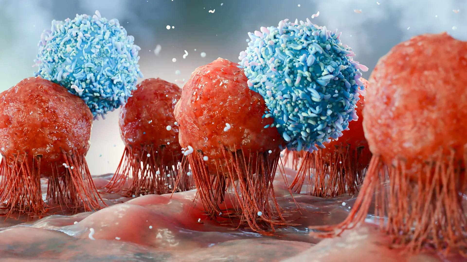
    CAR T cells were first developed as a cancer treatment, but their potential uses are expanding. In cancer therapy, a patient’s immune cells are modified so they can recognize and destroy tumor cells. For autoimmune diseases, scientists instead want to direct those engineered cells toward immune cells that help sustain the disease.

    “The identifying marker on many B cells, both abnormal B cells in cancers of the blood or lymphatic system and disease-driving B cells in rheumatoid arthritis, is the surface molecule CD19. You could think of it as a kind of ‘name tag’,” explains David Simon. “To enable CAR T cells to detect and eliminate the disease-causing cells, we equip patients’ own immune cells with a receptor that acts like a search sensor for CD19.”

    To produce this form of CD19 CAR T-cell therapy, doctors first collect T cells from the patient’s blood. T cells are immune cells that normally help recognize and destroy infected or abnormal cells.

    Scientists then genetically modify those T cells in the laboratory. The cells receive a chimeric, or artificial, antigen receptor known as a CAR, which is designed to bind specifically to CD19.

    Before receiving the modified cells, patients undergo a short course of preparatory chemotherapy. This temporarily lowers the number of certain immune cells, creating room for the CAR T cells to multiply and function effectively.

    The engineered cells are then returned to the patient in a single infusion. Once inside the body, they search for cells carrying CD19 and attack them. This temporarily removes all CD19-positive B cells, including long-lived disease-driving cells in the joints that can otherwise be difficult to reach. By clearing out those cells, the treatment may allow the immune system to reset.

    First Trial in Severe Rheumatoid Arthritis

    For the first clinical trial evaluating both the safety and effectiveness of CD19 CAR T-cell therapy in rheumatoid arthritis, the Charité team initially enrolled six patients with especially severe disease.

    The group included three women and three men between the ages of 31 and 69. During the previous decade, they had received as many as eight targeted or biologic therapies, but none had controlled their disease adequately.

    Researchers wanted to learn whether CAR T cells could reach the disease-driving B cells inside the joints and whether the approach could do so safely.

    The first phase of the COMPARE trial produced results the team considers highly encouraging.

    “Disease activity decreased markedly in all six patients. During follow-up of up to one year, three patients were in sustained remission without any medication for rheumatoid arthritis,” reports Gerhard Krönke, who leads the joint Clinical Rheumatology research group at Charité and the German Rheumatology Research Center (DRFZ), a Leibniz Institute. “This is particularly remarkable given that none of the established treatments had previously been able to relieve their symptoms adequately.”

    CAR T Cells Reached Hidden Disease Reservoirs

    The treatment appeared to do more than temporarily reduce inflammation in the joints. Researchers found that the modified immune cells also reached and eliminated disease-promoting B cells in deeper locations, including the bone marrow, lymph nodes, and joint tissue.

    During regular follow-up visits over the following 12 months, levels of the autoantibodies associated with rheumatoid arthritis dropped sharply.

    David Simon adds: “When the B-cell system later recovered, predominantly naïve B cells that had not yet been shaped by the disease returned. In contrast, the B cells directed against the body’s own tissues that had been present before treatment were no longer detectable in almost all patients, an indication that the treatment may indeed be able to reset the pathological immune memory.”

    Importantly, antibodies generated by earlier vaccinations, including those against chickenpox and tetanus, could still be detected. That suggests protective antibody memory was largely preserved even though the therapy caused a profound temporary depletion of B cells.

    Researchers still need to determine whether the therapy has any longer-term effects on the immune system.

    Promising Results, but the Therapy Remains Experimental

    The trial suggests that a single CAR T-cell treatment can, in some patients, produce a sustained period without symptoms or rheumatoid arthritis medication. This state of disease inactivity is known as remission.

    For carefully selected patients whose rheumatoid arthritis does not respond adequately to available treatments, researchers hope it may eventually be possible to directly reset pathological immune memory and stop the ongoing inflammation rather than continuously suppressing it with medication.

    However, CAR T-cell therapy for autoimmune diseases, including rheumatoid arthritis, is still experimental. Researchers do not yet have long-term experience with the treatment.

    Responses also differed among the six participants. Some did not achieve a complete response, and one patient’s disease returned after an initial period of medication-free remission.

    So far, however, the researchers consider the safety findings encouraging.

    “After the participants received the CD19 CAR T cells, we observed only a temporary, mild-to-moderate cytokine release syndrome (CRS) in all participants, which was readily manageable. There were no severe neurological complications or other serious adverse events, and infections were rare,” explains Dr. Marie Luise Hütter-Krönke, Medical Director of the Hematology Early Clinical Trial Unit at Charité’s Department of Hematology, Oncology and Cancer Immunology.

    A Larger Comparison Is Next

    The second phase of the trial will include ten additional patients. Researchers plan to compare CAR T-cell therapy with an already approved rheumatoid arthritis drug that also targets B cells.

    The comparison should help determine whether CAR T cells produce stronger or longer-lasting effects and whether they truly reset immune memory.

    If these results are confirmed in this phase and in future larger studies, CAR T-cell therapy could eventually offer another option for people with severe rheumatoid arthritis whose lives are significantly affected by the disease and who currently have no adequate treatment.

    About the Study

    The first of the two phases of the COMPARE study was designed to assess the safety and effectiveness of CD19 CAR T-cell therapy in treatment-refractory rheumatoid arthritis.

    Major contributors included researchers from Charité’s Department of Rheumatology and Clinical Immunology and the Department of Hematology, Oncology and Cancer Immunology at Campus Benjamin Franklin, along with scientists from the Cluster of Excellence ImmunoPreCept, the German Rheumatology Research Center (DRFZ), a Leibniz Institute, and the Fraunhofer Institute for Translational Medicine and Pharmacology ITMP.

    The study was initiated and designed at Charité and also received support from Kyverna Therapeutics. The immunotherapy company had no role in designing the study, collecting or analyzing the data, or presenting the results.

  • Long-term melatonin use linked to 90% higher heart failure risk

    Long-term melatonin use linked to 90% higher heart failure risk

    Long-term use of melatonin, a widely used sleep supplement, was linked to a higher risk of heart failure, hospitalization for heart failure, and death from any cause among people with chronic insomnia, according to preliminary research presented at the American Heart Association’s Scientific Sessions 2025.

    The findings do not prove that melatonin itself caused the increased risks. However, they raise new questions about the long-term safety of a supplement that many people view as a harmless or “natural” way to improve sleep.

    Why Melatonin Is So Widely Used

    Melatonin is a hormone made naturally by the pineal gland in the brain. It helps control the body’s sleep and wake cycle, also known as the circadian rhythm. Levels of the hormone typically rise when it gets dark and fall during daylight.

    Synthetic melatonin is chemically identical to the hormone produced by the body. It is commonly used for insomnia (difficulty falling and/or staying asleep) and jet lag.

    Melatonin supplements are available over the counter in many countries, including the U.S. In the U.S., over-the-counter supplements are not regulated, so each brand of supplement can vary in strength, purity, etc.

    For the new study, researchers divided participants according to their documented melatonin use. People with at least one year of melatonin use recorded in their electronic medical records were placed in the “melatonin group.” Those with no record of melatonin use anywhere in their medical records were assigned to the “non-melatonin group.”

    “Melatonin supplements may not be as harmless as commonly assumed. If our study is confirmed, this could affect how doctors counsel patients about sleep aids,” said Ekenedilichukwu Nnadi, M.D., lead author of the study and chief resident in internal medicine at SUNY Downstate/Kings County Primary Care in Brooklyn, New York.

    Researchers Looked for Long-Term Heart Risks

    Melatonin is often promoted as a safe sleep aid, but researchers say there is limited evidence about its cardiovascular safety when taken for extended periods.

    That uncertainty led the team to investigate whether long-term melatonin use might be associated with heart failure among people who already had chronic insomnia.

    Heart failure does not mean that the heart has completely stopped working. It develops when the heart cannot pump enough oxygen-rich blood to meet the body’s needs. According to the American Heart Association’s 2025 Heart Disease and Stroke Statistics, the condition affects about 6.7 million adults in the U.S.

    Researchers used data from the TriNetX Global Research Network, a large international database containing de-identified electronic health records.

    They examined five years of medical records for adults with chronic insomnia whose records showed melatonin use for more than a year. Those participants were matched with other people who also had insomnia but had no documented melatonin use.

    Anyone who had already been diagnosed with heart failure or who had been prescribed other sleep medications was excluded.

    Heart Failure Risk Was About 90% Higher

    The main analysis found a substantial difference between the two groups.

    Among adults with insomnia, people with documented long-term melatonin use (12 months or more) had about a 90% higher chance of developing heart failure during the following five years compared with matched nonusers. Heart failure occurred in 4.6% of the melatonin group compared with 2.7% of the comparison group.

    Researchers then performed another analysis designed to strengthen confidence that participants had actually been using melatonin over an extended period.

    When they limited the analysis to people who had filled at least two melatonin prescriptions at least 90 days apart, the association remained. That group had an 82% higher risk of heart failure. (Melatonin is only available by prescription in the United Kingdom.)

    Hospitalization and Death Were Also Higher

    A secondary analysis found even larger differences in some outcomes.

    People in the melatonin group were nearly 3.5 times as likely to be hospitalized for heart failure as those in the comparison group. The hospitalization rates were 19.0% and 6.6%, respectively.

    Deaths from any cause were also more common among people with documented melatonin use. During the five-year period, 7.8% of participants in the melatonin group died compared with 4.3% of those in the non-melatonin group, making the risk nearly twice as high.

    “Melatonin supplements are widely thought of as a safe and ‘natural’ option to support better sleep, so it was striking to see such consistent and significant increases in serious health outcomes, even after balancing for many other risk factors,” Nnadi said.

    The results also drew concern from sleep researcher Marie-Pierre St-Onge, Ph.D., C.C.S.H., FAHA, who was not involved in the study.

    “I’m surprised that physicians would prescribe melatonin for insomnia and have patients use it for more than 365 days, since melatonin, at least in the U.S., is not indicated for the treatment of insomnia. In the U.S., melatonin can be taken as an over-the-counter supplement and people should be aware that it should not be taken chronically without a proper indication,” said Marie-Pierre St-Onge, Ph.D., C.C.S.H., FAHA, chair of the writing group for the American Heart Association’s 2025 scientific statement, Multidimensional Sleep Health: Definitions and Implications for Cardiometabolic Health.

    St-Onge is a professor of nutritional medicine in the division of general medicine and director of the Center of Excellence for Sleep & Circadian Research in the department of medicine at Columbia University Irving Medical Center in New York City.

    Important Limitations Complicate the Findings

    The study comes with several significant limitations, particularly because melatonin is handled differently from one country to another.

    Some countries, such as the United Kingdom, require a prescription for melatonin, while others, including the United States, allow people to buy it over the counter. Researchers did not have access to participants’ locations because the medical data had been de-identified.

    Melatonin use was identified only when it appeared in electronic medical records. That means people in the U.S. or other countries who bought melatonin over the counter without having it entered into their medical records could have been incorrectly classified as nonusers.

    As a result, the melatonin and nonmelatonin groups may not perfectly reflect who was actually taking the supplement.

    There was another complication involving hospitalizations. The number of heart failure-related hospitalizations was higher than the number of newly diagnosed heart failure cases because hospitals may enter a variety of related diagnostic codes. Those records do not always include a code specifically identifying a new heart failure diagnosis.

    Researchers also did not have information about how severe each participant’s insomnia was or whether participants had other psychiatric disorders.

    Those missing factors are important because people with more severe insomnia, depression, anxiety, or other conditions may be more likely to use melatonin and could independently have a different cardiovascular risk.

    “Worse insomnia, depression/anxiety, or the use of other sleep-enhancing medicines might be linked to both melatonin use and heart risk,” Nnadi said. “Also, while the association we found raises safety concerns about the widely used supplement, our study cannot prove a direct cause-and-effect relationship. This means more research is needed to test melatonin’s safety for the heart.”

    What the Study Included

    • The analysis included 130,828 adults (average age of 55.7 years; 61.4% women) who had been diagnosed with insomnia.
    • The data came from TriNetX, established in 2013, a growing global network of real-world, de-identified patient information used for medical research.
    • Of the participants, 65,414 had been prescribed melatonin at least once and reported using it for at least one year.
    • Researchers also created a comparison group (control group) made up of people who had never been prescribed melatonin. These participants were matched to the melatonin group using 40 factors, including demographic characteristics, existing health conditions, and medications.
    • Anyone who had already been diagnosed with heart failure was excluded, as were people who had been prescribed other kinds of sleeping pills such as benzodiazepines.
    • The melatonin and comparison groups were matched for age, sex, race/ethnicity, heart and nervous system diseases, medications for heart and nervous system diseases, blood pressure, and body mass index.
    • Researchers then examined electronic medical records covering the five years after the groups were matched.
    • For the primary analysis, they searched for medical codes indicating an initial diagnosis of heart failure. Secondary outcomes included codes associated with hospitalization for heart failure or death.
    • The researchers also carried out what is known as a sensitivity analysis, a method used to check whether results remain similar when the study criteria are changed slightly.
    • For this additional test, participants in the melatonin group had to have filled at least two melatonin prescriptions at least 90 days apart. The association with higher heart failure risk remained, providing an additional check on the original findings.
    • Even so, the researchers emphasize that the study was observational. It identified an association between long-term documented melatonin use and serious health outcomes, but it cannot establish that melatonin directly caused those outcomes. Further research will be needed to determine whether long-term melatonin use itself affects cardiovascular health and, if so, why.
  • Common medications may change your gut for years

    Common medications may change your gut for years

    Medications can influence the community of microbes living in the human gut long after a person stops taking them, according to a large study led by researchers at the University of Tartu Institute of Genomics.

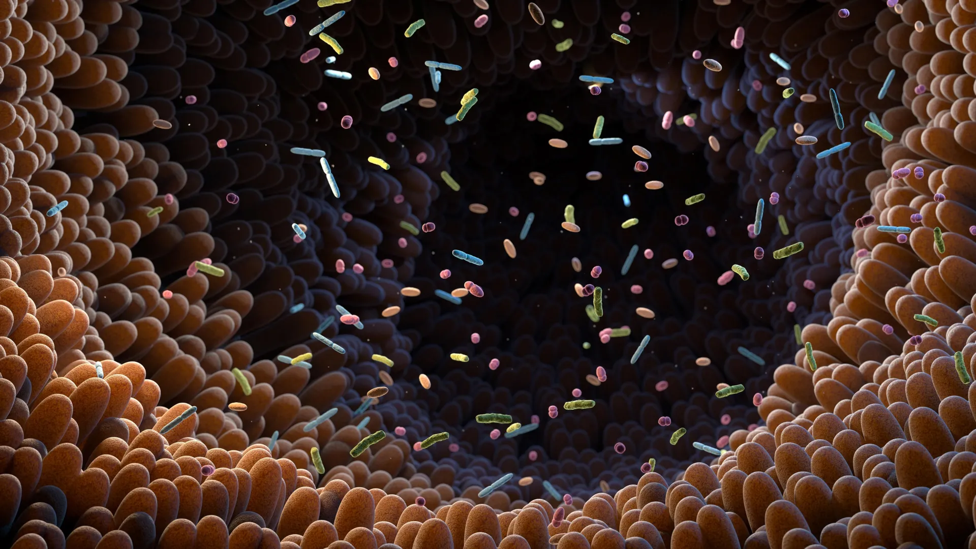
    The findings suggest that a person’s prescription history may help explain differences in the gut microbiome years later. The gut microbiome includes the vast community of bacteria and other microorganisms that live in the digestive tract and can influence digestion, metabolism, immune function, and other aspects of health.

    Drug Effects May Persist for Years

    Researchers analyzed stool samples and prescription records from more than 2,500 participants in the Estonian Biobank who were part of the Estonian Microbiome cohort. They found that most of the medications examined were associated with differences in the gut microbiome.

    For a substantial number of drugs, those differences could still be detected years after people had stopped taking the medication.

    The lasting effects were not limited to antibiotics, which are already well known for their ability to disrupt populations of gut bacteria. Antidepressants, beta-blockers, proton pump inhibitors, and benzodiazepines were also associated with distinctive microbial “fingerprints.”

    Beta-blockers are commonly used to treat conditions such as high blood pressure and certain heart problems. Proton pump inhibitors reduce stomach acid and are often prescribed for acid reflux and related conditions. Benzodiazepines are medications commonly used for anxiety and other disorders.

    “Most microbiome studies only consider current medications, but our results show that past drug use can be just as important as it is a surprisingly strong factor in explaining individual microbiome differences,” said Dr. Oliver Aasmets, lead author.

    The finding suggests that researchers studying connections between the microbiome and disease may need to look beyond the medications a person is currently taking. Drugs used months or even years earlier could still influence the microbial patterns seen in a stool sample.

    Anxiety Drugs Show Surprisingly Strong Effects

    One particularly striking finding involved benzodiazepines, which are commonly prescribed for anxiety. Their associations with the gut microbiome were comparable to those seen with broad-spectrum antibiotics.

    Broad-spectrum antibiotics are designed to act against many different types of bacteria, which is why they can produce substantial changes in the gut microbial community.

    The study also found that medications belonging to the same drug class did not necessarily affect the microbiome in the same way. Drugs that may be prescribed for similar conditions, such as diazepam and alprazolam, differed in how strongly they appeared to disrupt gut microbes.

    That distinction could be important because medications are often grouped together in microbiome research based on their drug class. The new results suggest that individual drugs may need to be considered separately.

    Follow-Up Samples Reveal Predictable Changes

    Researchers also examined follow-up stool samples from a smaller group of participants. These samples allowed them to observe what happened when people started or stopped certain medications.

    Those changes were accompanied by predictable shifts in gut microbes, providing evidence that the medications themselves may be responsible for at least some of the observed differences.

    Although the second time-point analysis involved a relatively small number of participants, researchers were able to confirm persistent effects linked to proton pump inhibitors, selective serotonin reuptake inhibitors and antibiotics, such as penicillins in combination and macrolides.

    Selective serotonin reuptake inhibitors are a widely used class of antidepressants. Macrolides are a group of antibiotics that includes drugs used to treat a range of bacterial infections.

    Medication History Could Matter in Microbiome Research

    The results add to growing evidence that the gut microbiome reflects more than a person’s current diet, lifestyle, health, and medication use. Past treatments may leave biological traces that remain detectable long after the prescription has ended.

    “This is a comprehensive systematic evaluation of long-term medication effects on the microbiome using real-world medical health records,” said Professor Elin Org, corresponding author. “We hope this encourages researchers and clinicians to factor in medication history when interpreting microbiome data.”

    Accounting for that history could help scientists more accurately distinguish microbiome changes associated with disease from changes caused by medications taken in the past.

  • Gut bacteria may unlock a hidden benefit of vegetables

    Gut bacteria may unlock a hidden benefit of vegetables

    Researchers at Karolinska Institutet have found that gut microbes can transform nitrate and iron from food into molecules that may help protect against cardiovascular and metabolic disease. The findings, published in Cell, uncover a previously unknown way the gut microbiota may influence important functions throughout the body.

    Nitrate is found naturally in many vegetables, especially beetroot and leafy greens such as spinach, rocket, and lettuce. Non-haem iron — the form of iron found in plant-based foods such as beans, whole grains and green vegetables — is also a common part of the diet.

    The researchers found that gut bacteria can combine nitrate and non-haem iron from food to produce compounds known as dinitrosyl iron complexes (DNICs). After they are formed, these molecules can be absorbed into the body and carried to different organs, particularly the liver and kidneys.

    Gut Microbes Are Essential for DNIC Formation

    The study drew on experiments involving mice, cells, bacteria and human samples. Using advanced analytical techniques, the researchers detected DNIC in several types of tissue.

    They also found that DNIC molecules were completely absent in germ-free mice. That result suggests that gut microbes play an essential role in producing them.

    “Our results show that gut bacteria can convert components in food into biologically active molecules that influence important bodily functions,” says Andrei L. Kleschyov, Senior Researcher at the Department of Physiology and Pharmacology, Karolinska Institutet, the study’s first and co-corresponding author.

    Higher DNIC Levels Improved Key Health Markers

    The researchers then tested what happened when DNIC levels were increased. They did this either by giving dietary supplements containing nitrate and iron or by administering synthetically produced DNIC.

    In an animal model of cardiovascular and metabolic disease, higher DNIC levels were associated with improvements in several measures of health.

    “Among other things, we observed lower blood pressure and improved vascular function, better blood sugar control and reduced fat accumulation in the liver. The results help to explain why a diet rich in vegetables, which contain both nitrate and iron, is linked to a lower risk of several diseases,” says Mattias Carlström, Professor of Cardiorenal Physiology at the Department of Physiology and Pharmacology, Karolinska Institutet, one of the study’s shared last authors together with Professor Jon Lundberg at the same department, and co-corresponding author.

    A Possible Link Between Vegetables and Disease Protection

    According to the researchers, the results reveal a previously unknown mechanism through which diet and specific gut bacteria may work together to support health.

    The findings could also help explain why diets rich in vegetables are associated with a lower risk of cardiovascular and metabolic diseases. However, the researchers caution that much of the work was carried out in experimental models, so additional research will be necessary to determine exactly how the process operates in humans.

    Researchers Now Want to Study DNIC in Humans

    The next goal is to develop reliable ways to measure DNIC levels in people. Researchers also want to learn more about how the molecules are produced, how they move through the body, and how they influence different physiological processes.

    Another major question is whether diet or changes to the gut microbiota could be used to alter DNIC levels and potentially help prevent disease.

    The study was carried out in collaboration with the University Medical Centre Hamburg-Eppendorf and the Johannes Gutenberg University Medical Centre Mainz in Germany. The research was funded by, amongst others, the Swedish Research Council, the Swedish Heart-Lung Foundation, the Novo Nordisk Foundation, the European Research Council (ERC), the Knut and Alice Wallenberg Foundation, Diabetes Wellness Sweden and the Karolinska Institute. The researchers state that they have no conflicts of interest.

  • This yeast supplement may boost cancer-fighting immunity

    This yeast supplement may boost cancer-fighting immunity

    Researchers at Trinity College Dublin and University College Dublin (UCD) have found that a yeast-based dietary supplement can strengthen immune cells involved in fighting cancer in mice.

    In experiments with obese laboratory mice, the researchers found that adding the supplement to the animals’ diets changed the way immune cells developed and produced stronger cancer-fighting responses.

    Obesity can interfere with immune function, making it more difficult for the body to respond effectively to tumors. The new findings suggest that a yeast-based supplement could potentially help restore some of that lost immune activity.

    The research was published in the scientific journal Cell Reports. The study was led by Frederick Sheedy, Associate Professor in Immunology in Trinity’s School of Biochemistry and Immunology, and Helen Roche, Professor in Nutrigenomics, UCD School of Public Health, Physiotherapy and Sports Science and Director of the UCD Conway Institute.

    Training Immune Cells Through Diet

    Dr. Anna Ledwith, postdoctoral researcher in Prof. Roche’s group and first author of the research paper, said: “We wanted to investigate whether a common dietary supplement, yeast beta-glucan, could reprogram early-stage immune cells in the bone marrow to produce long-lasting, enhanced anti-tumor immune responses.

    “Mice were fed a standard or high-fat diet supplemented with yeast beta-glucan for 4-12 weeks, and then their immune system was challenged by different types of cancer cells: colorectal, skin, and breast cancer. The study also tested whether yeast supplementation could overcome immune dysfunction caused by obesity and whether protective effects persist after weight loss.”

    The researchers wanted to determine whether consuming yeast beta-glucan could produce a form of lasting immune training by changing early immune cells in the bone marrow. They also examined whether the supplement could counter the immune problems caused by obesity.

    Prof. Roche said the findings show for the first time that consuming yeast beta-glucan in the diet is enough to trigger trained immunity by altering bone marrow stem cells. Earlier studies achieved similar effects using injections.

    Commenting on the significance of the findings, Prof. Roche added: “This is the first demonstration that dietary delivery of yeast beta-glucan is sufficient to induce trained immunity through reprogramming of bone marrow stem cells. Previous research required injections.

    “Crucially, this dietary intervention restores anti-tumor innate immunity in obese mice and reverses long-term immune memory defects that persist even after weight loss, a major unmet clinical challenge.”

    Reversing Immune Problems Linked to Obesity

    Obesity affects millions of people around the world and can significantly disrupt immune function, increasing susceptibility to both infections and cancer. These immune changes can continue even after a person loses weight.

    That persistence is an important challenge because weight loss does not necessarily erase all of obesity’s long-term effects on the immune system. In the mouse experiments, yeast beta-glucan helped reverse immune memory problems that remained after weight loss.

    Cancer, meanwhile, continues to be one of the leading causes of death worldwide. Researchers are therefore looking for safe, widely available approaches that could help restore immune function and potentially work alongside existing cancer treatments.

    Yeast beta-glucan is already sold as a dietary supplement and has a strong safety record. That makes it a practical candidate for future studies in people.

    A Potential Addition to Cancer Treatment

    Looking to the future, Professor Sheedy said: “This research paves the way for dietary intervention studies in people living with obesity, chronic infections and other immunocompromised populations. The yeast beta-glucan used, WellmuneTM, from Kerry Group, is already food-grade and commercially available, facilitating rapid clinical trials.

    “Ultimately, a simple dietary supplement could help boost the immune system’s cancer-fighting ability, complementing existing treatments such as chemotherapy and immunotherapy, with the potential to improve vaccine responses and resistance to infection.”

    The researchers say the findings could now support dietary intervention studies involving people with obesity, chronic infections, and other conditions associated with weakened immune function.

    Because the specific yeast beta-glucan used in the study is already commercially available as a food-grade supplement, researchers believe it could move relatively quickly into clinical testing. Future studies will need to determine whether the immune benefits seen in mice can also be reproduced in humans.

  • This weight-loss hormone may protect the liver even without weight loss

    This weight-loss hormone may protect the liver even without weight loss

    Researchers at McMaster University have uncovered an unexpected role for a naturally occurring hormone already known for reducing appetite and supporting weight loss. The hormone appears to protect the liver from inflammation through a previously unknown signaling pathway, a finding that could point toward new approaches for treating advanced fatty liver disease.

    The study, published in Cell Metabolism on August 10, 2026, found that GDF15 can reduce liver inflammation and slow the development of liver scarring even when weight loss does not occur. The result challenges the long-held view that the hormone’s benefits are primarily tied to its effects on appetite and body weight.

    A New Target for Advanced Fatty Liver Disease

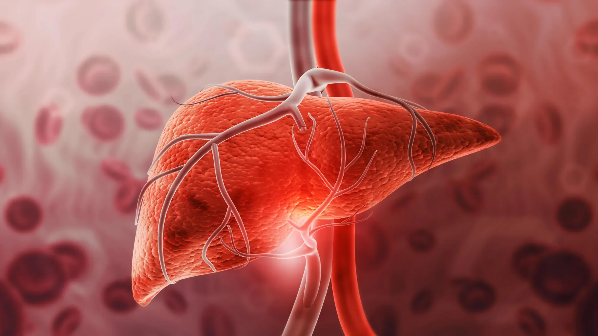
    Millions of people around the world are affected by metabolic dysfunction-associated steatohepatitis (MASH), an advanced form of fatty liver disease that can eventually lead to cirrhosis, liver cancer, and liver failure. New weight loss medications have helped improve outcomes for many patients, but inflammation in the liver can remain even after substantial weight loss.

    By identifying a biological pathway that directly regulates this inflammation, the new findings suggest that future therapies could potentially target liver inflammation alongside existing treatments focused on weight and liver fat.

    “Our findings show that GDF15 does much more than regulate appetite and body weight,” says Gregory Steinberg, professor in McMaster University’s Department of Medicine, co-director of the Centre for Metabolism, Obesity and Diabetes Research (MODR), and senior author of the study. “We discovered that GDF15 activates a natural brain-to-liver signaling pathway that helps suppress liver inflammation and reduce fibrosis. This changes how we think about the hormone and suggests it may be part of the body’s own defense system against chronic liver injury.”

    How GDF15 Sends a Protective Signal to the Liver

    To explore how GDF15 affects advanced liver disease, the team used mouse models designed to closely reproduce human MASH. Researchers combined genetic, pharmacological, genomic, and spatial transcriptomics techniques to examine what happens when the hormone becomes active.

    They found that GDF15 initiates signaling from the brain through the nervous system, ultimately causing the release of glucocorticoids. These steroid hormones are important for metabolism, immune activity, and the body’s response to stress.

    The glucocorticoids then help suppress inflammation in the liver. Importantly, researchers observed these protective effects regardless of changes in food consumption, body weight or liver fat. GDF15 also appeared to slow the progression of liver fibrosis, the accumulation of scar tissue that occurs as liver disease becomes more advanced.

    “GDF15 helps reprogram liver cells to reduce inflammation and scarring by advanced spatial technology,” says Dongdong Wang, first and corresponding author of the study and assistant professor in McMaster’s Department of Medicine. “Instead of causing liver damage, GDF15 appears to help calm the liver’s immune system. It shifts immune cells into a more protective and less active state, helping reduce inflammation and prevent damage to the liver.”

    A Second Surprising Role for GDF15

    Research published in 2023 by Steinberg and Wang showed that GDF15 helps the body maintain calorie burning during weight loss. The latest findings reveal a very different function for the same hormone: protecting the liver through a previously unknown anti-inflammatory pathway.

    Researchers say that, taken together, the two studies could help shape future therapies that specifically address liver inflammation. This inflammation is a major factor driving the progression of MASH and remains difficult to treat.

    Steinberg, an executive member of NexusHealth at McMaster and chief scientific officer, shareholder and co-founder of Espervita Therapeutics, recently co-authored preclinical research describing a promising drug candidate for advanced liver disease.

    That research focused on a potential therapeutic compound. In contrast, the new study reveals a biological pathway that the body naturally uses to control liver inflammation, providing additional clues that could help guide the development of future treatments.

    Combining Weight Loss With Inflammation Control

    “Current therapies largely focus on reducing body weight and liver fat,” says Steinberg. “Our work suggests there may be value in combining those approaches with therapies that directly target inflammation. By understanding how the body naturally protects the liver, we can identify new opportunities to develop more effective treatments for people living with MASH.”

    The study also included collaborators Rune E. Kuhre and Sebastian B. Jørgensen of Novo Nordisk A/S. The research was funded by the Natural Sciences and Engineering Research Council of Canada (NSERC), the Canadian Institutes of Health Research (CIHR), and Diabetes Canada. Novo Nordisk provided research support and supplied the GDF15 hormone used in the study.