Category: Research and Discovery

  • The Ozempic and Wegovy mistake sending thousands to poison control

    The Ozempic and Wegovy mistake sending thousands to poison control

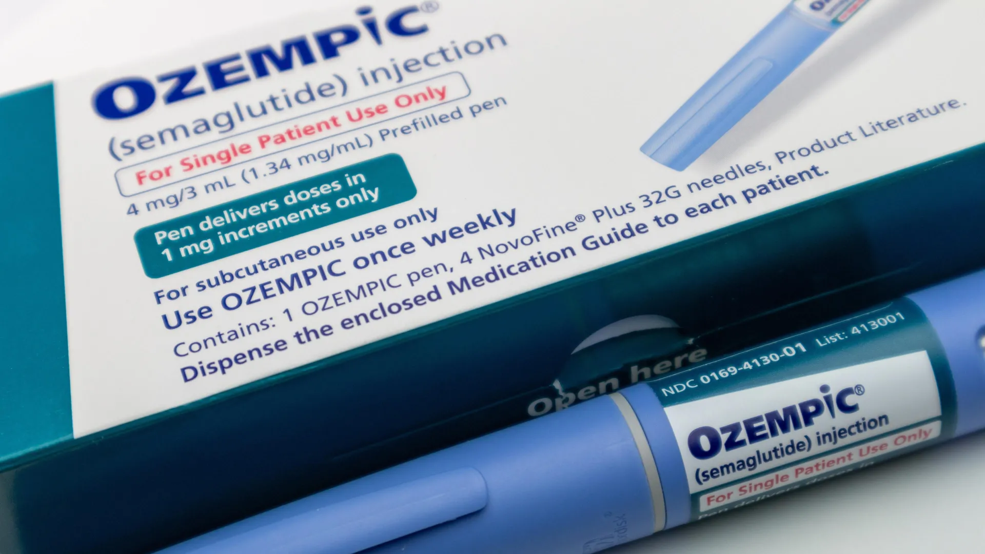
    The explosive growth in semaglutide (Ozempic and Wegovy) We use after its FDA approval for chronic weight management in 2021 was accompanied by another trend that drew researchers’ attention: a dramatic increase in calls to poison control centers across the United States.

    Jordan Miller, then an undergraduate student at UT San Antonio, wanted to find out whether the surge in poison control cases was directly connected to the FDA’s expanded approval of semaglutide or if the timing was merely coincidental.

    Originally introduced as a treatment for type 2 diabetes, Glucagon-Like Peptide-1 Receptor Agonists (GLP-1RAs) quickly gained widespread popularity after the FDA approved semaglutide for weight management. As demand for the drug accelerated, poison control centers also began reporting a steep increase in calls, with semaglutide standing out far above other medications.

    “One of them was this quite odd category of semaglutide,” said David Han, Miller’s research mentor and Romo Endowed Professor in the UT San Antonio Department of Statistics & Data Science. “We suspected that the call volume was skyrocketing because of the misuse and mishandling of this drug and that it may be attributed to the FDA approval of this drug for weight management.”

    Working with Han through the Provost’s Undergraduate Research Fellowship, Miller collaborated with Long School of Medicine researchers Robert S. Miller, Pharm.D, senior specialist in poison information, and Shawn M. Varney, MD, professor in the Department of Emergency Medicine and medical director of the South Texas Poison Center, to analyze national poison control data.

    Miller later presented the research at UT San Antonio’s Los Datos conference, where the project earned first place.

    Poison Control Calls Climbed Dramatically

    Before 2021, poison control centers nationwide typically handled between 1,000 and 1,500 GLP-1RA related cases each year. After the middle of 2021, that number nearly doubled, and by 2023 poison centers had recorded more than 8,000 GLP-1RA related calls.

    Although most incidents involved accidental dosing or therapeutic mistakes rather than intentional misuse, the sheer size of the increase surprised the research team.

    “In that figure that tracks the increase by specific drug, I wasn’t expecting semaglutide to be so incredibly dominant,” Jordan Miller recalled. “I figured that it would lead the pack, but it was staggering. On the other hand, it makes sense with all the media attention.”

    Han said the project illustrates how data science can uncover meaningful public health trends instead of simply producing statistics.

    “This work demonstrates the quantified impact of these drugs on public health,” he said. “Statistics, data science, analytics, machine learning and AI are meant to help people. We use them to transform data from any field into meaningful insight and informed action. Without that focus, it becomes hollow — numbers without real impact.”

    A Research Question That Paid Off

    The project began after Miller decided to ask a professor about possible research opportunities, a step she nearly skipped.

    “You lose nothing by asking,” she said. “If you have a professor, you really get along with or admire, you lose nothing by asking them what they’re working on or if they have space for a research assistant. I got really lucky with Dr. Han saying, ‘I’m here to help — you pick what you want to work on.’”

    The team’s analysis showed that the FDA’s approval of semaglutide for weight management marked a clear turning point. Both the number and type of poison control calls changed significantly after approval, reflecting the rapid expansion of semaglutide use beyond people with diabetes to a much larger population seeking weight loss.

    “When the GLP-1[RA] drugs are being sold to diabetic patients, that’s a completely different story versus when the drug is used for weight management,” Han explained. “So, we had to quantify this evidence to show that it stemmed from the FDA approval and how to contain the risk. We need to better educate the public because how this drug behaves in our body and its long-term safety are not yet fully understood.”

    Preventable Dosing Mistakes Drove Many Cases

    The researchers found that many of the poison control cases could have been avoided with better patient education. Semaglutide is designed to be injected once a week, not every day. Patients also begin treatment with a low dose that is gradually increased over time.

    The data showed that two of the most common mistakes were taking the medication daily instead of weekly and starting immediately with the highest dose instead of following the recommended step by step schedule.

    “Can you imagine something you’re supposed to trickle up to, and you’re going full blast and seven times more often than you’re supposed to?” Jordan Miller said.

    According to both Miller and Han, improving education at every step of the prescribing process, from the doctor’s office to the pharmacy counter, could help prevent many of these medication errors.

    The research was featured as the cover story in Significance, the flagship magazine of the Royal Statistical Society and the American Statistical Association. The findings were also published in the Journal of Medical Toxicology, the official journal of the American College of Medical Toxicology.
  • Tiny silica particles wiped out aggressive prostate cancer in mice

    Tiny silica particles wiped out aggressive prostate cancer in mice

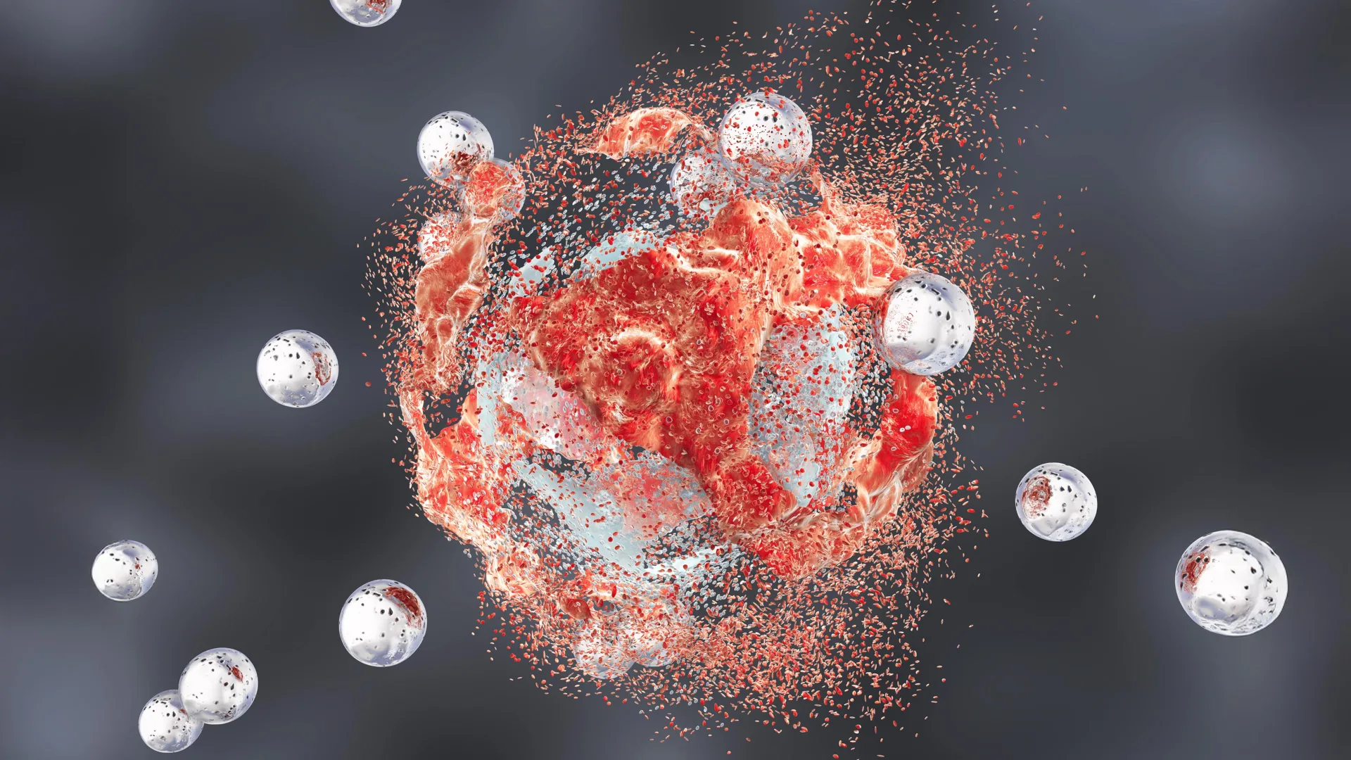
    Researchers have developed tiny silica nanoparticles that can directly destroy prostate tumors while also awakening the body’s immune system to fight cancer, according to a new preclinical study led by scientists at Weill Cornell Medicine and the Cornell Duffield College of Engineering. In mouse models of aggressive prostate cancer, the targeted particles produced several complete tumor remissions, offering encouraging evidence that the approach could eventually advance to human clinical trials.

    Made from amorphous silica, a form of silicon dioxide found naturally in foods and the fossilized remains of microscopic organisms, the engineered nanoparticles appear to attack prostate cancer in multiple ways at once.

    Tiny nanoparticles with a dual cancer fighting strategy

    The nanoparticles, known as ultrasmall fluorescent core shell silica nanoparticles or Cornell Prime dots (C’ dots), were originally created to improve medical imaging. They have already advanced into late stage clinical trials for image guided surgery and other therapeutic uses.

    More recently, researchers discovered that the particles themselves can selectively damage cancer cells while leaving healthy cells largely unharmed.

    In the new study, published June 15 in Cancer Research, a journal of the American Association for Cancer Research, the team tested the nanoparticles in mice with aggressive prostate cancer. They found that the particles made tumor cells highly vulnerable to a form of self destruction while also transforming the tumor environment from an immune resistant “cold” state into an immune active “hot” state. This shift could significantly improve the effectiveness of existing immunotherapies.

    “We’re very encouraged by these results; a treatment that directly induces tumor-cell death while transforming the immune microenvironment, as this does, would represent a new clinical paradigm,” said senior author Dr. Michelle Bradbury, the Endowed Professor of Imaging Research in Radiology and director of the Molecular Imaging Innovations Institute at Weill Cornell Medicine and a neuroradiologist at NewYork-Presbyterian/Weill Cornell Medical Center.

    The work is part of a long running collaboration between Dr. Bradbury’s laboratory and the laboratory of co corresponding author Dr. Ulrich Wiesner, the Spencer T. Olin Professor in the Department of Materials Science and Engineering and a professor in the Department of Design Tech in the College of Architecture, Art, and Planning. The research received support in part from the Parker Institute for Cancer Immunotherapy at Weill Cornell Medicine.

    How the silica particles kill cancer cells

    One of the most unusual findings involved a process called “ferroptosis,” a specialized form of cell death driven by overwhelming oxidation inside cells. During ferroptosis, oxidation damages critical molecules, particularly the fatty molecules that make up cell membranes, eventually causing the cells to break down.

    Scientists do not yet fully understand how the nanoparticles trigger this process. However, evidence suggests the particles, which were initially designed to carry imaging agents, can collect positively charged iron ions from the bloodstream and transport them into tumor cells. Once inside, those iron ions may fuel the intense oxidation that drives ferroptosis.

    Reawakening the immune system

    Beyond directly killing tumor cells, the nanoparticles also reshaped the immune environment surrounding the cancer.

    The researchers observed that T cells, macrophages, and other immune cells near the tumors shifted from inactive or immune suppressing states into active cancer fighting cells. The nanoparticles also made tumors much more responsive to approved immunotherapy drugs. At the same time, they disrupted metabolic processes across several types of cells within the tumor microenvironment, further slowing tumor growth.

    To ensure the treatment reached prostate cancer cells, the team attached a targeting molecule that recognizes PSMA, a protein found on the surface of prostate tumor cells. Although some particles briefly accumulated in other organs such as the spleen, the researchers found no signs of toxicity outside the tumors.

    “It seems unreal — how is it possible that rather than a single pathway we see all these effects happening simultaneously and only in tumors and not in healthy tissues?” Dr. Wiesner said. “I have to wonder whether ultrasmall silica’s very early and ubiquitous presence in the environment and foods like leafy greens or cereal grains has given it a connection to biology that we’re only beginning to glimpse.”

    Combination therapy produced the strongest results

    The most dramatic findings came from survival studies involving mice with aggressive prostate cancer.

    On their own, both the C’ dots and immunotherapy modestly improved survival compared with no treatment. However, combining the nanoparticles with an immune checkpoint blockade therapy produced complete or nearly complete remissions and indefinite survival in four out of ten mice.

    Adding a third treatment called CSF-1R blockade, which targets tumor associated macrophages, increased the number of complete remissions to five out of ten mice.

    “We think there’s nothing else out there that has such a strong and durable tumor growth suppressing effect,” Dr. Bradbury said.

    “One of the most intriguing aspects of this work is the convergence of direct tumor cell killing with broad immune remodeling,” said study co author Dr. Jedd Wolchok, the Meyer Director of the Sandra and Edward Meyer Cancer Center, professor of medicine at Weill Cornell Medicine, director of the Parker Institute for Cancer Immunotherapy at Weill Cornell Medicine Meyer Cancer Center and an oncologist at NewYork-Presbyterian/Weill Cornell Medical Center. “By creating conditions that support a more effective antitumor immune response, these particles may help unlock the full potential of immunotherapy in prostate cancer, where durable responses have historically been difficult to achieve.”

    Next step is human clinical trials

    Dr. Bradbury also recognized the work of the study’s co first authors, Drs. Nabil Siddiqui, Li Zhang, and Gabriel DeLeon, who led many of the biological, mechanistic, and translational studies, along with graduate students Nada Naguib and Rachel Lee from Dr. Wiesner’s laboratory, whose careful synthesis and characterization of the nanoparticles were essential to the project.

    “This study reflects years of collaborative effort across multiple laboratories and would not have been possible without the dedication, creativity and perseverance of this tremendous research team that helped drive the science forward,” she said.

    The research team is continuing to investigate these ultrasmall core shell silica particles as a potential new class of cancer therapies capable of influencing inflammatory, immune, and metabolic pathways at the same time. Their long term goal is to evaluate the safety and effectiveness of the treatment in human clinical trials.

    Drs. Michelle Bradbury and Ulrich Wiesner are inventors on patents related to the technology described in this study.

    The study was funded by the Department of Defense (PC220534); the National Cancer Institute, part of the National Institutes of Health, through grant numbers R01CA253658, R01CA243085, U54CA199081, the Cancer Center Support Grant (P30 CA008748), and Cycle for Survival/Parker Institute funding.

  • Scientists finally crack nature’s secret for building better cancer drugs

    Scientists finally crack nature’s secret for building better cancer drugs

    Scientists have uncovered how bacteria naturally manufacture multiple versions of powerful cancer drugs, solving a mystery that has puzzled researchers for decades. The discovery could help speed the development of new treatments for cancers that are still difficult to treat.

    For years, scientists have hoped to harness bacterial enzymes to create new drug variants through a process known as combinatorial biosynthesis. However, progress has been limited because researchers did not fully understand how the enzymes coordinate their work.

    Published in Nature Communications, the new study reveals how bacterial enzymes communicate with one another to assemble a family of closely related anti-cancer compounds. That family includes Romidepsin (Istodax), an FDA-approved treatment for certain blood cancers. By uncovering this natural “mix and match” system and reproducing its underlying principles in the laboratory, the researchers have established a new strategy for designing future cancer therapies.

    “For decades, we’ve known that bacteria can naturally produce multiple versions of powerful anti-cancer drugs, yet we had no idea how they achieved this,” said first author Dr. Munro Passmore, Research Fellow, Department of Chemistry, University of Warwick. “This work finally cracks that code. We’ve identified how the different enzymes communicate and cooperate to produce these drug variants, something that has eluded researchers because the system is so elegantly economical. It’s the breakthrough we needed to actually engineer these drugs ourselves.”

    Tiny Molecular Connectors Reveal Nature’s Drug-Making Strategy

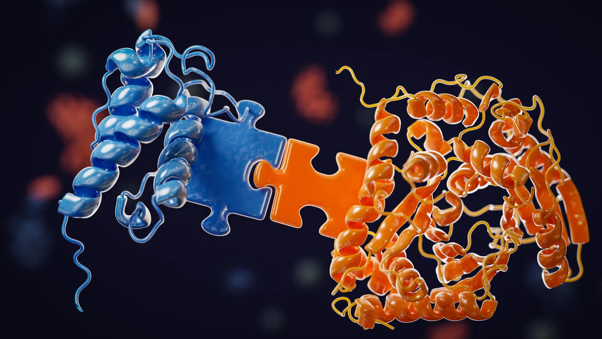
    The researchers discovered that small molecular regions known as ‘docking domains’ serve as connectors between the core drug-building machinery and the enzymes responsible for adding different components. These docking domains share a conserved connection point that allows them to interact with multiple enzyme partners.

    This flexible design explains how bacteria can create a variety of related drug molecules while still maintaining the precision needed for the compounds to remain effective.

    The study also sheds light on how these natural drug-producing systems evolved. According to the researchers, the newly identified compound most likely developed from a related drug-producing pathway through gene duplication and recombination over time.

    Prof. Greg Challis, Monash Warwick Alliance Professor of Sustainable Chemistry, University of Warwick and Monash University concludes: “This research gives us a blueprint to do what nature does, but better and faster. By reverse-engineering nature’s evolutionary logic, we can now design synthetic pathways that generate new anti-cancer drug candidates with properties optimized for clinical use, such as superior potency, improved selectivity, fewer side effects. Our immediate goal is to build an expanded library of candidates for various cancers where new treatments are urgently needed. This discovery is moving us from understanding how the systems work to building new ones.”

    How the Discovery Could Improve Cancer Drug Development

    The work focuses on a class of anti-cancer medicines known as HDAC inhibitors. These drugs block histone deacetylases, enzymes that help regulate which genes are switched on or off inside cells. Romidepsin (Istodax) is an FDA-approved HDAC inhibitor used to treat T-cell lymphomas.

    A chemically related compound called FR-901375 has been known for decades, but scientists had never identified the biological pathway bacteria use to produce it. This study finally fills in that missing piece.

    Like other HDAC inhibitors in its family, FR-901375 belongs to a group of complex cyclic molecules called depsipeptides. These compounds are assembled from amino acid building blocks along with a conserved hydroxy acid pharmacophore, all connected through a combination of peptide and ester bonds.

    Inside bacteria, these molecules are built by massive protein complexes called PKS-NRPS hybrids, which combine the activities of polyketide synthase (PKS) and nonribosomal peptide synthetase (NRPS). The new research shows that the key to this assembly process is the docking domains, which act like molecular connectors that allow one part of the production line to recognize and pass its product to the next. This mechanism is what enables combinatorial biosynthesis and allows bacteria to naturally generate multiple drug variants.

    How the Researchers Solved the Mystery

    To uncover how this system works, the team combined structural biology, biochemistry, genetics, and computational modeling.

    Their work included:

    • Bioinformatic searches of public databases that identified the FR-901375 biosynthetic gene cluster in Pseudomonas chlororaphis subsp. piscium, with the findings confirmed by mass spectrometry analysis of extracted metabolites.
    • In vitro reconstitution experiments using purified protein domains that demonstrated productive enzyme-enzyme interactions, verified with intact protein mass spectrometry.
    • AlphaFold computational modeling to predict protein complex structures, followed by carbene footprinting mass spectrometry to experimentally map the interaction sites.
    • Site-directed mutagenesis experiments that confirmed the importance of the predicted binding residues.
    • Gene deletion studies in bacterial strains showing that the docking domains are essential for the system to function in vivo.
    • Comparative analysis of biosynthetic gene clusters from multiple HDAC inhibitor-producing bacteria, revealing evolutionarily conserved features shared across these natural drug-making systems.
  • Creatine doesn’t just build muscle. It may also help fight cancer

    Creatine doesn’t just build muscle. It may also help fight cancer

    Creatine is widely known as a supplement used by athletes and bodybuilders to improve strength and performance. Now, new research from UCLA suggests it may have another surprising role: helping the immune system mount a stronger attack against cancer.

    The study, published in iScience, found that creatine boosts the activity of dendritic cells, specialized immune cells that detect tumors and activate the killer T cells responsible for destroying cancer. The findings, based on experiments in mice and human cells, build on previous research from the same laboratory showing that creatine also enhances the function of cancer-fighting T cells.

    Creatine May Strengthen Cancer Immunotherapy

    Many of today’s cancer immunotherapies are designed to activate killer T cells, but only about 20% to 40% of patients experience meaningful benefits. The UCLA team believes that improving the function of dendritic cells, which coordinate and direct those T cells, could make immunotherapy effective for more people.

    “Immunotherapy has shown remarkable promise, but it only works for a subset of patients,” said Lili Yang, the study’s senior author, a professor of microbiology, immunology and molecular genetics and a member of the Eli and Edythe Broad Center of Regenerative Medicine and Stem Cell Research at UCLA. “What this study shows is that creatine doesn’t just help the T cells fighting cancer — it also energizes the entire infrastructure supports and guides them. That makes creatine a promising supplement to holistically support the immune response that modern immunotherapies depend on.”

    Scientists Uncover Creatine’s Role in Dendritic Cells

    To understand how creatine influences the immune system, researchers first examined the activity of metabolic genes in dendritic cells that had entered tumors in mice. They discovered that the gene responsible for producing the creatine transporter, a protein that carries creatine into cells, was much more active in tumor-infiltrating dendritic cells than in dendritic cells found in healthy tissue.

    The team then engineered dendritic cells that lacked the creatine transporter. Without the ability to take up creatine, the cells survived less effectively, became less active, and were far less capable of preparing T cells to recognize and attack tumors. When these creatine-deficient dendritic cells were grown alongside T cells in laboratory experiments, the T cells multiplied less and produced fewer signaling molecules needed to mount an effective anti-cancer response.

    Creatine Slowed Tumor Growth in Mice

    Researchers also tested whether increasing creatine levels could produce the opposite effect.

    Daily creatine injections in mouse models of melanoma significantly slowed tumor growth while increasing both the number and activity of dendritic cells that had entered tumors. The treated dendritic cells also released higher levels of chemical signals that attracted additional immune cells into the tumor environment.

    Using metabolomics analyses, the scientists found that creatine supplementation increased intracellular ATP levels in dendritic cells. ATP serves as the primary energy source that powers nearly every cellular process. By boosting these energy reserves, creatine helped maintain the inflammatory signaling pathways required for dendritic cell activation.

    The researchers compared creatine’s role to that of a rechargeable battery, allowing dendritic cells to store and release energy as needed, even while competing with rapidly growing tumor cells for limited nutrients.

    Potential Benefits for Cancer Vaccines

    The team also explored creatine’s effects on human immune cells.

    In laboratory experiments, creatine enhanced the activation of human monocyte-derived dendritic cells, which are commonly used to develop dendritic cell cancer vaccines. It also improved those cells’ ability to stimulate human T cells against a cancer-associated target.

    The findings suggest that adding creatine during the production of dendritic cell vaccines could potentially make those therapies more effective.

    “The potential we see here is that creatine could be used in two complementary ways: as a supplement to enhance the immune response of patients already receiving immunotherapy, and as a tool to improve the quality of dendritic cell-based vaccines before they’re administered,” said James Elsten-Brown, a co-first author and graduate student in Yang’s lab.

    Overall, the results suggest creatine could strengthen the immune system’s anti-cancer defenses at multiple stages, beginning with the cells that detect cancer and initiate the body’s response.

    “Understanding how to metabolically support dendritic cells is about supporting the entire anti-tumor response, not just the killer T cells at the end of it,” said Elliot Kang, a co-first author of the study and former undergraduate student researcher in Yang’s lab.

    Human Trials Are Still Needed

    Despite the encouraging findings, the researchers caution that the work remains at an early stage. The experiments were conducted in mice and human cells grown in the laboratory, not in cancer patients, so the results should not be interpreted as evidence that creatine supplements improve cancer treatment in people.

    Although creatine monohydrate has been widely used for decades and is generally considered safe when taken at recommended doses, the researchers stress that anyone receiving cancer treatment should consult their physician before adding any supplement to their routine.

    The next step will be prospective clinical trials to determine whether creatine supplementation can improve outcomes for patients undergoing cancer immunotherapy.

    The experimental approaches described in the study have not been tested in humans or approved by the Food and Drug Administration as safe and effective for use in people.

    Funding for the research was provided by a UCLA Broad Stem Cell Research Center Rose Hills Foundation Innovator Grant; the UCLA Health Jonsson Comprehensive Cancer Center and UCLA Broad Stem Cell Research Center Ablon Scholars Program; and a Magnolia Council Senior Investigator Grant Award and a fellowship from the Tower Cancer Research Foundation.

    The potential therapeutic strategy identified in the study is also the subject of a patent application filed by the UCLA Technology Development Group on behalf of the Regents of the University of California.

  • New weight loss pill beats oral Ozempic in major trial

    New weight loss pill beats oral Ozempic in major trial

    A new type of daily pill has proven more effective for weight loss and blood sugar control for people with type 2 diabetes than its currently available counterparts, according to a recent trial. The drug, known as orforglipron, could be a game-changer in the rapidly expanding oral weight-loss drug market.

    The advent of the injectable weight-loss drug semaglutide, known better by its brand names Wegovy (for weight loss) and Ozempic (for type 2 diabetes), marked a distinct shift in the weight-loss drugs market when it became available just a few years ago.

    Semaglutide is a class of glucagon-like peptide-1 (GLP-1) medication. These drugs mimic the gut hormone GLP-1, which is released soon after eating. This hormone signals fullness to the brain, slowing digestion and stimulating the release of insulin. By replicating the action of this hormone, GLP-1 drugs have proven highly effective at managing type 2 diabetes and promoting weight loss.

    Although semaglutide is widely used, a key issue with the drug is that it needs to be injected into the belly, thighs or back of the arm. This can make it difficult for patients with needle phobia or who don’t want to self-inject due to the inconvenience.

    Another logistical issue with injectable GLP-1 drugs is that they require refrigeration throughout the supply chain. This can pose a challenge in low- and middle-income countries.

    It’s for these reasons that researchers and developers have started investigating the efficacy of oral versions of semaglutide.

    Based on current research, it appears that oral semaglutide is very effective. However, it must be taken on an empty stomach – and users must wait 30 minutes before eating or drinking.

    Alongside being expensive to produce, it also has poor bioavailability compared with injectable semaglutide. This means only about 1% of the ingested drug is absorbed and able to exert its effects.

    But a recent phase 3 clinical trial has shown that a new type of oral weight-loss pill may have overcome these issues, making it competitive with the current oral semaglutide products on the market.

    Oral weight-loss pill

    The recent 52-week phase 3 trial involved 1,698 adults with type 2 diabetes across six countries. It set out to compare current oral semaglutide products against orforglipron, which is also taken as a daily tablet.

    The primary measure researchers were looking for was a reduction in HbA1c. This blood test reflecting average blood sugar levels over three months is the standard indicator of diabetes control. Diabetes is present if HbA1c is 6.5% or more.

    From a baseline average HbA1c of 8.3%, it was found that after 52 weeks, orforglipron was able to reduce this value by an average of 1.71–1.91%. In comparison, oral semaglutide only reduced HbA1c by 1.47%.

    Not only did orforglipron meet the trial’s goals of proving it was as effective to oral semaglutide, it proved it was superior for lowering blood sugar. The participants who took orforglipron also lost more weight – an average of 6.1kg-8.2kg, compared with 5.3kg in those taking semaglutide.

    However, a key issue highlighted by the trial was one of tolerability.

    GLP-1 drugs can cause gastrointestinal side-effects such as nausea, vomiting, diarrhea and constipation. In this latest trial, around 59% of participants on orforglipron reported such symptoms, compared with 37–45% on semaglutide.

    The reason for this difference may be the more prominent, daily peak drug concentrations with orforglipron. The consequence was that around 10% of orforglipron participants discontinued treatment due to adverse effects. Just 4-5% of those taking semaglutide discontinued treatment.

    No head-to-head trials have been done of injectable GLP-1 versus orforglipron. However, the weight loss seen in this study of people with type 2 diabetes is broadly comparable with that previously observed with injectable GLP-1.

    Market implications

    The trial’s results show that orforglipron, which was developed by Eli Lilly, can be considered one of semaglutide’s most credible challengers.

    Another remarkable thing about orforglipron is that it belongs to a new category of drugs called small-molecule drugs. This means it’s a synthetic chemical compound small enough to be absorbed directly through the gut wall. There, it’s able to act on GLP-1 receptors, even though it isn’t of a similar structure to a GLP-1 hormone.

    Oral semaglutide, on the other hand, is a peptide drug. This means the structure of its amino acids (one of the building blocks of protein) closely resembles that of the natural GLP-1 hormone.

    As a small-molecule drug, orforglipron is cheaper and simpler to manufacture than peptide-based drugs such as semaglutide.

    And as with oral semaglutide, it requires no refrigeration. This gives it a logistical advantage over injectable GLP-1 formulations – a potentially important consideration for expanding access in low- and middle-income countries, where cold chain infrastructure is unreliable.

    It remains to be seen, however, how orforglipron will perform against oral semaglutide in the broader market.

    Although this latest trial has shown it is superior for controlling blood sugar and aiding weight loss, its higher rate of side-effects and treatment discontinuation may temper enthusiasm. In a crowded and competitive market, long-term adherence – shaped as much by tolerability as by efficacy – is probably a critical differentiator.

    Orforglipron is still undergoing trials in patients with obesity but without diabetes.The Conversation

  • A hidden weakness in deadly cancers could lead to powerful new treatments

    A hidden weakness in deadly cancers could lead to powerful new treatments

    Researchers at UCLA have identified a hidden weakness in some of the most aggressive and difficult-to-treat cancers, raising hopes for a new way to attack tumors that have resisted treatment for decades.

    Small cell neuroendocrine cancers can develop in the lung, prostate, and ovary. These fast-growing tumors tend to spread early and have remained among the hardest cancers to treat successfully.

    A key feature of these cancers is the loss of a gene called RB. Under normal conditions, RB helps keep cell growth under control. When the gene is missing, cancer cells multiply rapidly and become resistant to many targeted therapies.

    Now, a new study published in Proceedings of the National Academy of Sciences suggests that losing RB also creates an unexpected vulnerability that researchers believe could become a powerful treatment target.

    A Hidden Dependency in Deadly Cancers

    The UCLA team discovered that cancer cells lacking RB become heavily dependent on a protein called E2F3 to survive. In laboratory experiments, blocking E2F3 prevented tumor growth through a process known as “synthetic lethality.”

    In simple terms, cancer cells can survive without RB, but they cannot survive when both RB and E2F3 are lost. Removing E2F3 alongside the missing RB exposes a critical weakness that researchers believe could be exploited with future therapies.

    “Discovering a vulnerability like this opens the door to thinking about entirely new treatment strategies,” said study senior author Dr. Owen N. Witte, who holds the Presidential Chair in Developmental Immunology in the Department of Microbiology, Immunology, and Molecular Genetics and is a member of the UCLA Health Jonsson Comprehensive Cancer Center. “That’s especially important because there has not been a major change in how we treat these cancers for decades. When I first encountered these tumors as a medical student more than 50 years ago, the survival statistics were essentially the same as they are today.”

    Building Better Models To Study Small Cell Cancer

    Progress against small cell neuroendocrine cancers, especially those that develop in the prostate, has been slowed by the lack of realistic laboratory models. Without them, scientists have struggled to identify the genes these tumors rely on and uncover their biological weaknesses.

    To overcome that challenge, the UCLA researchers engineered normal human prostate cells with five major cancer-causing genetic changes, including the loss of RB and TP53. The cells were grown into organoids and then used to produce tumors in mice, creating models that closely resemble human small cell prostate cancer.

    The work builds on more than a decade of research by Witte’s laboratory to develop specialized models of small cell neuroendocrine prostate cancer.

    CRISPR Screens Reveal a Shared Weakness

    Using these models, the team performed genome-wide CRISPR screens that examined thousands of genes to determine which ones were essential for cancer cell survival.

    The researchers identified nearly 1,400 genes that play important roles in keeping the cancer cells alive. Among the most significant discoveries was that small cell cancers from different organs all shared a strong dependence on E2F3.

    When the scientists reduced E2F3 levels in RB-deficient cancer cells, the tumors stopped dividing, could no longer form clusters, and in some cases died completely.

    “It’s not that the two genes do the same thing,” said Witte, who is also the founding director emeritus of the UCLA Broad Stem Cell Research Center and co-director of the Parker Institute of Cancer Immunotherapy Center at UCLA. “But the combination of what they do together becomes essential for the cancer cell. Losing one gene may not matter much, but losing both has a dramatic effect on tumor growth.”

    “These new model systems allowed us to uncover a genetic vulnerability that would have been very difficult to find otherwise,” added first author Dr. Evan Abt, an assistant professor of Molecular and Medical Pharmacology at the David Geffen School of Medicine at UCLA.

    Existing FDA-Approved Drugs May Offer a Shortcut

    Because no medicines currently target E2F3 directly, the researchers looked for another way to exploit the cancer’s weakness.

    They found that blocking a metabolic pathway involved in producing DNA building blocks by inhibiting an enzyme called DHODH lowered E2F3 levels and slowed tumor growth.

    That finding is especially intriguing because DHODH inhibitors, including leflunomide and teriflunomide, are already FDA-approved to treat autoimmune diseases. Repurposing existing medications could potentially speed the development of new therapies for patients with these cancers.

    “What’s exciting is that our findings open the door to applying existing drugs in a new way,” Abt said. “By understanding how these cancers depend on E2F3, we can start to think about strategies that might work much more quickly in patients.”

    Although the research remains in its early stages, the findings provide important new insight into how these aggressive cancers survive and point toward a promising new direction for future treatments.

    Other UCLA authors are Liang Wang, Grigor Varuzhanyan, Jack Freeland, Tian He, Guadalupe M. Peña-Garcia, Lauryn Ruegg, Jami McLaughlin, Donghui Cheng, Nikolas G. Balanis, Chia-Chun Chen, Sanaz Memarzadeh, Caius G. Radu and Thomas G. Graeber.

  • Nearly half of kidney transplant patients never even get started

    Nearly half of kidney transplant patients never even get started

    Nearly half of Americans with kidney failure who are referred for a kidney transplant never begin the evaluation process required to be considered for a donor organ, according to a new nationwide study. Even more striking, fewer than one in five complete the evaluation and secure a place on the transplant waitlist.

    Researchers say much attention has been given to patients after they reach the waitlist, but far less is known about what happens before that point and why so many people never make it there.

    Major Barriers to Kidney Transplant Waitlisting

    The study, led by researchers at NYU Langone Health, analyzed data from 720,348 patients referred for kidney transplantation. The findings revealed significant disparities in who advances through the process.

    Patients who were unmarried, had severe obesity, or lived in rural communities were less likely to begin or complete a transplant evaluation and ultimately reach the waitlist. Older adults, Spanish speakers, and people with lower incomes faced even greater challenges. Patients receiving care at smaller transplant centers or programs located in the Southern United States were also less likely to move forward.

    Overall, just 19% of referred patients completed the evaluation process and were placed on the waitlist, while 48% never started the evaluation at all.

    “Our findings suggest that a substantial proportion of people who need a new kidney fall out of the process long before they reach the waitlist, let alone make it to the operating room,” said study lead author Conor Donnelly, MD. “Which transplant center you go to, where you live, and even whether you are married all appear to influence your chances of moving forward to the waitlist for a new kidney.”

    Donnelly is a resident and PhD student in the Department of Surgery at NYU Grossman School of Medicine.

    Why the Evaluation Process Can Be Difficult

    According to Donnelly, the complexity of the transplant evaluation process may explain much of the variation seen in the study.

    After receiving a referral, patients must complete an extensive medical assessment designed to evaluate their overall health. This often includes blood tests, chest imaging, cancer screenings, and other examinations. The process may require multiple appointments over several months while patients continue attending regular dialysis treatments.

    Only after completing these requirements and receiving approval can a patient be added to the transplant waitlist.

    Researchers noted that smaller transplant centers may have fewer resources and available transplant opportunities, which could lead them to be more selective when evaluating candidates. They also pointed out that patients who are unmarried or have limited social support may face greater difficulties arranging transportation and attending repeated appointments.

    These factors may help explain why patients living in urban areas, where transplant centers are often more accessible, were generally more likely to continue through the process.

    Largest Study of Kidney Transplant Dropout Rates

    Published online June 20 in the Journal of the American Society of Nephrology, the research is the largest and most detailed study to date examining where patients leave the kidney transplant pathway before reaching the waitlist, according to the authors.

    The findings are also being presented at the American Transplant Congress, the annual meeting jointly organized by the American Society of Transplantation and the American Society of Transplant Surgeons.

    To conduct the analysis, researchers used Epic Cosmos, a database containing more than 300 million electronic health records from over 1,850 hospitals, including more than one-third of US transplant centers.

    The team examined adults referred for kidney transplantation between 2014 and 2025. Each patient was tracked through four stages: referral, evaluation, waitlist, and transplant.

    Social and Geographic Factors Influence Outcomes

    Using statistical modeling, the researchers evaluated how factors such as age, sex, medical history, and geographic location affected the likelihood of advancing from one stage to the next.

    The team also studied social vulnerability, which reflects challenges related to living conditions and access to care. Examples include poverty, unstable housing, and limited transportation options, all of which can make navigating complex medical systems more difficult.

    “These results demonstrate that finding ways to reduce barriers to both evaluation and waitlisting could help expand much-needed access to kidney transplantation,” said study co-senior author Allan B. Massie, PhD, an associate professor in the Departments of Surgery and Population Health at NYU Grossman School of Medicine. “Providing patients with better education and support to help them navigate the complex and sometimes grueling process would be a good start.”

    “Our findings highlight the need to better support patients in progressing from referral to the waitlist, where many possibly eligible individuals are not ultimately listed,” said study co-senior author Michal A. Mankowski, PhD.

    Mankowski, an assistant professor in the Department of Surgery at NYU Grossman School of Medicine, said future research will apply a similar approach to other types of organ transplantation, where the path to the waitlist can differ significantly.

    Additional NYU Langone researchers involved in the project included Suhani Patel, MPH; Syed Ali Husain, MD, MPH; Sommer E. Gentry, PhD; Bonnie E. Lonze, MD, PhD; Sunjae Bae, MD, PhD; Babak J. Orandi, MD, PhD; Mara A. McAdams DeMarco, PhD; and Dorry L. Segev, MD, PhD. Other collaborators included Rachel Patzer, PhD, MPH, of Indiana University in Indianapolis and David Axelrod, MD, of University Hospitals in Cleveland.

    Dr. Orandi has served on an advisory board for the pharmaceutical company Boehringer Ingelheim. NYU Langone Health is managing the terms and conditions of this relationship in accordance with its policies and procedures.

    NYU Langone Health funded the study.

  • USC scientists just unlocked an endless supply of cancer-fighting immune cells

    USC scientists just unlocked an endless supply of cancer-fighting immune cells

    Scientists at USC Stem Cell have developed a new method for creating a renewable and expandable supply of immune cell precursors that could help advance cancer immunotherapy and other treatments.

    Published in the journal Cell, the study focuses on granulocyte-monocyte progenitors (GMPs), a type of progenitor cell that produces macrophages and several other immune cells. Macrophages play a key role in defending the body against infections and have attracted growing interest as potential tools for treating cancer.

    The researchers showed that GMPs can be expanded extensively in the laboratory and genetically modified to recognize cancer cells while also boosting broader immune responses.

    “The study establishes a scalable and engineerable GMP platform for cellular immunotherapy and introduces concepts that we believe could have broad implications for both cancer immunotherapy and stem cell biology,” said the paper’s corresponding author Qi-Long Ying, MD, PhD, professor of stem cell biology and regenerative medicine at the Keck School of Medicine of USC.

    One of the study’s most significant findings relates to self-renewal, a characteristic traditionally associated with stem cells. Self-renewal allows cells to repeatedly divide while maintaining their identity. Scientists generally have not considered progenitor cells to possess this long-term capability.

    “The prevailing view has been that long-term self-renewal in the blood system is primarily a property of the hematopoietic stem cells that can generate any type of blood or immune cell,” said Ying. “We found that, under the right conditions, GMPs can also self-renew, dividing extensively while keeping their identity and ability to produce functional immune cells. That gives us a scalable starting point for engineering cell therapies for cancer, infectious disease and potentially many other conditions.”

    Why Macrophage Precursors Matter

    Macrophages are appealing candidates for cancer immunotherapy because they naturally enter tumors, consume cancer cells, and help organize immune responses. While T-cell therapies have achieved major success against blood cancers, macrophage-based therapies may offer particular advantages against solid tumors.

    However, mature macrophages have several drawbacks as therapeutic products. They are difficult to grow in large numbers outside the body, challenging to genetically engineer, and can be damaged during freezing and storage. They also tend to accumulate in organs such as the lungs and liver instead of spreading widely throughout the body.

    To overcome these obstacles, first author Shi Yue, MD, and colleagues in the Ying Lab focused on GMPs, which sit earlier in the developmental pathway that produces macrophages.

    Using a carefully defined chemical cocktail, the team prevented GMPs from maturing into other immune cell types and succeeded in maintaining and expanding them over long periods in the laboratory.

    Even after extended growth, the cells preserved their molecular and cellular characteristics and continued to generate functional macrophages and other immune cells.

    Researchers in the laboratory of Ravi Majeti, MD, PhD, at Stanford University independently reproduced the long-term maintenance and genetic engineering of GMPs, providing additional support for the platform’s reliability and potential therapeutic value.

    Majeti, Director of the Institute for Stem Cell Biology and Regenerative Medicine at Stanford University, noted: “This method for the expansion and engineering of GMPs opens the door to numerous translational applications, much like T cell expansion and engineering. We have already demonstrated engineering of these cells to drive multiple potent functions, and there is a lot more to be explored.”

    Engineering GMPs To Fight Cancer

    Beyond their ability to grow long term in the laboratory, GMPs can also be genetically engineered for use as immunotherapies.

    In this study, the researchers equipped GMPs with a chimeric antigen receptor, or CAR, enabling the cells to recognize a specific marker found on cancer cells. The team also added a second signal designed to activate nearby immune cells that help stimulate tumor-fighting T cells and strengthen the body’s natural defenses.

    Importantly, this additional signal remains effective even when donor and recipient cells are immunologically mismatched. That raises the possibility of creating off-the-shelf therapies produced in advance from donor cells and used in many patients, rather than generating a custom treatment for each individual.

    After expanding and engineering both mouse and human GMPs, the researchers tested them in mice. The cells successfully settled into the bone marrow and other blood-forming tissues, where they continuously generated engineered macrophages and additional immune cells.

    Because the GMPs maintained an ongoing supply of these cells from the bone marrow, they avoided the rapid loss that has limited mature macrophage therapies, including those evaluated in recent clinical trials.

    In mice with blood cancers and solid tumors, CAR-engineered GMPs slowed disease progression. GMPs carrying both the CAR and the additional immune-activating signal produced even stronger benefits.

    Potential Beyond Cancer

    The platform may have applications that extend beyond oncology.

    The researchers tested the approach in mice with chronic granulomatous disease, an inherited immune disorder. GMP treatment restored the animals’ ability to fight bacterial infections, demonstrating the potential of the technology for immune deficiencies as well.

    “Our study suggests that the future of immunotherapy may depend not only on designing better CAR receptors, but also on choosing the right developmental stage of the cell,” said Ying.

    About the Study

    The paper in Cell is titled “Expansion and CAR engineering of granulocyte-monocyte progenitors for cellular immunotherapy.”

    In addition to Ying, Yue and Majeti, additional authors are: Zheng Guo, Crystal Pan, Xueyuan A. Jing, Tai Nguyen, Jiaqi Tang, Yanpui Chan, Humberto Contreras-Trujillo, Du Jiang, Xue Yan, Hang Xiang, Xugeng Liu, Xiao Wang, Ziyuan Wang, Natalie Shu, Daniel B. McKim, Rong Lu and Chao Zhang from USC; and Litao Tao and Celia Bloom from Creighton University; Asiri Ediriwickrema and Sebastian Koschade from Stanford University School of Medicine; and Yingxiao Shi from Harvard Medical School and the Dana-Farber Cancer Institute.

    This work was supported by the Chen Yong Foundation of the Zhongmei Group, a sponsored research project from Myelogene Inc., the L.K. Whittier Foundation, the Eli and Edythe Broad Innovation Award, the Ming Hsieh Institute for Research on Engineering Medicine for Cancer Award, the USC SBIR/STTR Planning Award, the Xia Research Fund, and the Wu & Jiang Research Fund. Majeti reports support from the Ludwig Institute for Cancer Research, and Guo was supported by the California Institute for Regenerative Medicine Predoctoral Training Fellowship.

    Disclosures

    Ying, Yue, Jing, Guo, Majeti, Zhang, Nguyen and Tang are co-inventors on patents related to this study, filed by USC and licensed to Myelogene Inc. Ying, Yue, Zhang and Majeti are co-founders of Myelogene Inc. Majeti is on the Advisory Boards of Kodikaz Therapeutic Solutions, Pheast Therapeutics, Prelude Therapeutics, Mubadala Capital, Aculeus Therapeutics, Sequentify, BMS and Bectas Therapeutics. Majeti is also a co-founder and equity holder of Pheast Therapeutics.

  • FDA-approved drug may finally help immunotherapy defeat rare liver cancer

    FDA-approved drug may finally help immunotherapy defeat rare liver cancer

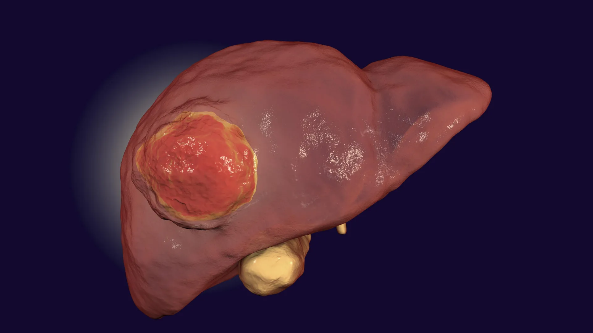
    A rare and aggressive form of liver cancer has long resisted immunotherapy, a treatment that helps the body’s own immune system attack cancer cells. Now, researchers have identified a potential way to overcome that resistance using an FDA-approved drug that is already available for another medical condition.

    The findings suggest a possible new treatment strategy for fibrolamellar carcinoma, a rare liver cancer that primarily affects children and young adults.

    Fibrolamellar carcinoma makes up about 2% of all liver cancer cases. There is currently no cure, and the disease is often discovered only after it has spread to other parts of the body, leaving many patients with limited treatment options and poor survival prospects.

    How Fibrolamellar Carcinoma Evades the Immune System

    The study, published in the journal Gastroenterology, sheds light on why immunotherapy has struggled against this cancer.

    Researchers found that fibrolamellar tumors alter their surrounding environment in a way that prevents immune T cells from reaching the cancer. Instead of moving into the tumor and attacking cancer cells, the T cells become trapped elsewhere. This process, known as T-cell exclusion, effectively keeps the immune system from carrying out its normal cancer-fighting role.

    The team also discovered that AMD3100, a drug already approved by the U.S. Food and Drug Administration for another disorder, can disrupt this process. By preventing tumors from trapping T cells, the drug allows those immune cells to reach and attack the cancer.

    “Our results provide among the first indications of why a type of immunotherapy called immune checkpoint inhibition hasn’t worked well in these patients, and even if this particular drug isn’t the end-all-be-all, it teaches us that this T-cell exclusion phenomenon is an important one to tackle in fibrolamellar carcinoma,” said Praveen Sethupathy ’03, professor of physiological genomics and chair of the Department of Biomedical Sciences in the College of Veterinary Medicine.

    Sethupathy served as co-senior author of the study alongside Dr. Venu Pillarisetty, a surgical oncologist at the University of Washington.

    Advanced Technology Reveals the Tumor Environment

    To better understand what was happening inside these tumors, researchers used a powerful technique known as single-nucleus transcriptomics.

    This technology allowed the team to isolate the nucleus of individual cells within tumor tissue and determine which genes were active in each cell. The approach provided an unprecedented view of the tumor microenvironment and the interactions taking place inside it.

    “It wasn’t until we were able to use this technology that the picture of the tumor microenvironment began to clear up for us,” said Andreas Stephanou, a co-first author on the study and a Cornell graduate student co-mentored by Sethupathy and Iwijn de Vlaminck, associate professor in the Meinig School of Biomedical Engineering in the Duffield College of Engineering.

    Why Immunotherapy Sometimes Fails

    Immune checkpoint inhibitors work by activating the body’s own T cells and encouraging them to move into tumors where they can destroy cancer cells.

    These therapies have produced major benefits in several cancers, including liver, lung, kidney, and bladder cancers, as well as melanoma. However, many other cancers, including pancreatic, prostate, and brain cancers, often do not respond well to these treatments.

    The researchers say that features of the tumor microenvironment, including T-cell exclusion, may help explain why some cancers remain resistant to immune checkpoint inhibitors.

    The Role of Fibrous Bands in Tumors

    Fibrolamellar carcinoma gets its name from the thick fibrous bands that run throughout the tumors.

    “Despite all of the recent advances in the study of this cancer, we still haven’t pinpointed how these fibrous bands contribute, if at all, to the tumor’s progression,” Stephanou said.

    The researchers found that these bands are produced by stellate cells, which are normal liver cells that become altered by the cancer. Once changed, the stellate cells release fibrous proteins that build the characteristic bands within the tumor.

    Using single-cell technology, the team discovered that these altered stellate cells also send signals to nearby T cells. Those signals direct the immune cells away from cancer cells and toward the fibrous bands, where the T cells become trapped.

    AMD3100 Restores Immune Cell Access

    “So, then we asked, what if we were to block this signaling in T cells with a compound?” Sethupathy said.

    To test that idea, researchers in the Pillarisetty laboratory at the University of Washington used slices of patient tumor tissue and treated them with AMD3100.

    The results showed that the drug successfully guided T cells back into the center of the tumors. When AMD3100 was combined with immune checkpoint inhibition, T-cell activation increased even further, resulting in a significant rise in tumor cell death.

    The researchers are now seeking liver cancer specialists interested in launching clinical trials to evaluate the treatment approach in patients.

    “A compelling feature of this work is that AMD3100 is already FDA-approved, which can reduce risks and potentially speed up timelines for clinical trials in fibrolamellar carcinoma,” Sethupathy said.

    Co-first authors of the study were Jason Carter and Lindsey Dickerson, both members of the Pillarisetty laboratory at the University of Washington. Bo Shui, a senior research associate in the Sethupathy laboratory, was also a co-author.

    The research was supported by funding from the Fibrolamellar Cancer Foundation.

  • One of the world’s most popular weedkillers may be fueling deadly superbugs

    One of the world’s most popular weedkillers may be fueling deadly superbugs

    Antimicrobial resistance (AMR) contributes to an estimated 1.1 million to 1.4 million deaths worldwide each year. While this growing threat is typically linked to the overuse and misuse of antibiotics, new research suggests another factor may also be playing a role: certain weedkillers.

    Scientists have found evidence that glyphosate, one of the world’s most widely used herbicides, may help select for bacteria that are resistant to multiple antibiotics.

    “Here we show that the most common species of multidrug-resistant bacteria from hospitals are not only resistant to multiple antibiotic classes, but also to high concentrations of the weedkiller glyphosate,” said Dr. Daniela Centrón, a researcher at the Institute of Medical Microbiology and Parasitology in Buenos Aires and the senior author of the study published in Frontiers in Microbiology.

    “These results suggest that weedkillers — which, unlike antibiotics, are widely applied in agricultural environments — may have the unintended side-effect of selecting for AMR among bacterial communities within the soil.”

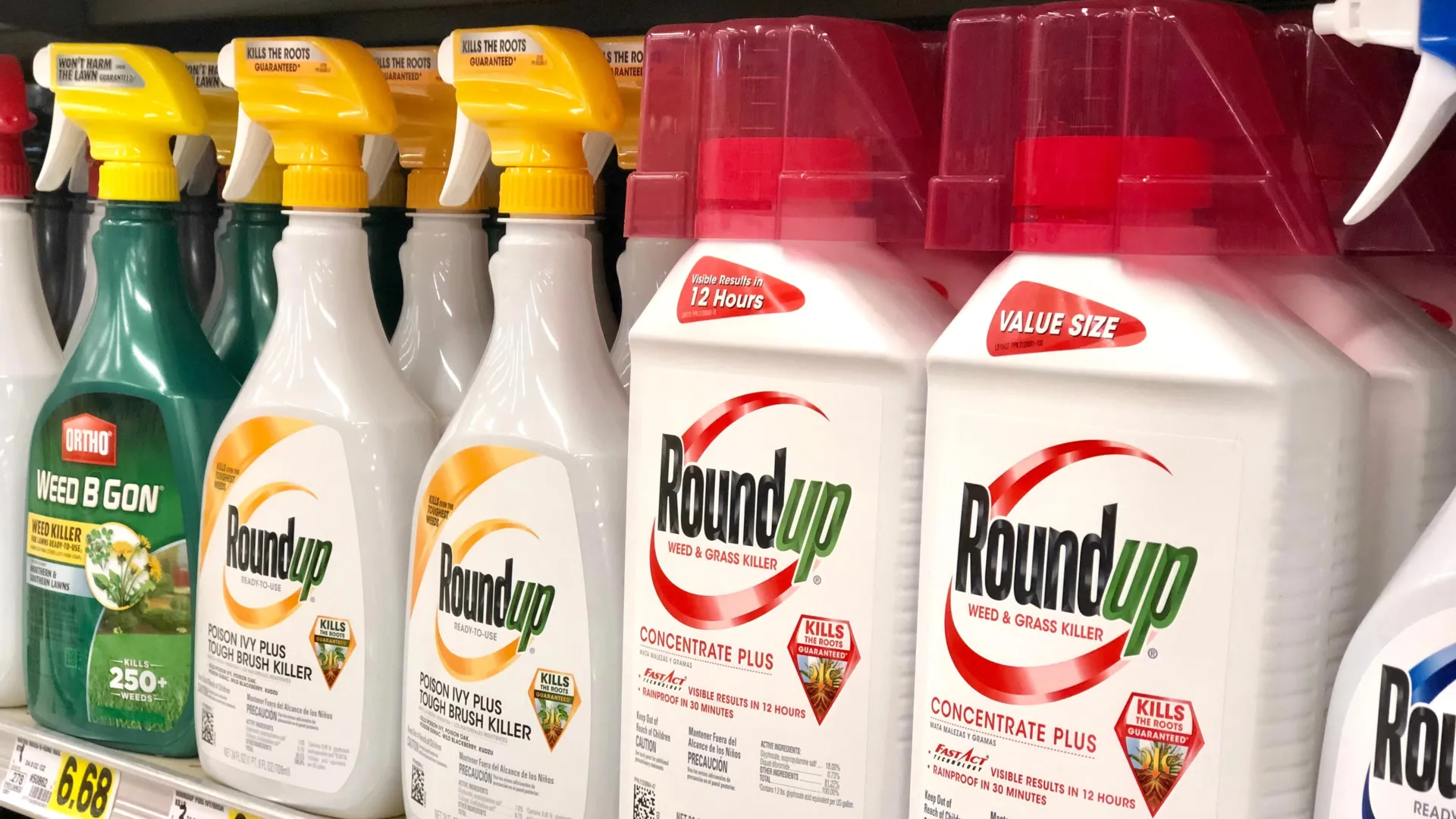
    For decades, Roundup was closely associated with glyphosate, the herbicide first registered in the United States in 1974. Today, the answer is more complicated. Home-use Roundup products found in many hardware and garden stores have been reformulated without glyphosate and may contain ingredients such as triclopyr, fluazifop, and diquat. But glyphosate remains in professional and agricultural Roundup products used in farming, landscaping, and other commercial settings.

    Testing Bacteria From Nature, Farms, and Hospitals

    To investigate the connection, Centrón and her colleagues analyzed 68 bacterial strains collected in 2018 and 2020 from sediment in a protected nature reserve in the Paraná delta, a wetland region north of Buenos Aires. Although herbicides have never been applied inside the reserve, glyphosate is commonly used in nearby agricultural areas.

    The researchers examined how resistant each strain was to 16 commonly used antibiotics, including ampicillin combined with sulbactam, meropenem, tetracycline, and vancomycin. They also tested resistance to pure glyphosate and glyphosate-based herbicides, which are among the most widely used weedkillers worldwide.

    The findings were then compared with 19 bacterial strains obtained from local hospitals, including multidrug-resistant species. An additional 15 strains came from feedlots and agricultural soils affected by herbicide use.

    Hospital Superbugs Also Resist Glyphosate

    The hospital strains showed widespread antimicrobial resistance. Individual strains were resistant to between one and 16 of the antibiotics tested. Particularly concerning was the finding that 74% were resistant to carbapenems, a class of broad-spectrum antibiotics often reserved as a last line of defense against serious infections.

    All of the hospital-derived strains were also highly resistant to glyphosate and glyphosate-based herbicides.

    “This means that if these bacteria enter the environment through untreated wastewater from hospitals, they could go on to thrive in agricultural areas where glyphosate is used,” said first author Dr. Camila Knecht from Dr. Centrón’s research group.

    The 68 strains collected from the Paraná delta represented 15 different genera, including Acinetobacter, Pseudomonas, Exiguobacterium, and Chryseobacterium. Every one of them displayed at least some resistance to glyphosate and glyphosate-based herbicides, despite the fact that these chemicals had never been applied within the reserve itself.

    Among the environmental strains, Enterobacter species tolerated the highest glyphosate concentrations, surviving levels of up to 80 milligrams per milliliter. In contrast, Bacillus species, which are commonly found in soil, were especially sensitive. Their growth was inhibited at concentrations as low as 2.5 milligrams per milliliter. High glyphosate resistance was also observed in strains isolated from hospital infections that showed extreme drug resistance.

    Resistant Bacteria Share Similar Genetic Backgrounds

    The researchers then constructed a genetic “family tree” using all 102 bacterial strains included in the study. They found that bacteria with the greatest glyphosate resistance were often closely related, regardless of whether they originated from hospitals, farms, or the Paraná delta.

    For example, the same bacterial genera showed glyphosate resistance across all three environments.

    “In the environment, the use of glyphosate leads to the evolution of resistant bacteria in impacted soils, whereas the use of antibiotics favors their evolution in hospitals. Bacteria carrying antibiotic resistance genes can spread and breed between those two niches in both directions and in multiple ways, with the water cycle playing a key role in transmission,” concluded coauthor Dr. Jochen A Müller, a group leader at Karlsruhe Institute of Technology.

    Concerns About Glyphosate and Public Health

    Glyphosate has long been the subject of scientific and regulatory debate. Research has shown that it can harm arthropods (in particular bees), and the International Agency for Research on Cancer classifies it as a probable human carcinogen.

    Several European countries have already restricted some uses of the herbicide. France, Belgium, and the Netherlands have banned glyphosate for household applications, while Germany currently prohibits its use in public spaces.

    Based on the findings, the researchers argue that pesticide regulations should take antibiotic resistance into account before products reach the market.

    “Policies for the use of any pesticide, as well as its metabolites, should stipulate the requirement for co-selection testing with antibiotics before marketing. Labels should include a warming that genes for antibiotic resistance can spread from glyphosate-contaminated soils to hospitals through untreated water,” counseled Centrón.