Category: Scientists

  • MIT scientists discover hidden 3D genome loops that survive cell division

    MIT scientists discover hidden 3D genome loops that survive cell division

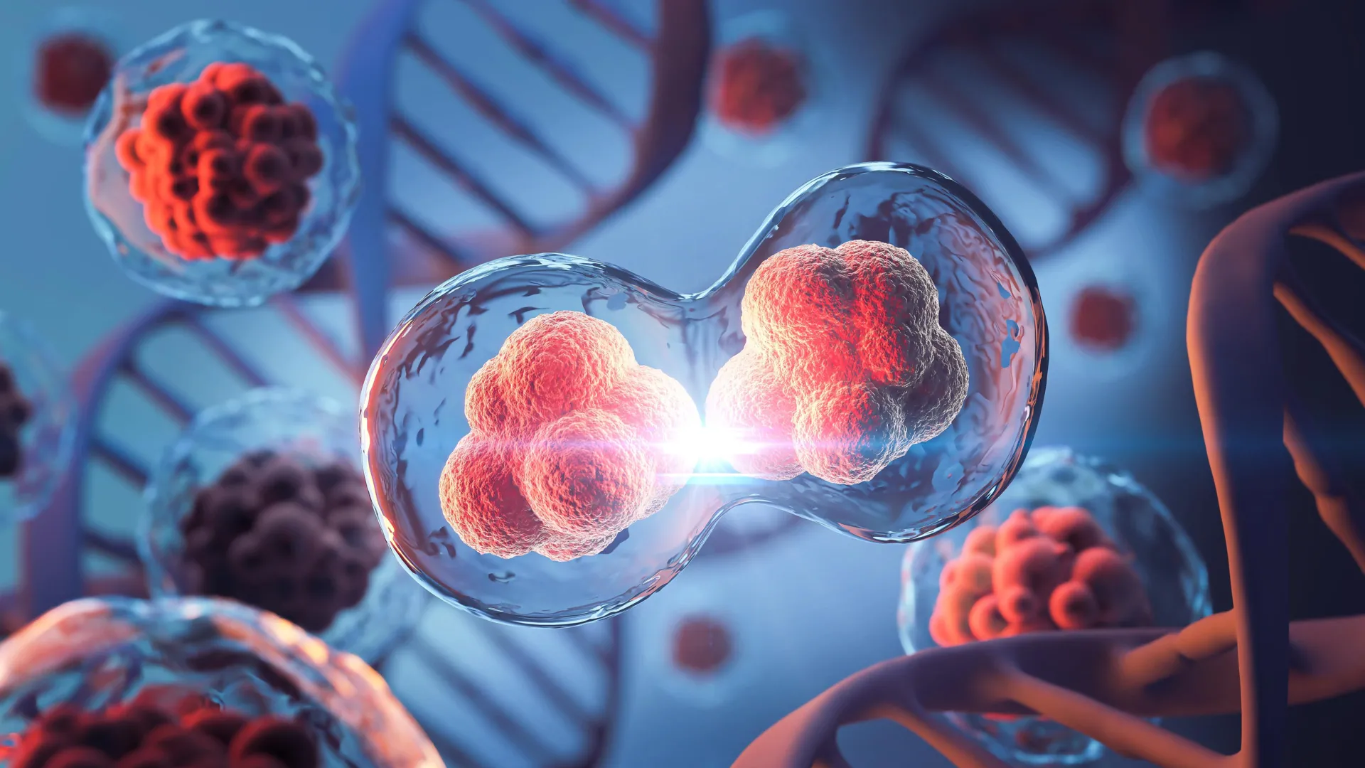
    Before a cell can split into two, it must first copy all of its chromosomes so each new cell inherits a complete set of genetic material. For years, researchers believed that as this process unfolded, the genome’s intricate three-dimensional shape temporarily vanished.

    After division, scientists thought, the DNA would slowly rebuild its complex, folded structure, which is key to regulating which genes are active in a given cell.

    Now, new research from MIT reveals that this long-accepted model isn’t quite right. Using a powerful, high-resolution genome mapping method, the team found that small 3D loops — connections between regulatory DNA elements and genes — remain intact even during cell division, a stage known as mitosis.

    “This study really helps to clarify how we should think about mitosis. In the past, mitosis was thought of as a blank slate, with no transcription and no structure related to gene activity. And we now know that that’s not quite the case,” says Anders Sejr Hansen, an associate professor of biological engineering at MIT. “What we see is that there’s always structure. It never goes away.”

    DNA Loops That Help Cells “Remember”

    The researchers also discovered that these DNA loops actually strengthen as chromosomes condense in preparation for division. This tightening brings distant regulatory elements closer together, encouraging them to stick. According to the team, this could allow cells to “remember” which genetic interactions existed before division and reestablish them afterward.

    “The findings help to bridge the structure of the genome to its function in managing how genes are turned on and off, which has been an outstanding challenge in the field for decades,” says Viraat Goel PhD ’25, the lead author of the study.

    Hansen and Edward Banigan, a research scientist at MIT’s Institute for Medical Engineering and Science, are senior authors of the study, published in Nature Structural and Molecular Biology. Co-authors include MIT professors Leonid Mirny and Gerd Blobel of the University of Pennsylvania’s Perelman School of Medicine.

    Mapping the Hidden Architecture of DNA

    Over the last two decades, scientists have uncovered that DNA in the cell nucleus organizes itself into 3D loops. Many of these loops allow genes to interact with distant regulatory regions — sometimes millions of base pairs apart — while others form during mitosis to pack chromosomes tightly.

    Most of this mapping has relied on a tool called Hi-C, a technique co-developed by MIT researchers and Job Dekker at the University of Massachusetts Chan Medical School. Hi-C works by cutting DNA into small fragments and linking together those that are near each other in the cell nucleus before sequencing them to identify which regions interact.

    While effective, Hi-C lacks the resolution needed to detect fine-scale interactions between genes and regulatory sequences known as enhancers. Enhancers are short DNA segments that activate genes by binding to promoters, the regions where transcription begins.

    A Breakthrough Tool: Region-Capture Micro-C

    In 2023, Hansen and colleagues developed a next-generation technique that can map genome structures with up to 1,000 times greater precision. The method, called Region-Capture Micro-C (RC-MC), uses a different enzyme to cut DNA into evenly sized fragments and focuses on a smaller portion of the genome. This allows researchers to create highly detailed 3D maps of targeted DNA regions.

    Using RC-MC, the team identified a new structural feature they named “microcompartments.” These are tiny, densely connected loops that form when nearby enhancers and promoters attach to one another.

    Earlier work had shown that microcompartments formed by a different mechanism than larger 3D genome structures, but the team didn’t yet understand how. To explore this, they decided to examine what happens to these structures as cells go through mitosis. During this phase, chromosomes compact dramatically to ensure they can be duplicated and evenly distributed between daughter cells. As this occurs, large genome domains known as A/B compartments and topologically associating domains (TADs) typically disappear.

    Unexpected Stability During Cell Division

    The researchers expected microcompartments to vanish as well. To test this, they monitored cells throughout the entire division cycle to see how these loops behaved before and after mitosis.

    “During mitosis, it has been thought that almost all gene transcription is shut off. And before our paper, it was also thought that all 3D structure related to gene regulation was lost and replaced by compaction. It’s a complete reset every cell cycle,” Hansen says.

    To their surprise, the loops didn’t disappear. In fact, they became even more pronounced as cells divided.

    “We went into this study thinking, well, the one thing we know for sure is that there’s no regulatory structure in mitosis, and then we accidentally found structure in mitosis,” Hansen says.

    Using their technique, the researchers also confirmed that larger structures such as A/B compartments and TADs do disappear during mitosis, as had been seen before.

    “This study leverages the unprecedented genomic resolution of the RC-MC assay to reveal new and surprising aspects of mitotic chromatin organization, which we have overlooked in the past using traditional 3C-based assays. The authors reveal that, contrary to the well-described dramatic loss of TADs and compartmentalization during mitosis, fine-scale “microcompartments” — nested interactions between active regulatory elements — are maintained or even transiently strengthened,” says Effie Apostolou, an associate professor of molecular biology in medicine at Weill Cornell Medicine, who was not involved in the study.

    Explaining a Mysterious Burst of Gene Activity

    The discovery may also explain a long-observed burst in gene transcription that occurs near the end of mitosis. Since the 1960s, scientists believed transcription stopped entirely during cell division. However, studies in 2016 and 2017 revealed a brief spike of gene activity before it shuts down again.

    In their new study, the MIT team found that during mitosis, microcompartments are more likely to be found near the genes that spike during cell division. They also discovered that these loops appear to form as a result of the genome compaction that occurs during mitosis. This compaction brings enhancers and promoters closer together, allowing them to stick together to form microcompartments.

    Once formed, the loops that constitute microcompartments may activate gene transcription somewhat by accident, which is then shut off by the cell. When the cell finishes dividing, entering a state known as G1, many of these small loops become weaker or disappear.

    “It almost seems like this transcriptional spiking in mitosis is an undesirable accident that arises from generating a uniquely favorable environment for microcompartments to form during mitosis,” Hansen says. “Then, the cell quickly prunes and filters many of those loops out when it enters G1.”

    Because chromosome compaction can also be influenced by a cell’s size and shape, the researchers are now exploring how variations in those features affect the structure of the genome and in turn, gene regulation.

    “We are thinking about some natural biological settings where cells change shape and size, and whether we can perhaps explain some 3D genome changes that previously lack an explanation,” Hansen says. “Another key question is how does the cell then pick what are the microcompartments to keep and what are the microcompartments to remove when you enter G1, to ensure fidelity of gene expression?”

    The research was funded in part by the National Institutes of Health, a National Science Foundation CAREER Award, the Gene Regulation Observatory of the Broad Institute, a Pew-Steward Scholar Award for Cancer Research, the Mathers Foundation, the MIT Westaway Fund, the Bridge Project of the Koch Institute and Dana-Farber/Harvard Cancer Center, and the Koch Institute Support (core) Grant from the National Cancer Institute.

  • Scientists just found a hidden factor behind Earth’s methane surge

    Scientists just found a hidden factor behind Earth’s methane surge

    Roughly two-thirds of all emissions of atmospheric methane — a highly potent greenhouse gas that is warming planet Earth — come from microbes that live in oxygen-free environments like wetlands, rice fields, landfills and the guts of cows.

    Tracking atmospheric methane to its specific sources and quantifying their importance remains a challenge, however. Scientists are pretty good at tracing the sources of the main greenhouse gas, carbon dioxide, to focus on mitigating these emissions. But to trace methane’s origins, scientists often have to measure the isotopic composition of methane’s component atoms, carbon and hydrogen, to use as a fingerprint of various environmental sources.

    A new paper by researchers at the University of California, Berkeley, reveals how the activity of one of the main microbial enzymes involved in producing methane affects this isotope composition. The finding could change how scientists calculate the contributions of different environmental sources to Earth’s total methane budget.

    “When we integrate all the sources and sinks of carbon dioxide into the atmosphere, we kind of get the number that we’re expecting from direct measurement in the atmosphere. But for methane, large uncertainties in fluxes exist — within tens of percents for some of the fluxes — that challenge our ability to precisely quantify the relative importance and changes in time of the sources,” said UC Berkeley postdoctoral fellow Jonathan Gropp, who is first author of the paper. “To quantify the actual sources of methane, you need to really understand the isotopic processes that are used to constrain these fluxes.”

    Gropp teamed up with a molecular biologist and a geochemist at UC Berkeley to, for the first time, employ CRISPR to manipulate the activity of this key enzyme to reveal how these methanogens interact with their food supply to produce methane.

    “It is well understood that methane levels are rising, but there is a lot of disagreement on the underlying cause,” said co-author Dipti Nayak, UC Berkeley assistant professor of molecular and cell biology. “This study is the first time the disciplines of molecular biology and isotope biogeochemistry have been fused to provide better constraints on how the biology of methanogens controls the isotopic composition of methane.”

    Many elements have heavier or lighter versions, called isotopes, that are found in small proportions in nature. Humans are about 99% carbon-12 and 1% carbon-13, which is slightly heavier because it has an extra neutron in its nucleus. The hydrogen in water is 99.985% hydrogen-1 and 0.015% deuterium or hydrogen-2, which is twice as heavy because it has a neutron in its nucleus.

    The natural abundances of isotopes are reflected in all biologically produced molecules and variations can be used to study and fingerprint various biological metabolisms.

    “Over the last 70 years, people have shown that methane produced by different organisms and other processes can have distinctive isotopic fingerprints,” said geochemist and co-author Daniel Stolper, UC Berkeley associate professor of earth and planetary science. “Natural gas from oil deposits often looks one way. Methane made by the methanogens within cow guts looks another way. Methane made in deep sea sediments by microorganisms has a different fingerprint. Methanogens can consume or ‘eat’, if you will, a variety of compounds including methanol, acetate or hydrogen; make methane; and generate energy from the process. Scientists have commonly assumed that the isotopic fingerprint depends on what the organisms are eating, which often varies from environment to environment, creating our ability to link isotopes to methane origins.”

    “I think what’s unique about the paper is, we learned that the isotopic composition of microbial methane isn’t just based on what methanogens eat,” Nayak said. “What you ‘eat’ matters, of course, but the amount of these substrates and the environmental conditions matter too, and perhaps more importantly, how microbes react to those changes.”

    “Microbes respond to the environment by manipulating their gene expression, and then the isotopic compositions change as well,” Gropp said. “This should cause us to think more carefully when we analyze data from the environment.”

    The paper will appear Aug. 14 in the journal Science.

    Vinegar- and alcohol-eating microbes

    Methanogens — microorganisms that are archaea, which are on an entirely separate branch of the tree of life from bacteria — are essential to ridding the world of dead and decaying matter. They ingest simple molecules — molecular hydrogen, acetate or methanol, for example — excreted by other organisms and produce methane gas as waste. This natural methane can be observed in the pale Will-o’-the-wisps seen around swamps and marshes at night, but it’s also released invisibly in cow burps, bubbles up from rice paddies and natural wetlands and leaks out of landfills. While most of the methane in the natural gas we burn formed in association with hydrocarbon generation, some deposits were originally produced by methanogens eating buried organic matter.

    The isotopic fingerprint of methane produced by methanogens growing on different “food” sources has been well established in laboratory studies, but scientists have found that in the complexity of the real world, methanogens don’t always produce methane with the same isotopic fingerprint as seen in the lab. For example, when grown in the lab, species of methanogens that eat acetate (essentially vinegar), methanol (the simplest alcohol), or molecular hydrogen (H2) produce methane, CH4, with a ratio of hydrogen and carbon isotopes different from the ratios observed in the environment.

    Gropp had earlier created a computer model of the metabolic network in methanogens to understand better how the isotope composition of methane is determined. When he got a fellowship to come to UC Berkeley, Stolper and Nayak proposed that he experimentally test his model. Stolper’s laboratory specializes in measuring isotope compositions to explore Earth’s history. Nayak studies methanogens and, as a postdoctoral fellow, found a way to use CRISPR gene editing in methanogens. Her group recently altered the expression of the key enzyme in methanogens that produces the methane — methyl-coenzyme M reductase (MCR) — so that its activity can be dialed down. Enzymes are proteins that catalyze chemical reactions.

    Experimenting with these CRISPR-edited microbes — in a common methanogen called Methanosarcina acetivorans growing on acetate and methanol — the researchers looked at how the isotopic composition of methane changed when the enzyme activity was reduced, mimicking what is thought to happen when the microbes are starved for their preferred food.

    They found that when MCR is at low concentrations, cells respond by altering the activity of many other enzymes in the cell, causing their inputs and outputs to accumulate and the rate of methane generation to slow so much that enzymes begin running both backwards and forwards. In reverse, these other enzymes remove a hydrogen from carbon atoms; running forward, they add a hydrogen. Together with MCR, they ultimately produce methane (CH4). Each forward and reverse cycle requires one of these enzymes to pull a hydrogen off of the carbon and add a new one ultimately sourced from water. As a result, the isotopic composition of methane’s four hydrogen molecules gradually comes to reflect that of the water, and not just their food source, which starts with three hydrogens.

    This is different from typical assumptions for growth on acetate and methanol that assume no exchange between hydrogen derived from water and that from the food source.

    “This isotope exchange we found changes the fingerprint of methane generated by acetate and methanol consuming methanogens vs. that typically assumed. Given this, it might be that we have underestimated the contribution of the acetate-consuming microbes, and they might be even more dominant than we have thought,” Gropp said. “We’re proposing that we at least should consider the cellular response of methanogens to their environment when studying isotopic composition of methane.”

    Beyond this study, the CRISPR technique for tuning production of enzymes in methanogens could be used to manipulate and study isotope effects in other enzyme networks broadly, which could help researchers answer questions about geobiology and the Earth’s environment today and in the past.

    “This opens up a pathway where modern molecular biology is married with isotope-geochemistry to answer environmental problems,” Stolper said. “There are an enormous number of isotopic systems associated with biology and biochemistry that are studied in the environment; I hope we can start looking at them in the way molecular biologists now are looking at these problems in people and other organisms — by controlling gene expression and looking at how the stable isotopes respond.”

    For Nayak, the experiments are also a big step in discovering how to alter methanogens to derail production of methane and redirect their energy to producing useful products instead of an environmentally destructive gas.

    “By reducing the amount of this enzyme that makes methane and by putting in alternate pathways that the cell can use, we can essentially give them another release valve, if you will, to put those electrons, which they were otherwise putting in carbon to make methane, into something else that would be more useful,” she said.

    Other co-authors of the paper are Markus Bill of Lawrence Berkeley National Laboratory and former UC Berkeley postdoc Rebekah Stein, and Max Lloyd, who is a professor at Penn State University. Gropp was supported by a fellowship from the European Molecular Biology Organization. Nayak and Stolper were funded, in part, by Alfred B. Sloan Research Fellowships. Nayak also is an investigator with the Chan-Zuckerberg Biohub.

  • Scientists optimize biohybrid ray development with machine learning

    Scientists optimize biohybrid ray development with machine learning

    The Harvard John A. Paulson School of Engineering and Applied Sciences (SEAS) and NTT Research, Inc., a division of NTT, announced the publication of research showing an application of machine-learning directed optimization (ML-DO) that efficiently searches for high-performance design configurations in the context of biohybrid robots. Applying a machine learning approach, the researchers created mini biohybrid rays made of cardiomyocytes (heart muscle cells) and rubber with a wingspan of about 10 mm that are approximately two times more efficient at swimming than those recently developed under a conventional biomimetic approach.

    A team led by Harvard SEAS Postdoctoral Fellow John Zimmerman and including NTT Research Medical and Health Informatics Scientist Ryoma Ishii, Harvard SEAS Tarr Family Professor of Bioengineering and Applied Physics Kevin Kit Parker, and members of the Harvard SEAS Disease Biophysics Group led by Parker demonstrated this research in a new paper published in Science Robotics titled, “Bioinspired Design of a Tissue Engineered Ray with Machine Learning.”

    “This research seeks to answer a fundamental question in the development of biohybrid robots, in this case the marine ray: How do we select fin geometries to operate under novel working environments while preserving natural scaling laws in terms of swimming speed and efficiency,” said Ishii, who also works as a visiting scientist for Harvard University. “Our research indicates the application of ML-DO, inspired by protein engineering, offers a more efficient and less computationally intensive path forward in automating the creation of muscular structure-function relationships.”

    Limitations of the Biomimetic Approach

    In biomimetic design, the conventional approach to biohybrids, engineers form functional devices by recreating existing biological structures. That approach, however, has limits. For biohybrid lifeforms that resemble batoid fishes (skates and rays), for example, there is a wide range of natural aspect ratios and fin morphologies. Which ones do you mimic? Also, biomimetics may neglect the natural biomechanical and hydrodynamic forces that govern how fast an organism can swim based on its size and body kinematics, leading to inefficient muscle mass and limited swimming speeds.

    In that light, the motivating question in this study became: How to select fin geometries that operate under novel working environments while preserving natural scaling laws in terms of swimming speed and efficiency?

    The Design Breakthroughs of Machine Learning

    The multi-disciplinary and iterative nature of the problem required computationally intensive modeling, but the team believed that directed optimization by machine learning (ML-DO) would enable an efficient search for fin designs that maximized their relative swimming speeds. They based their hypothesis in part on a trial function that demonstrated an approximately 40 percent improvement of ML-DO over other leading methods in recognizing known high-rank sequences. Testing the assumption involved three steps: 1) developing an algorithm for expressing a multitude of different fin geometries; 2) describing a generalized ML-DO approach for searching within a large discontinuous configuration space; and 3) using this methodology to identify biohybrid fin geometries for high-performance swimming with smooth and orderly flow.

    The ML-DO-driven results included a quantitative exploration of fin structure-function relationships and reconstruction of general trends in open-sea batoid morphology, as well as a winning design: Fins with large aspect ratios and fine tapered tips, which preserved their utility across multiple length-scales of swimming. On that basis, the team built biohybrid mini-rays out of engineered cardiac muscle tissue, which were capable of self-propelled swimming at the millimeter length scale and demonstrated improved swimming efficiencies approximately two times greater than observed in previous biomimetic designs.

    Looking Ahead

    While promising, researchers note that additional work is needed to completely match natural scaling laws. While the devices presented in this study demonstrated greater efficiency than other recent biomimetic designs, they were still slightly less efficient on average than naturally occurring marine lifeforms.

    In the future, researchers expect to continue the development of biohybrid robotics for use cases including remote sensors, probes for dangerous working environments and as therapeutic delivery vehicles. Researchers believe that the ML-DO-informed approach better mimics the selective pressures of evolution, enabling them to better understand how biological tissues are shaped — both in a healthy physiology as well as the maladaptive pathophysiology of disease. Additionally, this research advances scientific understanding of 3D organ biofabrication, such as a biohybrid heart.

    “The Harvard Biophysics Group entered into a joint research agreement with NTT Research two years ago aiming to fundamentally interrogate our understanding of cardiac physiology, with an eye towards advancing the development of biohybrid devices, including biohybrid robotics and biohybrid human heart,” Parker said. “This paper indicates the positive progress our shared research has achieved so far, and I am excited to see what the future of our collaboration has in store.”

    In 2022, NTT Research and Harvard SEAS announced a three-year joint research agreement to engineer a model of the human heart, study fundamental laws of muscular pumps and apply joint findings to the development of a cardiovascular bio digital twin model.

  • Scientists predict what will be top of the crops in UK by 2080 due to climate change

    Scientists predict what will be top of the crops in UK by 2080 due to climate change

    While climate change is likely to present significant challenges to agriculture in coming decades, it could also mean that crops such as chickpeas, soyabeans and oranges are widely grown across the UK, and home-produced hummus, tofu and marmalade are a common sight on our supermarket shelves by 2080.

    A new study led by the UK Centre for Ecology & Hydrology (UKCEH) in collaboration with the University of East Anglia (UEA) predicts that future warmer temperatures in this country would be suitable for a variety of produce such as oranges, chickpeas and okra that are traditionally grown in warmer parts of the world.

    Scientists investigated the future suitability for over 160 existing and new food crops in different regions of the UK under warming scenarios of 2 and 4 degrees Celsius compared to pre-industrial times.

    The modelling study, part of the OpenCLIM research project, is the most comprehensive research of its kind to date and is also the first to show how suitability for certain produce will vary across the UK. It provides mapped projections for every 1km square in the UK, giving valuable information to the farming and food sectors on the future opportunities and challenges of cultivating new crops here.

    Need for resilience

    Lead author Dr John Redhead, a Spatial Ecologist at UKCEH, said: “Our climate is expected to change substantially over coming decades at a time when there will be rising demand for food due to population growth. It is therefore essential that arable farming becomes more resilient; one possible solution is growing different crops that are more suited to the new local conditions.”

    Climate change is already having a major impact on UK agriculture, either affecting crop plants or the ability of farmers to manage them effectively. There have been several years of record low yields caused by extreme weather, often in combination, such as a wet winter followed by a particularly dry spring, while new agricultural pests and diseases are becoming established or increasing with climate change.

    Winners and losers

    Many new crops highlighted in the UKCEH-led study — funded by UK Research & Innovation (UKRI) and published in the journal Climate Resilience and Sustainability – are not only more tolerant of hotter, drier summers but also benefit from milder, wetter winters.

    The research looked at whether future climate scenarios would be more or less suitable for crops, compared with recent average UK temperatures, estimated at being around 0.5 degrees Celsius above pre-industrial times. The key findings were:

    • Climate change results in increased suitability for many current and potential new crops across much of the UK.
    • However, some key produce is likely to become harder to grow in the South East and East Anglia, the UK’s most productive arable regions. The climate in these areas is expected to become less suitable for wheat and strawberries under the 2 degrees of warming scenario, while 4 degrees of warming would result in reduced suitably for other major crops including onions and oats.
    • There would be substantial increases in suitability for a broad range of crops not currently widely grown in the UK — such as sunflower, durum wheat, soybeans, cow peas, chickpeas, citrus fruit and okra — as well as wine grapes.

    Diversity offers multiple benefits

    The study authors say the majority of global food supply relies on a relatively small number of crops, and greater diversity would boost food security by improving climate resilience.

    There would also be potentially positive impacts on wider biodiversity and people’s diets. Legumes such as chickpeas and soybeans, which have recently had their first commercial UK harvests, are important protein sources. This would support shifts from heavy meat consumption to a more balanced diet and lower carbon footprint. Legumes also add nitrogen to the soil, reducing the reliance on fertilisers.

    Risks and challenges

    While a changing climate across the UK is expected to support a range of new crops, the study shows the largest increases in suitability will be in the southwest and Scottish borders due to the benefits of rising temperatures not being cancelled out by more restricted water in the summer, especially under 4 degrees of warming.

    However, many areas outside the South East and East Anglia have small field sizes, variable topography and are far from the current food processing and supply chains, limiting potential shifts in production to these regions. There are also economic risks investing in new agronomic practices and technology, and potential environmental risks in introducing crops to new locations, including interactions with pollinators, wild crop relatives and pests.

    Dr Redhead said: “Clearly, it’s unlikely to be feasible just to switch large-scale food production from Britain’s agricultural heartlands of southeastern England to Scotland, for example. However, climate change is happening now, and its impacts will increase by 2080, so whatever action is taken will involve big challenges in terms of where our food comes from and the way our agricultural landscapes are managed.”

    Solutions

    Methods to cope with the challenges highlighted in the study include:

    • Further research into the viability of the crops that are identified as ‘winners’.
    • Changing our agricultural supply and distribution networks to better support farmers growing new crops in new locations.
    • Adopting new agricultural systems such as paludiculture (wet farming) and indoor vertical farming.
    • Breeding and growing more heat- or drought-resilient varieties of existing staple crops.

    No-one can definitively say whether a certain crop will be possible or profitable in 2080, but the study addresses the gap in knowledge about climatic suitability of crops in particular regions, which is currently limiting uptake of new produce.

    Study co-author Professor Rachel Warren of the Tyndall Centre for Climate Change Research at UEA said: “Without such data, agricultural systems are likely to be ‘locked in’ to current crops, with adaptations failing to keep pace with climate change or relying on practices that exacerbate its impacts, such as heavy irrigation that would increase water scarcity.

    “Major changes to agricultural systems and diets can take decades to implement and so our long-term projections provide important information well ahead of time for farmers, supermarkets, researchers, policymakers and the public on the opportunities, challenges and trade-offs involved in adapting to the impacts of climate change.”

    The study authors say their horizon-scanning approach on how climate change affects suitability of crops could be transferable to other countries.

  • Scientists ‘turn up the heat’ on understanding coffee wilt disease which threatens our favorite daily brew

    Scientists ‘turn up the heat’ on understanding coffee wilt disease which threatens our favorite daily brew

    Scientists, including those from Imperial College London, University of Oxford and CABI, have ‘turned up the heat’ on how repeated outbreaks of coffee wilt disease threatened arabica and robusta varieties of our favourite daily coffee brew.

    The scientists, who present their findings in the journal PLoS Biology, say the fungal pathogenFusarium xylarioides continues to pose a significant threat to coffee production and incomes across sub-Saharan Africa.

    Their work supports earlier findings, based on DNA markers and crossing experiments which suggested that F. xylarioides is a species complex containing distinct, host-specific populations.

    The scientists confirm the presence of genetically differentiated lineages with different host-specificity for arabica and robusta coffee and show that the fungal pathogen F. xylarioides repeatedly took up “horizontally transferred” segments of DNA from a related fungal Fusarium taxa which contributed to successive outbreaks of the disease.

    Primary source of income for more than 12 million households

    Coffee is a primary source of income for more than 12 million households in Africa and contributes a significant proportion of tax revenue in a number of these countries, with Ethiopia, for example, recording an export value of $762.8m annually. Yet repeated outbreaks of coffee wilt disease have continued to decimate coffee production in east and central Africa since the 1920s, affecting yields and farmer incomes.

    As part of this study, the scientists compared the genomes of 13 historical strains of F. xylarioides spanning six decades from CABI’s Culture Collection, to identify the evolutionary process behind these repeated outbreaks of coffee wilt disease.

    Dr Matthew Ryan, Senior Research Lead, Biological Resources at CABI and a co-author of the paper, co-supervised lead author of the research, Dr Lily Peck, of Imperial College London, together with Professor Tim Barracloughfrom the University of Oxford.

    Crucial to understand genetic structure and evolutionary potential

    Dr Peck said, “To improve the disease management of fungal pathogens such as Fusarium xylarioides, it is crucial to understand their genetic structure and evolutionary potential.

    “We found that F. xylarioides comprised at least four distinct lineages: one host-specific to arabica, one to robusta and two historic lineages isolated from various coffee species.

    “The presence/absence of large genomic regions across these lineages showed that horizontal transfers of effector genes, namely genes that are important in establishing successful infection, contributed to establishing host specificity.”

    Dr Peck also said that multiple transfers into F. xylarioides populations matched different parts of the Fusarium oxysporum mobile pathogenicity chromosome and were enriched in effector genes and transposons.

    Transposons, also known as “jumping genes,” are DNA sequences that can move around in genomes. They are found in many organisms, including bacteria, yeasts, humans, mice, fruit flies and frogs.

    Shown by transcriptomics to be highly expressed during infection

    Effector genes and other carbohydrate-active enzymes important in the breakdown of plant cell walls were shown by transcriptomics to be highly expressed during infection of arabica coffeeby the pathogenic arabica strains.

    Professor Barraclough said, “Widespread sharing of specific transposons between F. xylarioides and F. oxysporum, and the correspondence of a putative horizontally transferred region to a Starship (recently identified large mobile elements which are involved in fungal horizontal gene transfers), reinforce the inference of horizontal transfers and suggest that mobile elements were involved.

    “Our results support the hypothesis that horizontal gene transfers contributed to the repeated emergence of coffee wilt disease.”

    Potentially lucrative export of various coffee varieties

    Dr Ryan said the research findings potentially show what is happening more widely in relation to fungi and gives scientists the potential to further build on Dr Peck’s study in the future.

    “The fact that horizontal gene transfers between two different species of Fusarium contributed to the repeated emergence of coffee wilt disease is a key mechanism to understand what is happening more widely in fungi — to what extent, we just do not know,” he said.

    The scientists suggest that horizontal transfer could have occurred in a ‘shared niche,’ as both F. xylarioides and F. oxysporum are soil-borne pathogens and have been isolated from the roots and wood of coffee wilt-diseased trees in Ethiopia and central Africa, and from the roots of a banana tree in central Africa.

    They add that knowledge of horizontal transfer mechanisms and putative donor taxa might help to design future intercropping strategies that minimize the risk of transfer of effector genes between closely related Fusarium taxa.

  • Scientists preserve DNA in an amber-like polymer

    Scientists preserve DNA in an amber-like polymer

    In the movie “Jurassic Park,” scientists extracted DNA that had been preserved in amber for millions of years, and used it to create a population of long-extinct dinosaurs.

    Inspired partly by that film, MIT researchers have developed a glassy, amber-like polymer that can be used for long-term storage of DNA, whether entire human genomes or digital files such as photos.

    Most current methods for storing DNA require freezing temperatures, so they consume a great deal of energy and are not feasible in many parts of the world. In contrast, the new amber-like polymer can store DNA at room temperature while protecting the molecules from damage caused by heat or water.

    The researchers showed that they could use this polymer to store DNA sequences encoding the theme music from Jurassic Park, as well as an entire human genome. They also demonstrated that the DNA can be easily removed from the polymer without damaging it.

    “Freezing DNA is the number one way to preserve it, but it’s very expensive, and it’s not scalable,” says James Banal, a former MIT postdoc. “I think our new preservation method is going to be a technology that may drive the future of storing digital information on DNA.”

    Banal and Jeremiah Johnson, the A. Thomas Geurtin Professor of Chemistry at MIT, are the senior authors of the study, published in the Journal of the American Chemical Society. Former MIT postdoc Elizabeth Prince and MIT postdoc Ho Fung Cheng are the lead authors of the paper.

    Capturing DNA

    DNA, a very stable molecule, is well-suited for storing massive amounts of information, including digital data. Digital storage systems encode text, photos, and other kind of information as a series of 0s and 1s. This same information can be encoded in DNA using the four nucleotides that make up the genetic code: A, T, G, and C. For example, G and C could be used to represent 0 while A and T represent 1.

    DNA offers a way to store this digital information at very high density: In theory, a coffee mug full of DNA could store all of the world’s data. DNA is also very stable and relatively easy to synthesize and sequence.

    In 2021, Banal and his postdoc advisor, Mark Bathe, an MIT professor of biological engineering, developed a way to store DNA in particles of silica, which could be labeled with tags that revealed the particles’ contents. That work led to a spinout called Cache DNA.

    One downside to that storage system is that it takes several days to embed DNA into the silica particles. Furthermore, removing the DNA from the particles requires hydrofluoric acid, which can be hazardous to workers handling the DNA.

    To come up with alternative storage materials, Banal began working with Johnson and members of his lab. Their idea was to use a type of polymer known as a degradable thermoset, which consists of polymers that form a solid when heated. The material also includes cleavable links that can be easily broken, allowing the polymer to be degraded in a controlled way.

    “With these deconstructable thermosets, depending on what cleavable bonds we put into them, we can choose how we want to degrade them,” Johnson says.

    For this project, the researchers decided to make their thermoset polymer from styrene and a cross-linker, which together form an amber-like thermoset called cross-linked polystyrene. This thermoset is also very hydrophobic, so it can prevent moisture from getting in and damaging the DNA. To make the thermoset degradable, the styrene monomers and cross-linkers are copolymerized with monomers called thionolactones. These links can be broken by treating them with a molecule called cysteamine.

    Because styrene is so hydrophobic, the researchers had to come up with a way to entice DNA — a hydrophilic, negatively charged molecule — into the styrene.

    To do that, they identified a combination of three monomers that they could turn into polymers that dissolve DNA by helping it interact with styrene. Each of the monomers has different features that cooperate to get the DNA out of water and into the styrene. There, the DNA forms spherical complexes, with charged DNA in the center and hydrophobic groups forming an outer layer that interacts with styrene. When heated, this solution becomes a solid glass-like block, embedded with DNA complexes.

    The researchers dubbed their method T-REX (Thermoset-REinforced Xeropreservation). The process of embedding DNA into the polymer network takes a few hours, but that could become shorter with further optimization, the researchers say.

    To release the DNA, the researchers first add cysteamine, which cleaves the bonds holding the polystyrene thermoset together, breaking it into smaller pieces. Then, a detergent called SDS can be added to remove the DNA from polystyrene without damaging it.

    Storing information

    Using these polymers, the researchers showed that they could encapsulate DNA of varying length, from tens of nucleotides up to an entire human genome (more than 50,000 base pairs). They were able to store DNA encoding the Emancipation Proclamation and the MIT logo, in addition to the theme music from “Jurassic Park.”

    After storing the DNA and then removing it, the researchers sequenced it and found that no errors had been introduced, which is a critical feature of any digital data storage system.

    The researchers also showed that the thermoset polymer can protect DNA from temperatures up to 75 degrees Celsius (167 degrees Fahrenheit). They are now working on ways to streamline the process of making the polymers and forming them into capsules for long-term storage.

    Cache DNA, a company started by Banal and Bathe, with Johnson as a member of the scientific advisory board, is now working on further developing DNA storage technology. The earliest application they envision is storing genomes for personalized medicine, and they also anticipate that these stored genomes could undergo further analysis as better technology is developed in the future.

    “The idea is, why don’t we preserve the master record of life forever?” Banal says. “Ten years or 20 years from now, when technology has advanced way more than we could ever imagine today, we could learn more and more things. We’re still in the very infancy of understanding the genome and how it relates to disease.”

    The research was funded by the National Science Foundation.