Category: Scientists

  • Scientists discover how gut bacteria “train” the intestine to fight inflammation

    Scientists discover how gut bacteria “train” the intestine to fight inflammation

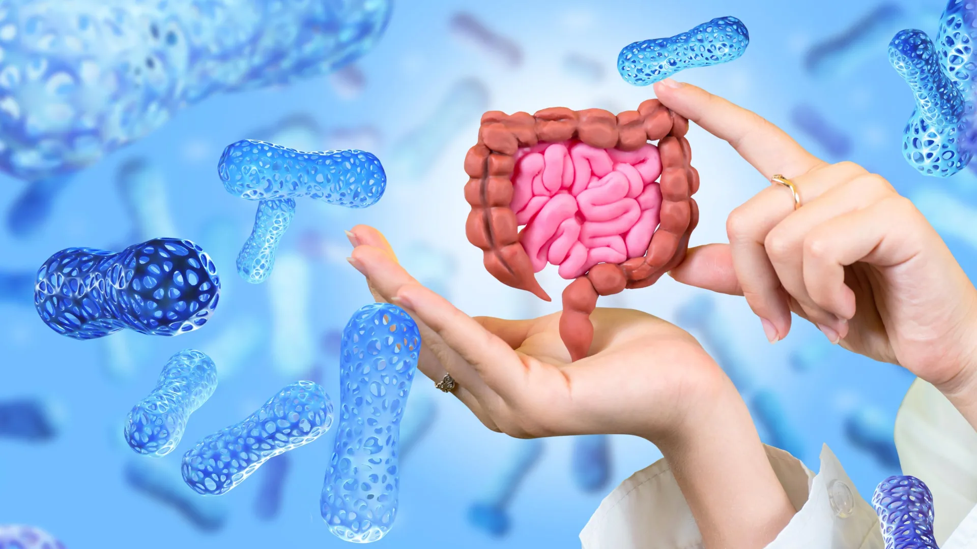
    New Northwestern Medicine research published in Nature Communications suggests that a substance produced when gut bacteria digest dietary fiber may leave a durable molecular mark on cells lining the intestine. That imprint appears to promote immune tolerance and may help protect against inflammatory bowel disease-like conditions even after exposure to the metabolite has ended.

    Yingzi Cong, PhD, the Stanley Gradowski Professor of Gastroenterology and a professor of Microbiology-Immunology and of Pathology, was senior and co-corresponding author of the study.

    The researchers say the results support the idea that beneficial compounds made by gut microbes can effectively “train” the intestinal lining, helping it preserve immune tolerance over time.

    “Our laboratory has long been interested in how the gut microbiota regulates immune responses at intestinal mucosal surfaces,” said first and co-corresponding author of the study Tianming Yu, PhD, research assistant professor of Medicine in the Division of Gastroenterology and Hepatology. “Short-chain fatty acids (SCFAs), which are produced by gut bacteria during the fermentation of dietary fiber, are known to have anti-inflammatory effects in the intestine.”

    How Butyrate May Influence Gut Immunity

    One major question, however, has remained unresolved, Yu said.

    “A large proportion of butyrate in the gut is rapidly absorbed and metabolized by intestinal epithelial cells (IECs), which limits the amount of free butyrate that can directly reach underlying immune cells,” Yu said. “This led us to ask whether butyrate might act through IECs to regulate intestinal immunity.”

    To investigate, the researchers supplied mice with butyrate in their drinking water for a set period and then ended the treatment. Two weeks after the butyrate had been withdrawn, CD4+ T-cells were still producing elevated levels of IL-10, an important anti-inflammatory cytokine that helps control intestinal inflammation.

    The treated mice were also more resistant to chemically induced colitis. Compared with untreated animals, they lost less weight, had lower levels of inflammatory markers, and developed less severe tissue damage. According to the study, this protection depended on IL-10 signaling.

    The researchers also determined that the lasting effect did not result from changes to the gut microbiome itself. In experiments involving germ-free mice, which lack all microbes, butyrate still created a persistent immune-regulating environment.

    “We found that oral butyrate treatment induces a sustained immunoregulatory response in the intestine, characterized by increased IL-10 production in CD4+ T-cells and protection from intestinal inflammation even after butyrate treatment is stopped,” Yu said. “This effect was also observed in germ-free mice, suggesting that butyrate can establish a lasting intestinal environment that does not depend on continuous microbial stimulation.”

    Intestinal Cells May Store a Lasting Biological Signal

    Yu and his colleagues next focused on intestinal epithelial cells (IECs), which form the physical boundary between the body and the microbiome. In laboratory experiments, epithelial cells exposed to butyrate caused a strong increase in IL-10 production in both mouse and human T-cells.

    “We further found that conditioned medium from butyrate-treated IECs strongly induced IL-10-producing CD4+ T-cells in both mouse and human T-cell culture systems,” Yu said. “These findings suggest that IECs secrete some immunoregulatory factors after butyrate treatment, and these factors can act on T-cells.”

    The team then used metabolomic analysis to search for molecules that might be carrying this signal. One likely candidate was N1-acetylspermidine. The compound increased IL-10 production in T-cells and appeared to account for part of the immune-regulating activity generated by epithelial cells exposed to butyrate, Yu said.

    “Mechanistically, we found that butyrate acts on IECs and induces sustained transcriptional and epigenetic activation of Sat1, an acetylpolyamine biosynthetic enzyme,” Yu said. “This promotes production of the metabolite N1-acetylspermidine, which contributes to the ability of butyrate-treated IEC-conditioned medium to induce IL-10 production in CD4⁺ T-cells.”

    The results challenge the traditional idea that intestinal epithelial cells are primarily temporary responders whose main role is to serve as a barrier. Instead, the findings suggest that these cells may retain a longer-lasting molecular record of beneficial signals from the microbiome.

    “The significance of this work is that it identifies a mechanism by which a microbiota-derived metabolite can create durable epithelial T-cell crosstalk,” Yu said. “The intestinal epithelium is often viewed as a short-lived barrier that responds rapidly to luminal stimuli. Our findings suggest that it can also retain a lasting imprint of a microbial metabolite signal. The study also introduces the idea that beneficial microbial metabolites may ‘train’ the IECs to maintain immune tolerance over time.”

    Potential Implications for Inflammatory Bowel Disease

    More research will be necessary to confirm the findings and determine whether the same mechanism operates in people, but Yu said the work could eventually have implications for inflammatory bowel disease.

    “One important next step is to determine how this epithelial metabolic pathway operates in human intestinal disease, especially in patients with inflammatory bowel disease,” Yu said. “We are interested in testing whether the butyrate-Sat1-N1-acetylspermidine pathway is altered in human IECs and whether it correlates with immune regulation or disease activity.”

    The researchers also intend to investigate other metabolites that may contribute to the effect. N1-acetylspermidine by itself did not fully account for all of the immune-regulating activity observed in the experiments.

    Over the longer term, Yu said the findings may expand scientists’ understanding of the relationship among diet, metabolites produced by gut microbes and the intestinal lining.

    “Many studies have focused on how inflammation can leave harmful memory in epithelial cells, but our findings suggest that beneficial microbial metabolites may also establish protective epithelial programs,” he said. “Understanding how diet, microbiota-derived metabolites and inflammation shape intestinal epithelial memory could open new directions for restoring intestinal immune tolerance in inflammatory bowel disease.”

    Wenjing Yang, MD, PhD, research assistant professor of Medicine in the Division of Gastroenterology and Hepatology, was co-first author of the study.

    Additional Feinberg co-authors include Suxia Yao, MD, research associate professor of Medicine in the Division of Gastroenterology and Hepatology; and Parambir Dulai, MD, associate professor of Medicine in the Division of Gastroenterology and Hepatology.

    Cong, Yu, Yang, Yao, and Dulai are members of the Center for Human Immunobiology.

    The study was supported by National Institutes of Health grants DK135193, DK124132 and DK145439.
  • Scientists turn probiotic bacteria into tiny drug factories for pancreatic cancer

    Scientists turn probiotic bacteria into tiny drug factories for pancreatic cancer

    Cancer immunotherapy has dramatically changed how doctors treat many forms of cancer, yet pancreatic cancer has remained particularly resistant to these advances. A major obstacle is the environment that develops around pancreatic tumors. These tumors often create a “cold” tumor microenvironment that blocks immune cells from launching an effective attack.

    Researchers at the University of Chicago have now developed a new approach that could help overcome this problem. In a study published in Science Advances, the team used BifidoSumIL-2, an engineered strain of Bifidobacterium longum, a probiotic bacterium naturally found in the gut, to carry an immune-stimulating treatment directly into tumors.

    In animal models, the therapy slowed the growth of pancreatic tumors by selectively activating T cells that fight cancer. Its effects became even stronger when researchers combined it with chemotherapy, radiotherapy or immunotherapy. The findings suggest that BifidoSumIL-2 could eventually provide a new way to improve how pancreatic tumors respond to treatment.

    Using Bacteria to Deliver Cancer Therapy

    “A big unmet medical need has been pancreatic cancer, and so that was going to be our mountain to climb,” said Ralph Weichselbaum, MD, the Daniel K. Ludwig Distinguished Service Professor and Chair of Radiation and Cellular Oncology at the University of Chicago.

    BifidoSumIL-2 was created to release a modified version of interleukin-2 (IL-2) once it reaches a tumor. IL-2 is a potent immune signaling molecule that activates T cells involved in attacking cancer. Conventional IL-2 treatment, however, can produce serious side effects and can also stimulate immune cells that actually weaken the antitumor response.

    The researchers sought to avoid these problems by using SumIL-2, a modified form of IL-2 engineered to more precisely activate cancer-fighting T cells while reducing stimulation of regulatory T cells. They then placed SumIL-2 inside Bifidobacterium longum so that the therapeutic molecule could be concentrated within tumors instead of throughout the body.

    Developing the treatment required scientists from several disciplines to work together, including specialists in microbiology, synthetic biology, oncology, and immunology.

    “This was a highly interdisciplinary effort,” said Mark Mimee, PhD, Assistant Professor of Microbiology at the University of Chicago. “We had to bring together people who understand bacteria, people who understand tumors, and people who understand the immune system to make something like this possible.”

    Why Bifidobacterium Can Target Tumors

    Bifidobacterium offered the researchers an unusual advantage as a delivery system. The bacterium grows in anaerobic environments, meaning places with very little oxygen. Low oxygen levels are common inside many solid tumors, including pancreatic tumors, while healthy tissues generally contain more oxygen and are therefore less suitable for the bacteria.

    “Bifidobacterium is an obligate anaerobe, so it doesn’t grow in the presence of oxygen,” Mimee said. When the bacteria are injected systemically, they are cleared from healthy tissues with abundant oxygen. Inside the low-oxygen regions of tumors, however, they can become active.

    That preference allows the engineered bacteria to function as microscopic drug factories inside tumors. Once there, they produce SumIL-2 where the treatment is needed rather than broadly throughout the body. Researchers also noted that Bifidobacterium has shown a favorable safety profile in preclinical models and is already well known as a probiotic organism. It is commonly present in yogurt and is generally recognized as a safe, off-the-shelf probiotic.

    Engineering the organism was not simple.

    “Bifidobacterium is not the easiest organism to work with,” Mimee said. “It’s anaerobic, it grows slowly, and the genetic tools for manipulating it are much more limited compared to model bacteria like E. coli. A lot of the work was just figuring out how to reliably engineer it.”

    Stronger Results With Combination Treatments

    Tests in animal models showed that BifidoSumIL-2 preferentially gathered inside tumors, stimulated immune activity, and slowed the growth of pancreatic cancer. It also changed the tumor microenvironment in a potentially beneficial way by increasing the activity of cancer-fighting CD8+ T cells.

    The results improved further when BifidoSumIL-2 was paired with established cancer treatments. Combining the bacterial therapy with chemotherapy, radiation therapy, or anti-PD-L1 immunotherapy led to better tumor control and longer survival than the individual treatments achieved on their own.

    “This combination potential is one of the study’s most important findings; BifidoSumIL-2 not only works by itself — it works with radiotherapy, chemotherapy, and immunotherapy,” Weichselbaum said.

    Despite the encouraging findings, BifidoSumIL-2 has not been tested in humans. Future research will need to examine its long-term safety, the possibility of effects outside the intended tumor, how long the immune response lasts, and whether the bacteria might eventually be given orally instead of through injection. The researchers also want to investigate whether the strategy can be combined with newer pancreatic cancer treatments, including KRAS inhibitors.

    The Growing “Bugs as Drugs” Approach

    The research adds to growing interest in a strategy known as “bugs as drugs.” By engineering probiotic bacteria to seek out tumors and produce therapies directly inside them, scientists may be able to concentrate powerful immune treatments where they are most useful while reducing unwanted effects elsewhere in the body.

    The study, “Engineered probiotic Bifidobacterium for tumor-targeted pancreatic cancer therapy” was supported by funds from the Ludwig Foundation and the National Institutes of Health.

    Additional authors include Jaehyun Lee, Kaiting Yang, Christina Nowicki, Wei Liu, Emile Naccasha, and Hua Liang from the University of Chicago; Zhichen Sun from the University of Texas Southwestern, Dallas; and Yang-Xin Fu from Tsinghua University, Beijing, China.

    UChicago Medicine and the Biological Sciences Division continue to be at the forefront of cancer care and research. In April 2027, UChicago Medicine will open the AbbVie Foundation Cancer Pavilion, Chicago’s first freestanding cancer pavilion, to bring advanced diagnostics, innovative treatments, translational discoveries, and comprehensive support to patients and the community.

  • Scientists turn DNA into a memory device that uses 100x less power

    Scientists turn DNA into a memory device that uses 100x less power

    DNA serves as the genetic blueprint for every living organism, but it is also an extraordinarily dense way to store information. A single gram can hold about 215 million gigabytes of data. Bringing that remarkable storage capacity into electronics could lead to more efficient data centers, faster processing and systems capable of handling increasingly complex information.

    The challenge has been finding a way to make biological DNA function effectively alongside electronic materials. Penn State researchers have now developed an approach designed to overcome that incompatibility.

    The work, published in Advanced Functional Materials and the subject of a patent application, relies on two key components. One is synthetic DNA, made from commercially available, chemically engineered molecules arranged into short genetic sequences tailored for specific electronic requirements. The other is crystalline perovskite, a semiconductor already used in technologies including solar cells, lasers and data storage devices.

    “Biology and electronics are different domains,” said Kavya S. Keremane, co-corresponding author and postdoctoral researcher in materials science and engineering at Penn State. “Bridging these two fields required developing an entirely new materials platform that allows them to function seamlessly together. By combining the information storage capabilities of DNA with the exceptional electronic properties of perovskite semiconductors, we created a bio-hybrid system that fundamentally changes how low-power memory devices can be designed.”

    Building a Low Power Memory Device

    Using these materials, the team created a memory resistor, known as a “memristor,” that operates with very little energy. Unlike ordinary resistors, which maintain a set resistance to electrical current in devices ranging from cell phones to space shuttles and lose their stored information when power disappears, memristors can preserve a record of previous electrical activity. They can remember the direction in which current previously flowed even after the power source is removed.

    That ability allows information to be stored and processed in the same place, resembling the way neurons function in the brain. Such an arrangement could support more simultaneous and sophisticated forms of data processing. According to the researchers, however, practical commercial systems would still require enough storage capacity and electrical power to become costly and inefficient without DNA’s ability to pack enormous amounts of information into a very small space while consuming little energy.

    “As the demand for artificial intelligence (AI) grows, we need a new strategy for low-power, high-storage devices,” said Bed Poudel, co-corresponding author and research professor of materials science and engineering at Penn State.

    Poudel said AI and other emerging technologies are expected to depend increasingly on neuromorphic computing, which is designed to operate more like the human brain. Such systems can evaluate multiple inputs at once while making decisions informed by previous experiences and future priorities.

    “Usually, it takes more power to store more information. Our device, however, consumes 100 times less power and the storage capacity is higher than traditional storage devices, like flash drives.”

    Engineering DNA to Conduct Electricity

    To construct the device, the researchers added silver nanoparticles to a layer of customized DNA sequences — specially designed to be of certain compositions and lengths — that was integrated with thin films of perovskite.

    This technique, called “doping,” involves introducing a small amount of another material to produce specific properties. In this case, adding the silver nanoparticles allowed the DNA to conduct electricity while also helping its molecular units line up in a more orderly arrangement.

    Synthetic DNA offered another important advantage over natural DNA. Unlike natural DNA — long, entangled strands that behave like wet spaghetti when handled — short and rigid pieces of synthetic DNA can be arranged with much greater precision at extremely small scales.

    According to co-author Neela H. Yennawar, research professor and director of the Penn State Huck Institutes of the Life Sciences’ Biomolecular Interactions Core Facility, molecularly engineered DNA can provide structural organization, adjustable electrical conductivity and functional control that natural DNA cannot achieve when incorporated into thin films.

    “We can computationally determine exactly which sequences we need and how long they should be, and then we can rationally design them with synthetic DNA,” Yennawar said. “These structures can be systematically doped with silver and other ions and engineered to interface seamlessly with perovskites — transforming DNA from a biological macromolecule into a programmable, multifunctional nanomaterials platform.”

    DNA and Perovskite Work Better Together

    When combined, the silver-doped DNA and perovskite formed bio-hybrid pathways that directed the flow of electrical current through the device.

    The researchers found that electrons moved reliably when they applied less than 0.1 volt — for comparison, standard U.S. outlets have 120 volts — and the device responded predictably when the direction of the current was changed.

    The carefully designed DNA structures, combined with the perovskite, also helped make the device unusually stable. According to the team, it continued operating consistently at temperatures approaching 250 degrees Fahrenheit and remained functional at room temperature for more than six weeks, substantially exceeding the performance standards of existing perovskite-based memory storage devices.

    The researchers also reported that the new system could perform the same memory function as comparable technologies while consuming only one-tenth as much power. That level of efficiency could make the approach especially attractive for future electronics designed to handle large amounts of information with lower energy demands.

    “Using just the DNA or just perovskite alone did not produce near as robust a result as the combination,” Keremane said. “It’s this combination that enables a very high memory storage density that requires very little power.”

    A New Direction for Bio-Inspired Electronics

    The team now plans to improve the technology further and explore additional uses for bio-inspired electronic systems.

    “Nature has the solution — we just have to find it and apply it,” Poudel said. “This work of integrating DNA into electronics to do amazing things gives a glimpse into what is possible.”

    In addition to Keremane, Yennawar and Poudel, other Penn State co-authors include co-corresponding author Luyao Zheng, postdoctoral research in materials science and engineering; Haodong Wu, doctoral student in materials science and engineering; Jiamao Zheng, who was a master’s student in materials science and engineering at the time of research and has since graduated from Penn State; Shashank Priya, who was a professor of materials science and engineering at the time of research; and Chiranth C. Ravi, who was a master’s student in the Huck Institutes of the Life Sciences at the time of research and has since graduated from Penn State. Abhinav Gorthy and co-corresponding author Rashmi Jha, chemical engineering and materials science, University of Minnesota, also contributed.

    The U.S. National Science Foundation, the National Institutes of Health, Penn State and the University of Minnesota supported this research.

  • Scientists tracked kids for 8 years — the screen time result was unexpected

    Scientists tracked kids for 8 years — the screen time result was unexpected

    A Finnish study has found an unexpected association between screen use and cognitive performance. Children who accumulated more screen time as they grew up tended to show better cognitive processing during adolescence. One of the researchers cautions against viewing screen time as entirely harmful and says the goal should be to balance physical activity with screen use that encourages active thinking.

    Low levels of physical activity among adolescents remain a significant public health concern. Sedentary behavior has been associated with poorer academic achievement, while physical activity is known to support brain function, particularly in adults. Much less is known, however, about how physical activity and sedentary behavior during childhood and adolescence relate to cognitive processing later in adolescence.

    These years are especially important because the brain is still developing. Factors that shape cognitive abilities during childhood and adolescence may have lasting effects that extend into adulthood, potentially influencing education and working life.

    Researchers examined how physical activity, sedentary behavior, and screen time from childhood through adolescence were related to cognitive processing in the teenage years. They also looked for differences between girls and boys and investigated whether the intensity of physical activity played a role.

    More Screen Time Linked to Better Cognitive Processing

    The results showed that greater screen time beginning in childhood was associated with better cognitive processing in adolescence.

    “The findings suggest that screen time can support children’s and adolescents’ cognitive processing. Presumably, the essential point here is what kind of things they do in their screen time. Teachers and parents should encourage children to use devices and screens in such ways that promote active thinking, problem-solving, creativity and learning,” states Doctoral Researcher Petri Jalanko from the University of Jyväskylä.

    The findings suggest that the type of screen activity may be important. Digital activities that involve learning, creativity, problem-solving, or other forms of active mental engagement could potentially help explain the association observed in the study.

    “We should not regard screen time solely as harmful but seek balance between physical activity and screen time that promotes active thinking,” Jalanko summarizes.

    Physical Activity Showed Different Links in Girls and Boys

    The relationship between physical activity and cognition was more complicated.

    Among girls, greater amounts of light intensity physical activity since childhood were associated with better working memory during adolescence. Among boys, greater participation in guided physical activity from childhood through adolescence was linked to better working memory.

    The researchers also found an unexpected pattern involving unsupervised exercise. Lower levels of self-reported unsupervised physical activity were associated with better cognitive processing in adolescence.

    By contrast, physical activity and sedentary time measured using a device that tracked heart rate and movement were not associated with cognitive processing. One possible explanation is that these sensors can measure movement and physiological activity but cannot determine exactly what a person is doing during periods of activity or inactivity.

    “Our study indicates that the connections of physical activity and sedentary behavior to cognitive processing are complex and depend on the sex, the type and assessment method of physical activity and sedentary time. Moreover, boys and girls may benefit from different types of physical activity in view of brain health,” Jalanko adds.

    More Research Is Needed to Determine Cause and Effect

    The researchers emphasize that the findings show associations rather than proving that screen time or particular types of physical activity directly cause changes in cognitive performance.

    “However, we need more intervention studies to find out causal relations and sex differences in this respect. Moreover, it is important to examine more specifically the effects of the intensity of physical activity on changes taking place in cognitive processing.”

    The results come from an eight-year follow-up of the PANIC study on children’s physical activity and nutrition. The current analysis included 124 girls and 136 boys with an average age of 15.8 years.

    Researchers measured physical activity and sedentary behavior using both survey questionnaires and a device that combined heart rate and movement measurements. Learning, attention, and working memory were evaluated using the CogState test battery.

    The research was published in the journal Pediatric Exercise Science.

  • Scientists find evidence for two origins of life on Earth

    Scientists find evidence for two origins of life on Earth

    Where and how did life first emerge on Earth? Those questions are at the heart of research at the Institute of Molecular Evolution at Heinrich Heine University Düsseldorf (HHU). Now, an international team led by biologists in Düsseldorf has reported new findings in Science Advances that shed light on the chemical reaction network used by the earliest cells to produce the basic ingredients of life, as well as the energy sources that powered those reactions. By tracing the origins of enzymes during the earliest split between bacteria and archaea, the researchers found evidence that free-living cells may have originated independently twice.

    About 4 billion years ago, Earth looked very different from the planet we know today. If it were possible to watch the earliest cells take shape, the scene may have involved two distinct forms of primitive cellular life.

    “We would see two very different kinds of cells emerging, pioneer bacteria and pioneer archaea, making their first attempts at life outside the confines of a hydrothermal vent,” says Natalia Mrnjavac, biologist at the University of Düsseldorf and lead author on the new publication in Science Advances.

    Mrnjavac and an international team examined genomes, protein structures and chemical reactions to investigate some of the earliest stages of microbial evolution, including the period before fully free-living cells existed.

    “These comparisons are giving us unprecedented insights into the phase of evolution when metabolism catalyzed by enzymes was arising from spontaneous reactions catalyzed by metals in the Earth’s crust,” says Düsseldorf biologist William Martin, senior author of the study.

    Reconstructing the Chemistry of Early Life

    Rather than focusing on only selected parts of early metabolism, the researchers examined the complete group of chemical reactions that cells use to manufacture key biological components (amino acids, RNA bases and vitamins) from materials that were available on the early Earth, including hydrogen gas, ammonia and CO2.

    Together, these 420 chemical reactions form the metabolic network known as metabolism. The reactions themselves are extremely ancient and are conserved across life to a degree comparable with the genetic code.

    What surprised the researchers was that the enzymes responsible for carrying out those reactions do not show the same degree of conservation between bacteria and archaea.

    Martin: “The surprise is that the enzymes that catalyze those reactions are not conserved across the evolutionary divide that separates bacteria and archaea. We found that the last universal ancestor of all cells, LUCA, possessed enzymes for only about half of the reactions of metabolism. The other half was catalyzed by metals in the environment where LUCA arose.”

    That finding suggests that the earliest metabolism depended much more heavily on the surrounding environment than modern cellular metabolism does.

    “Metals that naturally occur in hydrothermal vents can replace a surprisingly large number of enzymes in metabolism,” says Harun Tüysüz, inorganic chemist from the Max-Planck-Institut für Kohlenforschung and the IMDEA Materials Institute in Madrid, and co-author on the study.

    “The closer we look, the more clearly we can see that early biochemical evolution was a hybrid of enzymatic and metal catalysts,” says Joseph Moran from the University of Ottawa, Canada, an international leader in the use of metals to catalyze metabolic reactions, replacing enzymes and cofactors.

    From Metal Catalysts to Enzymes

    One of the study’s major advances was the reconstruction of four stages in the early evolution of biological catalysis.

    The process appears to have begun with reactions driven entirely by metals. That was followed by a stage in LUCA in which metals and enzymes worked together. Afterward, bacteria and archaea began moving along separate evolutionary paths. Within each lineage, newly evolved enzymes gradually replaced the inorganic catalysts supplied by the environment where metabolism had first developed.

    Importantly, the researchers identified examples in which bacteria and archaea appear to have independently developed different enzymes capable of performing the same essential metabolic task.

    “We can see cases where the ancestors of bacteria and archaea independently evolved structurally distinct enzymes to catalyze the same essential metabolic reaction,” says Mrnjavac, “such parallel inventions could have paved the way to the independent emergence of free-living bacteria and archaea.”

    That parallel evolution may have been crucial because it could have allowed the two lineages to become less dependent on the chemistry of hydrothermal vents and eventually survive as independent cells.

    How Early Metabolism May Have Been Powered

    Another major question concerns energy. Modern cells rely heavily on ATP to power metabolism, but ATP is itself a complex molecule produced with the help of enzymes. It would not simply have been freely available in ancient hydrothermal environments.

    The researchers therefore investigated what could have supplied energy before ATP-based metabolism existed.

    “We have identified a new source of energy at metabolic origin,” says Manon Schlikker from the Düsseldorf team.

    One possible answer involves palladium, a metal that occurs naturally in hydrothermal vents and has been known to chemists as an effective catalyst for roughly a century.

    The team found that phosphite, a form of phosphorus naturally found in hydrothermal vents, can react with organic compounds in the presence of palladium and produce reactions associated with metabolic phosphorylation.

    “When we react phosphite, a form of phosphorus that naturally occurs in hydrothermal vents, with organic compounds, we get metabolic phosphorylation reactions overnight in water. Phosphite and palladium replace ATP and enzymes; it’s amazing, and it makes early evolution a lot easier to grasp,” says Schlikker.

    The result provides a possible explanation for how some of the earliest metabolic reactions could have obtained the energy needed to proceed before modern biological energy systems evolved.

    Mapping 420 Reactions at the Origin of Metabolism

    The research is the first study specifically focused on the full reaction network known as metabolism.

    That network contains 420 highly connected reactions, and many of the same compounds participate in multiple parts of the system. Because of those connections, reconstructing how the network may have developed over time presents a difficult mathematical problem.

    Prof. Mike Steel, from the University of Canterbury in New Zealand, and Prof. Daniel Huson from the University of Tübingen brought expertise in analyzing complex networks to the project.

    They developed a method that allowed the researchers to arrange metabolic reactions from the simplest to the most complex. The resulting sequence may reflect, at least in part, the order in which those reactions appeared during the earliest stages of biological evolution.

    “The first question,” says Steel, “is whether or not a unique order exists for these reactions. Once we could prove that there is one, the algorithm to order them became tractable.”

    One Genetic Code, but Two Origins of Life

    Understanding how life began addresses one of humanity’s most fundamental questions: where we came from, where the first living systems appeared, and how Earth’s earliest cells were able to survive.

    According to the researchers, the broader implication of their findings is that bacteria and archaea did not make the transition to independent cellular life together. Instead, each lineage appears to have reached the free-living state separately.

    Martin: “The new data leave only one conclusion. The bacterial and archaeal lineages made the transition to the free-living state independently. Only free-living cells are alive. Let’s call it by name: we are looking at one origin of the genetic code, but two origins of life.”

    In addition to researchers from HHU, the international team included scientists from the Universities of Canterbury (New Zealand), Rostock, Constance, Ottawa (Canada), Strasbourg and Tübingen, the Max-Planck-Institute for Terrestrial Microbiology in Marburg and the Max-Planck-Institut für Kohlenforschung in Mülheim/Ruhr plus the IMDEA Materials Institute in Madrid (Spain).

    Background: Bacteria and Archaea

    Biologists broadly separate cellular life into two categories. Eukaryotes are advanced cells that contain a nucleus, while the more ancient cellular lineages without a nucleus are known as prokaryotes.

    Prokaryotes consist of the two primordial branches of life: Bacteria and Archaea. Many of these organisms are capable of surviving under extreme conditions, including very high temperatures and strongly acidic or alkaline environments.

    Many also live around hydrothermal vents on the ocean floor, environments that some theories identify as possible locations where life first arose.

  • Scientists say just 7 days of meditation can rewire your brain

    Scientists say just 7 days of meditation can rewire your brain

    A week of intensive meditation and other mind and body practices may be capable of producing measurable changes far beyond simple relaxation. In a recent study, researchers at the University of California San Diego found that a seven-day retreat was associated with changes in brain activity, blood chemistry, immune signaling, metabolism, and the body’s natural pain control systems.

    The researchers also found signs of increased neuroplasticity, which is the brain’s ability to adapt, reorganize, and form new connections. The findings, published in Communications Biology, offer new clues about how mental practices and conscious experiences may influence physical biology.

    Measuring the Biology of Meditation

    Meditation and related practices have been used in cultures around the world for thousands of years as tools for health and well-being. Scientists, however, are still working to understand exactly what happens inside the brain and body when people engage in these practices.

    The new research was part of a multi-million-dollar initiative supported by the InnerScience Research Fund. According to the researchers, it is the first study to comprehensively measure the biological effects of several mind and body practices delivered together during a short, intensive program.

    “We’ve known for years that practices like meditation can influence health, but what’s striking is that combining multiple mind-body practices into a single retreat produced changes across so many biological systems that we could measure directly in the brain and blood,” said senior study author Hemal H. Patel, Ph.D., professor of anesthesiology at UC San Diego School of Medicine and research career scientist at the Veterans Affairs San Diego Healthcare System. “This isn’t about just stress relief or relaxation; this is about fundamentally changing how the brain engages with reality and quantifying these changes biologically.”

    Inside the Seven-Day Retreat

    The study involved 20 healthy adults who participated in a 7-day residential program led by neuroscience educator and author Joe Dispenza, D.C. The retreat included daily lectures, approximately 33 hours of guided meditation, and group healing practices.

    Some of those healing activities relied on an “open-label placebo” approach. Unlike a traditional placebo, where participants may not know they are receiving an inactive treatment, people taking part in an open placebo know from the beginning that no active medical ingredient is involved. Researchers have studied this approach because expectations, social interaction, and the experience of participating in a treatment can sometimes produce measurable effects even when no active drug is given.

    Before and after the retreat, scientists examined participants’ brains using functional magnetic resonance imaging (fMRI). This type of scanning detects changes in blood flow associated with brain activity, allowing researchers to observe which areas and networks are more or less active.

    Blood samples were also collected so the team could examine changes involving metabolism, immune activity, molecular signaling, and other biological processes.

    Changes From the Brain to the Immune System

    The researchers reported several notable differences following the retreat.

    Brain activity became less pronounced in regions associated with internal mental chatter during meditation. The researchers interpreted this pattern as a sign of more efficient brain function.

    Evidence of increased neuroplasticity also emerged in laboratory experiments. When scientists exposed laboratory-grown neurons to blood plasma collected from participants after the retreat, the neurons developed longer branches and formed more connections. These changes are commonly associated with the ability of nerve cells to communicate and adapt.

    Metabolism changed as well. Cells exposed to plasma collected after the retreat showed increased glycolytic (sugar-burning) metabolism. Glycolysis is one of the main ways cells convert glucose into usable energy, and the researchers said the shift pointed toward a more flexible and adaptable metabolic state.

    The retreat also appeared to activate the body’s own pain control mechanisms. Blood concentrations of endogenous opioids increased after the program. Endogenous opioids are naturally produced chemicals that act on some of the same biological systems targeted by opioid pain medications, helping the body regulate pain without an external drug.

    Researchers also detected changes in immune signaling. Both inflammatory and anti-inflammatory signals increased at the same time, suggesting that the immune system was undergoing a more complicated adaptive response rather than simply becoming more or less active.

    Small RNA molecules and patterns of gene activity in the blood also changed, particularly in biological pathways associated with brain function. Small RNA molecules help control how genes are expressed and can influence the activity of cells throughout the body.

    Mystical Experiences and Brain Connectivity

    Researchers were also interested in the subjective experiences participants had while meditating.

    The volunteers completed the Mystical Experience Questionnaire (MEQ-30), which is designed to measure experiences involving feelings of unity, transcendence, and altered consciousness. Average scores increased significantly, from 2.37 before the retreat to 3.02 afterward.

    People who reported stronger mystical experiences also tended to show greater biological changes. In particular, higher questionnaire scores were associated with increased integration of activity between different regions of the brain.

    That relationship suggests that greater communication across brain networks may be associated with a stronger likelihood of experiencing profound changes in consciousness.

    Similarities to Psychedelic Brain States

    One of the most intriguing findings involved patterns of brain activity that resembled effects previously documented in people using psychedelic substances.

    The researchers said intensive meditation appeared capable of producing some of the same types of neural connectivity associated with psychedelic experiences, despite participants not taking psychedelic drugs.

    “We’re seeing the same mystical experiences and neural connectivity patterns that typically require psilocybin, now achieved through meditation practice alone,” added Patel. “Seeing both central nervous system changes in brain scans and systemic changes in blood chemistry underscores that these mind-body practices are acting on a whole-body scale.”

    Psilocybin is the psychoactive compound found in certain psychedelic mushrooms. Researchers have increasingly studied it because it can produce dramatic changes in perception, mood, and the way different brain networks communicate.

    The similarities observed in the meditation study do not mean that meditation and psychedelic drugs produce identical effects. Instead, the findings suggest that very different experiences may sometimes engage overlapping brain processes associated with altered states of consciousness.

    Possible Implications for Health and Pain

    The findings give researchers a potential biological framework for understanding how non-drug mind and body interventions might support health and well-being.

    Increased neuroplasticity could potentially support processes involved in mental health, learning, emotional regulation, and resilience to stress. Changes in immune activity could also prove important if future research shows that these effects are reliable and clinically meaningful.

    The increase in endogenous opioids is especially interesting for pain research. Because these chemicals are part of the body’s built-in pain-regulating system, the researchers said the combination of practices used during the retreat could eventually have implications for chronic pain management.

    However, the study was conducted in healthy adults rather than patients with diagnosed medical conditions. Controlled clinical trials will therefore be necessary before researchers can determine whether the observed biological changes translate into meaningful treatment benefits.

    The team is particularly interested in studying whether similar retreats could help people living with chronic pain, mood disorders, or conditions involving the immune system.

    What Researchers Still Need to Learn

    Another major unanswered question is which parts of the retreat were responsible for the observed effects.

    Participants experienced several interventions at the same time, including meditation, reconceptualization, and open-label placebo healing. Because those elements were combined, the current study cannot establish how much each individual practice contributed.

    Future research will examine the components separately and in different combinations. Scientists also want to determine how long the biological changes last and whether repeating the interventions can strengthen or maintain them over time.

    “This study shows that our minds and bodies are deeply interconnected — what we believe, how we focus our attention, and the practices we participate in can leave measurable fingerprints on our biology,” said first author Alex Jinich-Diamant, a doctoral student in the Departments of Cognitive Science and Anesthesiology at UC San Diego. “It’s an exciting step toward understanding how conscious experience and physical health are intertwined, and how we might harness that connection to promote well-being in new ways.”

    Additional coauthors of the study include Sierra Simpson, Juan P. Zuniga-Hertz, Ramamurthy Chitteti, Jan M. Schilling, Jacqueline A. Bonds, Laura Case, Andrei V. Chernov, Natalia Esther Amkie Stahl, Michael Licamele, Narin Fazlalipour and, Swetha Devulapalli, at UC San Diego; Joe Dispenza and Michelle A. Poirier at Metamorphosis LLC; Jacqueline Maree and Tobias Moeller-Bertram at VitaMed Research; and Leonardo Christov-Moore and Nicco Reggente at the Institute for Advanced Consciousness Studies.

    The research was supported by the InnerScience Research Fund and a Veterans Administration Research Career Scientist Award (BX005229).

    Disclosure: One co-author (Joe Dispenza) is employed by Encephalon, Inc., the company offering the retreat; all other authors declare no competing interests.

  • Scientists find a simple way to stop cavities without drilling

    Scientists find a simple way to stop cavities without drilling

    Untreated cavities send thousands of young children in the United States to emergency departments each year. Because hospital doctors usually cannot repair the underlying dental problem, some children continue to suffer from pain and infection, while others eventually need surgery under general anesthesia.

    A low-cost liquid known as silver diamine fluoride, or SDF, may offer a much simpler option. Dentists apply it directly to a cavity with a small sponge-tipped applicator, a process that takes only a few seconds for each tooth. The treatment can stop decay without drilling, injections, or sedation.

    A Long-Used Treatment Gains Stronger Evidence

    SDF has been used successfully in many countries for decades. In the United States, dentists have used it off-label since 2014, when it was approved as a medical device for reducing tooth sensitivity.

    However, researchers had not yet completed the large U.S. clinical trials needed to demonstrate its safety and effectiveness as a cavity treatment. That evidence would be necessary for the Food and Drug Administration to approve SDF as a drug for treating tooth decay.

    A clinical trial led by the University of Michigan has now supplied those data.

    The Phase III study, published in JAMA Pediatrics, included 830 children younger than age 6. Participants were recruited through dental offices, pediatric medical practices, Head Start and Early Head Start programs in Michigan, New York and Iowa.

    SDF Stopped Decay in Many Baby Teeth

    The researchers found that 38% SDF stopped tooth decay in more than half of the affected baby teeth when the treatment was applied every six months.

    Traditional cavity treatment usually involves removing damaged tooth material and placing a filling. SDF requires no removal of the tooth. Instead, the liquid is painted directly onto the decayed area.

    “This is a very effective and safe treatment — even in children as young as 1,” said Margherita Fontana, professor of dentistry at the University of Michigan School of Dentistry and the study’s lead investigator.

    Tooth decay is the most common chronic disease among children and affects more than 40% of children in the United States. Cavities that are not treated can lead to intense pain, infections, difficulty sleeping or eating, missed school days and repeated medical appointments.

    A Potential Option for Hard-to-Treat Patients

    Fontana said SDF could be particularly useful for very young children, older adults, people with developmental or physical disabilities, and patients with severe dental anxiety. It may also help people who have limited access to conventional dental care or cannot easily tolerate standard procedures.

    The treatment does have a visible drawback. The silver permanently turns the decayed part of the tooth dark.

    “If we want more children and families to benefit from this treatment, we need rigorous evidence showing both that it works and that it’s safe. From a public health perspective, if we want broader implementation across the United States, including in medical settings, we need carefully collected data in U.S. populations, and we now have that,” Fontana said.

    The research began in 2018 and continued despite disruptions caused by the COVID-19 pandemic.

    “In medicine, clinicians want high-quality evidence before changing practice,” Fontana said. “It is important to have data they can refer to because young children often see pediatricians years before they ever visit a dentist; broader acceptance could allow many more cavities to be treated while a referral to a dental home is successful, and before they become painful, infected, or require surgery.”

    Findings Could Support FDA Approval

    Fontana worked with researchers from New York University, the University of Iowa and Indiana University, as well as the NIH’s National Institute of Dental and Craniofacial Research. The institute provided more than $12 million to support the study.

    The trial generated the clinical evidence the manufacturer needs to submit a dental caries drug application to the FDA. Elevate Oral Care supplied Advantage Arrest 38% SDF, the product tested in the study.

    Amr Moursi, professor of pediatric dentistry at New York University College of Dentistry, said the findings could help expand access to the treatment.

    “Our results support FDA approval of SDF for managing arrest of tooth decay in young children. Removing SDF from off-label status would be an important innovation which could lead to increased utilization by providers, enhanced payments by insurers and more consistent product quality,” said Moursi, a co-principal investigator on the study.

    A Bridge or Long-Term Treatment

    For some children, applying SDF again every few months may control the cavity until the baby tooth falls out naturally. In adults, it could function as a long-term treatment or provide temporary protection until a restorative procedure becomes affordable or practical.

    “For almost anyone, this can arrest the decay and stop the infection and the pain it causes,” Fontana said. “This could benefit many people.”

  • Scientists revive a powerful antibiotic that superbugs had defeated

    Scientists revive a powerful antibiotic that superbugs had defeated

    Antibiotic resistance is becoming one of the most serious dangers facing modern medicine. As bacteria evolve, drugs that once worked reliably can lose their effectiveness. This makes common infections harder to treat and can increase the risks associated with routine surgery, cancer care, and other medical procedures.

    Researchers worldwide are searching for ways to stay ahead of these rapidly changing microbes. One promising strategy is not to invent a completely new antibiotic, but to help existing drugs work again. This is the idea behind antibiotic adjuvants, which are companion molecules that do not kill bacteria directly but instead restore the power of antibiotics.

    Building New Molecules to Speed Drug Discovery

    Professor John Moses and his team at Cold Spring Harbor Laboratory (CSHL) have spent years developing chemical reactions that can make the drug discovery process faster and more efficient.

    The researchers use a technique called diversity oriented clicking (DOC), which was created in the Moses laboratory. With this method, they have built a library containing more than 150 different compounds. Molecules from this collection have already contributed to research on both antibiotic resistance and cancer.

    Now, through a collaboration with Scripps Research, the library has helped scientists restore the effectiveness of vancomycin. This powerful antibiotic is commonly used against severe infections, including those caused by MRSA and Clostridium difficile (C. diff). Both pathogens can develop resistance and become “superbugs” that evade frontline drugs such as vancomycin. They can then spread through hospitals, nursing homes, and communities.

    Restoring Vancomycin Against Resistant Bacteria

    In the new study, scientists from the Moses laboratory at CSHL worked with Professor Howard Hang’s team at Scripps to identify a way to make vancomycin effective again.

    The researchers targeted a bacterial enzyme called secreted antigen A (SagA). They blocked the enzyme using a small molecule known as pghi-4, which was first discovered in the Moses laboratory in 2020.

    When drug-resistant E. faecium was treated with both vancomycin and pghi-4, the antibiotic regained its ability to kill the bacteria.

    For Moses, one of the most notable parts of the finding is that the research did not begin as a direct search for a new antibiotic.

    “This discovery came from fundamental chemical research,” he explains. “Reaction development led to the discovery of the first inhibitor of an important enzyme involved in antibiotic resistance. This is a process we’re constantly refining to both keep our library of molecules up to date and add more for collaborators to take advantage of in their research.”

    A Broader Strategy Against Superbugs

    By making the molecular library available to other researchers, the team hopes similar approaches could eventually lead to treatments for additional drug-resistant infections. These could include resistant forms of tuberculosis.

    “This work reflects a philosophy of chemistry that’s designed to accelerate drug discovery in its purest form,” says Moses. “By using reliable, robust, and intelligent chemical reactions, we can build new molecules more efficiently. That’s exactly the approach we used here.”

    As antibiotic resistance grows around the world, the findings show that important medical advances may come from rethinking the chemistry of drugs that already exist. A future treatment may begin not with a new antibiotic, but with a carefully designed molecule that helps an old one work again.

    Funding

    National Institutes of Health, National Cancer Institute, Australian Research Council, New York State Biodefense Commercialization Fund, F.M. Kirby Foundation, Starr Foundation

  • Scientists tested 39 sweeteners and found unexpected gut effects

    Scientists tested 39 sweeteners and found unexpected gut effects

    Commonly used sweeteners can directly interfere with the growth of bacteria that help support a healthy gut, according to laboratory research from the University of Cambridge.

    The strongest effect appeared when researchers combined isosteviol, a sweetener used by the food and beverage industry, with the antidepressant duloxetine. Together, the two compounds sharply reduced the growth of two important bacterial species associated with digestive health, blood sugar regulation, and immune function.

    The scientists caution that the experiments were conducted in a laboratory rather than in people. More research will therefore be needed to determine whether the bacterial changes lead to meaningful health effects under real-world conditions.

    Sweeteners May Not Be Biologically Inactive

    Sweeteners are found in countless everyday products, including soft drinks, candy, desserts, breakfast cereals, snacks, and some medications. They are commonly promoted as alternatives that provide sweetness with less sugar or fewer calories.

    However, growing evidence has linked sweetener consumption with conditions including type 2 diabetes, obesity, and cancer. These associations do not prove that sweeteners directly cause those diseases, and researchers are still working to understand the biological processes that might explain the connections.

    One possible factor is the gut microbiome, the enormous community of bacteria and other microorganisms living in the digestive system. These microbes help break down food, produce useful compounds, train the immune system, and influence metabolism. Changes in the number or balance of these organisms may affect health throughout the body.

    Despite the widespread use of sweeteners, relatively little research has examined whether they directly affect individual gut bacteria.

    Professor Kiran Patil from the Medical Research Council (MRC) Toxicology Unit at the University of Cambridge said: “Most of what we know about the potential impact of sweeteners on our health comes from animal research or from population studies. While these studies have indicated involvement of the microbiome in mediating the effect of sweeteners, it’s difficult to know how sweeteners act in the body — is it through direct interactions with our gut bacteria?”

    “Answering this is further complicated by the fact that we rarely ever take sweeteners by themselves — we take them with drinks, in snacks, or even in medication to mask bitterness,” added Dr. Sonja Blasche, a lead author of the study, also the MRC Toxicology Unit.

    Testing 39 Sweeteners Against Gut Bacteria

    For the study, published in Molecular Systems Biology, Dr. Blasche and her colleagues investigated how artificial and low-calorie sweeteners influence gut bacteria. They also examined whether those effects change when sweeteners are mixed with substances commonly encountered in foods, drinks, and medicines.

    The team grew 25 bacterial species separately in the laboratory. The selection included bacteria considered beneficial, neutral, or potentially harmful.

    Each species was then exposed to 39 commercially used sweeteners, including both natural and artificial varieties. The researchers monitored how quickly each bacterial culture multiplied and whether its growth slowed or stopped.

    About three-quarters of the sweeteners affected the growth of at least one bacterial species. Several reduced or completely halted the growth of bacteria associated with a healthy digestive system.

    These findings suggest that some sweeteners are not simply inactive substances that pass through the digestive tract without interacting with the organisms living there.

    More Than 100 Unexpected Interactions

    People rarely consume a sweetener in isolation. It may appear alongside caffeine in a beverage, flavoring in a dessert, or an active ingredient in a medication.

    To recreate some of that complexity, the researchers paired the sweeteners with substances including caffeine, vanillin (vanilla extract), advantame (an artificial sweetener), and eight commonly used drugs.

    The team identified more than 100 cases in which a sweetener’s effect changed when another compound was present. The combined effects became stronger in 34 cases and weaker in 68 cases.

    This means that the impact of a particular sweetener may depend partly on what else is consumed at the same time.

    Antidepressant Combination Stood Out

    The most dramatic result involved isosteviol and duloxetine, an antidepressant prescribed to treat depression, anxiety, and certain types of chronic pain.

    When used together, the compounds strongly suppressed Roseburia intestinalis and Parabacteroides merdae. Both species are considered important members of the gut microbiome and have been linked to digestive health and metabolic regulation.

    Duloxetine is widely used. More than 4.2 million patients in the US received prescriptions for the drug in 2023.

    Studying bacteria one species at a time can reveal direct effects, but the human gut is a crowded ecosystem in which microbes constantly interact. To better reflect those conditions, the scientists constructed a simplified microbial community containing all 25 bacterial species.

    They allowed the community to develop and then exposed it to different combinations of sweeteners and drugs. The team tracked which species became more abundant, which declined, and whether the community retained its overall variety.

    Gut Microbial Diversity Declined

    The combination of isosteviol and duloxetine reduced microbial diversity within the synthetic community. Greater diversity is generally considered a feature of a resilient and healthy gut microbiome, although the ideal microbial composition can vary between individuals.

    The combination also changed the community’s internal balance by allowing some bacterial species to flourish while others declined.

    Additional experiments suggested that these changes increased toxicity toward certain host cells. They also disrupted the activity of other cells involved in inflammation and immune responses.

    These results raise the possibility that interactions between sweeteners, medications, and microbes could influence more than digestion alone. However, the simplified laboratory system cannot fully reproduce the complexity of the human body.

    Dr. Blasche said: “Sweeteners are often marketed as metabolically neutral, but our study challenges this idea. We found that they can directly affect gut bacteria, particularly when mixed with other compounds such as medication and food additives. These common combinations could have unintended effects on our gut microbiome.”

    Human Studies Are Still Needed

    The researchers emphasize that the findings should not be interpreted as proof that sweeteners or the tested combinations cause harm in people.

    The experiments involved bacteria and cell models under controlled laboratory conditions. In the human digestive system, sweeteners may be absorbed, chemically altered, diluted, or broken down before reaching particular microbes. Diet, genetics, medication use, and the existing composition of a person’s microbiome could also change the outcome.

    Future studies will need to determine whether similar interactions occur in humans, what doses would be required, and whether any microbial changes produce measurable effects on health.

    Professor Patil, the study’s senior author, added: “Our study suggests that artificial sweeteners don’t just pass through the body passively — they can interact with gut microbes, and these effects can be amplified or altered by other substances like medications. These findings can help guide new studies towards understanding how sweeteners might influence health in unexpected ways.”

    The research was funded by the European Union’s Horizon 2020 program and the UK Medical Research Council.

  • Scientists discovered the brain doesn’t make decisions the way we thought

    Scientists discovered the brain doesn’t make decisions the way we thought

    Scientists at the University of Illinois Urbana Champaign have uncovered evidence that could reshape how researchers think about both the brain and artificial intelligence. Their findings suggest that decision making begins much earlier in the brain than traditional theories propose, offering fresh ideas for designing future AI systems that are more capable and far more energy efficient.

    Led by electrical and computer engineering professor Yurii Vlasov at The Grainger College of Engineering, the research was published in Proceedings of the National Academy of Science (PNAS). The study points to an unexpected role for early sensory brain regions in decision making, challenging the long accepted view that decisions emerge only after information moves through a strict hierarchy of brain regions.

    Rethinking How the Brain Makes Decisions

    The human brain is widely regarded as the most complex structure in the known universe. Scientists still do not fully understand how it works, which is why reverse engineering the brain was identified by the National Academy of Engineering in 2008 as one of the 14 grand challenges for engineering in the 21st century.

    For decades, many artificial intelligence systems, including convolutional neural networks, have been inspired by the idea that the brain processes information in a one way sequence. According to this traditional model, sensory information travels upward through increasingly complex brain regions until it reaches the frontal cortex, where decisions are made.

    Vlasov and other researchers have increasingly questioned whether that picture is complete.

    Instead, they are exploring a model based on natural intelligence, which has been refined through evolution over hundreds of millions of years. In this framework, the brain does not rely only on a step by step flow of information. Decision making also depends on interconnected feedback loops that allow information to move in both directions between brain regions.

    Because biological intelligence performs remarkably complex tasks while using far less energy than today’s AI systems, understanding this architecture could help guide the development of future artificial intelligence.

    “We want to learn from a billion years of evolution,” Vlasov said. “How is that biological intelligence organized architecturally? Can we learn from the architectural side of the brain and emulate that to make AI more effective, less power hungry, and more intelligent than it currently is? In the level of decision-making, that’s where current AI is lacking.”

    Early Brain Regions Show Decision Making Activity

    To investigate how these processes work, the research team focused on the brain’s earliest stages of sensing and perception.

    The scientists recorded neural activity in mice as they navigated a virtual reality corridor and made perceptual decisions. They found evidence of decision related activity in the primary somatosensory cortex (S1), one of the brain’s earliest sensory processing areas.

    Rather than simply passing information forward, S1 appeared to be influenced by higher brain regions through feedback loops. This top down regulation suggests that decision making involves continuous communication across multiple brain areas instead of a simple one direction flow of information.

    “The neural code of the brain is still mostly an unknown language,” Vlasov said. “But this systems-level understanding can be viewed as a potential impact on how more efficient artificial neural networks can be built — how the next generation of AI can be thought through. Maybe with these analogies that we learn from real brains, we can improve AI further.”

    What the Findings Could Mean for Future AI

    The researchers emphasize that the study does not provide a blueprint for building better artificial intelligence. Instead, it offers new insights into how the brain organizes decision making that could eventually inspire future AI architectures.

    Next, Vlasov and his team plan to investigate the timing of these brain signals in greater detail. They also intend to develop new technologies for measuring neural activity to better understand how feedback loops emerge and coordinate different levels of brain processing.

    “By looking at the fast temporal dynamics of neural activity, maybe we can understand better how these feedback loops are engaged in making decisions,” Vlasov said. “Maybe that’s the approach that potentially uncovers these currently unknown mechanisms — how these feedback loops are organized dynamically and how they form and shape different levels of processing. Maybe that can be implemented in new architectures for AI.”