Category: Scientists

  • Scientists discover the one nutrient beneficial parasites can’t live without

    Scientists discover the one nutrient beneficial parasites can’t live without

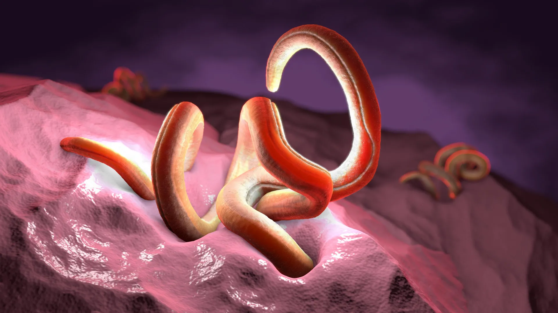
    Intestinal worms may help reduce inflammation, but new research suggests they can only do so when they have enough dietary fiber to thrive. Without sufficient fiber, these worms enter a hibernation-like state and lose their ability to provide anti-inflammatory benefits.

    The findings, published in Nature Communications, come from parasitologists at the Biology Centre of the Czech Academy of Sciences.

    Why Scientists Are Studying Intestinal Worms

    For most of human history, intestinal parasites were a normal part of the digestive system. In industrialized countries, however, they have become far less common because of improved hygiene and modern medicine. During that same period, autoimmune disorders and inflammatory bowel diseases have become increasingly common.

    This trend prompted researchers about two decades ago to investigate whether certain intestinal worms could be used to treat inflammatory conditions. The approach, known as helminth therapy, has shown promise, but its results have been inconsistent.

    “However, the results of helminth therapy have been inconsistent — sometimes the worms suppress inflammation, sometimes they do not. That’s why we focused on factors that may influence their effects in the gut,” says Kateřina Jirků from the Institute of Parasitology, Biology Centre CAS.

    The team’s research pointed to one major factor: the amount of dietary fiber in the host’s diet.

    How Fiber Changes Worm Behavior

    To better understand the connection, the researchers examined how an intestinal parasite responds to different levels of dietary fiber. They used the rat tapeworm Hymenolepis diminuta, a non-pathogenic species commonly used to study interactions among parasites, gut microorganisms, and the immune system because of its known anti-inflammatory properties.

    Their experiments revealed a striking difference between fiber-rich and fiber-poor diets.

    “We found that when the diet contains a high proportion of structural fiber, the tapeworm is not only in excellent condition but is also able to induce an anti-inflammatory response in the host. When fiber is lacking, the worm enters an energy-saving state resembling hibernation in mammals, and its anti-inflammatory effect disappears,” explains Jirků.

    When the animals consumed a low-fiber diet, the tapeworms were several times smaller, never reached sexual maturity, and failed to produce eggs. Genetic testing also showed widespread shifts in gene expression affecting the worms’ development, metabolism, and reproduction.

    Fiber Reshapes the Gut Microbiome

    The researchers also found that dietary fiber transformed the host’s gut microbiome.

    A fiber-rich diet encouraged the growth of bacteria associated with a healthy intestinal environment. In contrast, a Western-style diet reduced microbial diversity and allowed bacteria linked to dysbiosis to become more abundant. These microbial changes were mirrored by differences in the host’s immune response.

    According to the researchers, the study highlights how strongly diet influences the entire gut ecosystem, from parasites and microbes to the immune system itself.

    Health organizations generally recommend that adults consume about 25 to 30 grams of fiber each day, yet average intake across many Western countries falls below that level. Traditional populations, by comparison, are estimated to consume between 80 and 120 grams of fiber daily.

    Previous research has shown that too little dietary fiber can weaken the gut microbiome, which plays an important role not only in digestive health but also in immune function, brain health, and mental well-being. An unhealthy microbial balance has been associated with a higher risk of allergies, depression, anxiety, and neurodegenerative diseases, including Alzheimer’s disease.

  • Scientists finally crack nature’s secret for building better cancer drugs

    Scientists finally crack nature’s secret for building better cancer drugs

    Scientists have uncovered how bacteria naturally manufacture multiple versions of powerful cancer drugs, solving a mystery that has puzzled researchers for decades. The discovery could help speed the development of new treatments for cancers that are still difficult to treat.

    For years, scientists have hoped to harness bacterial enzymes to create new drug variants through a process known as combinatorial biosynthesis. However, progress has been limited because researchers did not fully understand how the enzymes coordinate their work.

    Published in Nature Communications, the new study reveals how bacterial enzymes communicate with one another to assemble a family of closely related anti-cancer compounds. That family includes Romidepsin (Istodax), an FDA-approved treatment for certain blood cancers. By uncovering this natural “mix and match” system and reproducing its underlying principles in the laboratory, the researchers have established a new strategy for designing future cancer therapies.

    “For decades, we’ve known that bacteria can naturally produce multiple versions of powerful anti-cancer drugs, yet we had no idea how they achieved this,” said first author Dr. Munro Passmore, Research Fellow, Department of Chemistry, University of Warwick. “This work finally cracks that code. We’ve identified how the different enzymes communicate and cooperate to produce these drug variants, something that has eluded researchers because the system is so elegantly economical. It’s the breakthrough we needed to actually engineer these drugs ourselves.”

    Tiny Molecular Connectors Reveal Nature’s Drug-Making Strategy

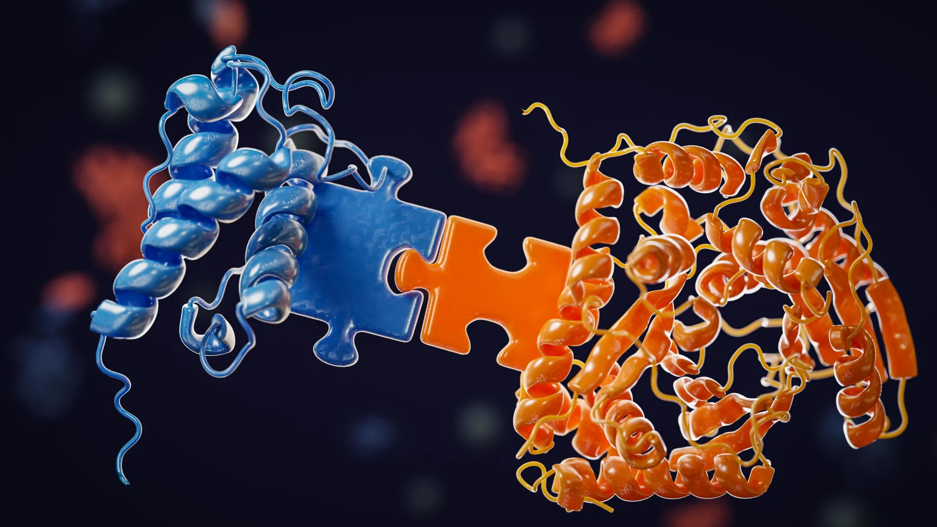
    The researchers discovered that small molecular regions known as ‘docking domains’ serve as connectors between the core drug-building machinery and the enzymes responsible for adding different components. These docking domains share a conserved connection point that allows them to interact with multiple enzyme partners.

    This flexible design explains how bacteria can create a variety of related drug molecules while still maintaining the precision needed for the compounds to remain effective.

    The study also sheds light on how these natural drug-producing systems evolved. According to the researchers, the newly identified compound most likely developed from a related drug-producing pathway through gene duplication and recombination over time.

    Prof. Greg Challis, Monash Warwick Alliance Professor of Sustainable Chemistry, University of Warwick and Monash University concludes: “This research gives us a blueprint to do what nature does, but better and faster. By reverse-engineering nature’s evolutionary logic, we can now design synthetic pathways that generate new anti-cancer drug candidates with properties optimized for clinical use, such as superior potency, improved selectivity, fewer side effects. Our immediate goal is to build an expanded library of candidates for various cancers where new treatments are urgently needed. This discovery is moving us from understanding how the systems work to building new ones.”

    How the Discovery Could Improve Cancer Drug Development

    The work focuses on a class of anti-cancer medicines known as HDAC inhibitors. These drugs block histone deacetylases, enzymes that help regulate which genes are switched on or off inside cells. Romidepsin (Istodax) is an FDA-approved HDAC inhibitor used to treat T-cell lymphomas.

    A chemically related compound called FR-901375 has been known for decades, but scientists had never identified the biological pathway bacteria use to produce it. This study finally fills in that missing piece.

    Like other HDAC inhibitors in its family, FR-901375 belongs to a group of complex cyclic molecules called depsipeptides. These compounds are assembled from amino acid building blocks along with a conserved hydroxy acid pharmacophore, all connected through a combination of peptide and ester bonds.

    Inside bacteria, these molecules are built by massive protein complexes called PKS-NRPS hybrids, which combine the activities of polyketide synthase (PKS) and nonribosomal peptide synthetase (NRPS). The new research shows that the key to this assembly process is the docking domains, which act like molecular connectors that allow one part of the production line to recognize and pass its product to the next. This mechanism is what enables combinatorial biosynthesis and allows bacteria to naturally generate multiple drug variants.

    How the Researchers Solved the Mystery

    To uncover how this system works, the team combined structural biology, biochemistry, genetics, and computational modeling.

    Their work included:

    • Bioinformatic searches of public databases that identified the FR-901375 biosynthetic gene cluster in Pseudomonas chlororaphis subsp. piscium, with the findings confirmed by mass spectrometry analysis of extracted metabolites.
    • In vitro reconstitution experiments using purified protein domains that demonstrated productive enzyme-enzyme interactions, verified with intact protein mass spectrometry.
    • AlphaFold computational modeling to predict protein complex structures, followed by carbene footprinting mass spectrometry to experimentally map the interaction sites.
    • Site-directed mutagenesis experiments that confirmed the importance of the predicted binding residues.
    • Gene deletion studies in bacterial strains showing that the docking domains are essential for the system to function in vivo.
    • Comparative analysis of biosynthetic gene clusters from multiple HDAC inhibitor-producing bacteria, revealing evolutionarily conserved features shared across these natural drug-making systems.
  • USC scientists just unlocked an endless supply of cancer-fighting immune cells

    USC scientists just unlocked an endless supply of cancer-fighting immune cells

    Scientists at USC Stem Cell have developed a new method for creating a renewable and expandable supply of immune cell precursors that could help advance cancer immunotherapy and other treatments.

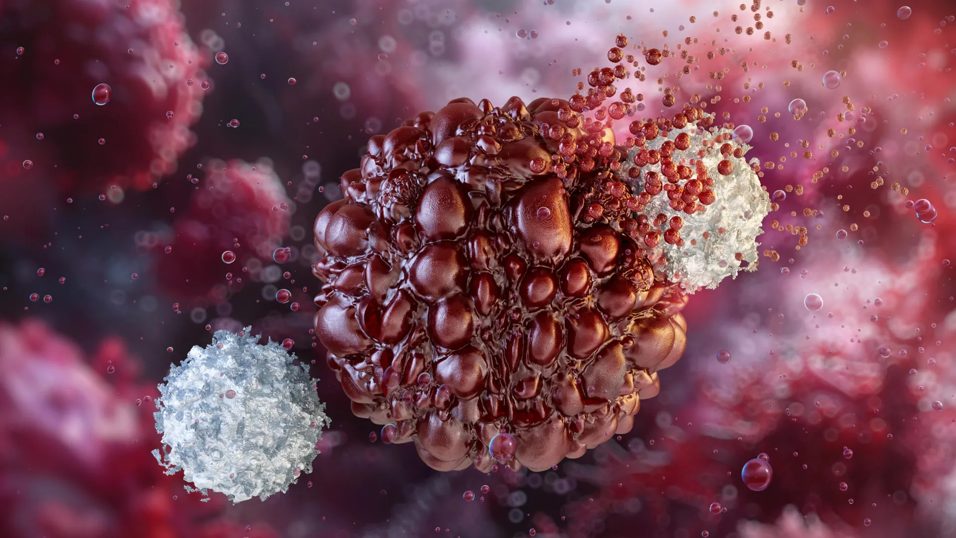
    Published in the journal Cell, the study focuses on granulocyte-monocyte progenitors (GMPs), a type of progenitor cell that produces macrophages and several other immune cells. Macrophages play a key role in defending the body against infections and have attracted growing interest as potential tools for treating cancer.

    The researchers showed that GMPs can be expanded extensively in the laboratory and genetically modified to recognize cancer cells while also boosting broader immune responses.

    “The study establishes a scalable and engineerable GMP platform for cellular immunotherapy and introduces concepts that we believe could have broad implications for both cancer immunotherapy and stem cell biology,” said the paper’s corresponding author Qi-Long Ying, MD, PhD, professor of stem cell biology and regenerative medicine at the Keck School of Medicine of USC.

    One of the study’s most significant findings relates to self-renewal, a characteristic traditionally associated with stem cells. Self-renewal allows cells to repeatedly divide while maintaining their identity. Scientists generally have not considered progenitor cells to possess this long-term capability.

    “The prevailing view has been that long-term self-renewal in the blood system is primarily a property of the hematopoietic stem cells that can generate any type of blood or immune cell,” said Ying. “We found that, under the right conditions, GMPs can also self-renew, dividing extensively while keeping their identity and ability to produce functional immune cells. That gives us a scalable starting point for engineering cell therapies for cancer, infectious disease and potentially many other conditions.”

    Why Macrophage Precursors Matter

    Macrophages are appealing candidates for cancer immunotherapy because they naturally enter tumors, consume cancer cells, and help organize immune responses. While T-cell therapies have achieved major success against blood cancers, macrophage-based therapies may offer particular advantages against solid tumors.

    However, mature macrophages have several drawbacks as therapeutic products. They are difficult to grow in large numbers outside the body, challenging to genetically engineer, and can be damaged during freezing and storage. They also tend to accumulate in organs such as the lungs and liver instead of spreading widely throughout the body.

    To overcome these obstacles, first author Shi Yue, MD, and colleagues in the Ying Lab focused on GMPs, which sit earlier in the developmental pathway that produces macrophages.

    Using a carefully defined chemical cocktail, the team prevented GMPs from maturing into other immune cell types and succeeded in maintaining and expanding them over long periods in the laboratory.

    Even after extended growth, the cells preserved their molecular and cellular characteristics and continued to generate functional macrophages and other immune cells.

    Researchers in the laboratory of Ravi Majeti, MD, PhD, at Stanford University independently reproduced the long-term maintenance and genetic engineering of GMPs, providing additional support for the platform’s reliability and potential therapeutic value.

    Majeti, Director of the Institute for Stem Cell Biology and Regenerative Medicine at Stanford University, noted: “This method for the expansion and engineering of GMPs opens the door to numerous translational applications, much like T cell expansion and engineering. We have already demonstrated engineering of these cells to drive multiple potent functions, and there is a lot more to be explored.”

    Engineering GMPs To Fight Cancer

    Beyond their ability to grow long term in the laboratory, GMPs can also be genetically engineered for use as immunotherapies.

    In this study, the researchers equipped GMPs with a chimeric antigen receptor, or CAR, enabling the cells to recognize a specific marker found on cancer cells. The team also added a second signal designed to activate nearby immune cells that help stimulate tumor-fighting T cells and strengthen the body’s natural defenses.

    Importantly, this additional signal remains effective even when donor and recipient cells are immunologically mismatched. That raises the possibility of creating off-the-shelf therapies produced in advance from donor cells and used in many patients, rather than generating a custom treatment for each individual.

    After expanding and engineering both mouse and human GMPs, the researchers tested them in mice. The cells successfully settled into the bone marrow and other blood-forming tissues, where they continuously generated engineered macrophages and additional immune cells.

    Because the GMPs maintained an ongoing supply of these cells from the bone marrow, they avoided the rapid loss that has limited mature macrophage therapies, including those evaluated in recent clinical trials.

    In mice with blood cancers and solid tumors, CAR-engineered GMPs slowed disease progression. GMPs carrying both the CAR and the additional immune-activating signal produced even stronger benefits.

    Potential Beyond Cancer

    The platform may have applications that extend beyond oncology.

    The researchers tested the approach in mice with chronic granulomatous disease, an inherited immune disorder. GMP treatment restored the animals’ ability to fight bacterial infections, demonstrating the potential of the technology for immune deficiencies as well.

    “Our study suggests that the future of immunotherapy may depend not only on designing better CAR receptors, but also on choosing the right developmental stage of the cell,” said Ying.

    About the Study

    The paper in Cell is titled “Expansion and CAR engineering of granulocyte-monocyte progenitors for cellular immunotherapy.”

    In addition to Ying, Yue and Majeti, additional authors are: Zheng Guo, Crystal Pan, Xueyuan A. Jing, Tai Nguyen, Jiaqi Tang, Yanpui Chan, Humberto Contreras-Trujillo, Du Jiang, Xue Yan, Hang Xiang, Xugeng Liu, Xiao Wang, Ziyuan Wang, Natalie Shu, Daniel B. McKim, Rong Lu and Chao Zhang from USC; and Litao Tao and Celia Bloom from Creighton University; Asiri Ediriwickrema and Sebastian Koschade from Stanford University School of Medicine; and Yingxiao Shi from Harvard Medical School and the Dana-Farber Cancer Institute.

    This work was supported by the Chen Yong Foundation of the Zhongmei Group, a sponsored research project from Myelogene Inc., the L.K. Whittier Foundation, the Eli and Edythe Broad Innovation Award, the Ming Hsieh Institute for Research on Engineering Medicine for Cancer Award, the USC SBIR/STTR Planning Award, the Xia Research Fund, and the Wu & Jiang Research Fund. Majeti reports support from the Ludwig Institute for Cancer Research, and Guo was supported by the California Institute for Regenerative Medicine Predoctoral Training Fellowship.

    Disclosures

    Ying, Yue, Jing, Guo, Majeti, Zhang, Nguyen and Tang are co-inventors on patents related to this study, filed by USC and licensed to Myelogene Inc. Ying, Yue, Zhang and Majeti are co-founders of Myelogene Inc. Majeti is on the Advisory Boards of Kodikaz Therapeutic Solutions, Pheast Therapeutics, Prelude Therapeutics, Mubadala Capital, Aculeus Therapeutics, Sequentify, BMS and Bectas Therapeutics. Majeti is also a co-founder and equity holder of Pheast Therapeutics.

  • Scientists are seriously asking if bees and ChatGPT are conscious

    Scientists are seriously asking if bees and ChatGPT are conscious

    You might think a honey bee foraging in your garden and a browser window running ChatGPT have nothing in common. But recent scientific research has been seriously considering the possibility that either, or both, might be conscious.

    There are many different ways of studying consciousness. One of the most common is to measure how an animal – or artificial intelligence (AI) – acts.

    But two new papers on the possibility of consciousness in animals and AI suggest new theories for how to test this – one that strikes a middle ground between sensationalism and knee-jerk skepticism about whether humans are the only conscious beings on Earth.

    A fierce debate

    Questions around consciousness have long sparked fierce debate.

    That’s in part because conscious beings might matter morally in a way that unconscious things don’t. Expanding the sphere of consciousness means expanding our ethical horizons. Even if we can’t be sure something is conscious, we might err on the side of caution by assuming it is – what philosopher Jonathan Birch calls the precautionary principle for sentience.

    The recent trend has been one of expansion.

    For example, in April 2024 a group of 40 scientists at a conference in New York proposed the New York Declaration on Animal Consciousness. Subsequently signed by over 500 scientists and philosophers, this declaration says consciousness is realistically possible in all vertebrates (including reptiles, amphibians and fishes) as well as many invertebrates, including cephalopods (octopus and squid), crustaceans (crabs and lobsters) and insects.

    In parallel with this, the incredible rise of large language models, such as ChatGPT, has raised the serious possibility that machines may be conscious.

    Five years ago, a seemingly ironclad test of whether something was conscious was to see if you could have a conversation with it. Philosopher Susan Schneider suggested if we had an AI that convincingly mused on the metaphysics of consciousness, it may well be conscious.

    By those standards, today we would be surrounded by conscious machines. Many have gone so far as to apply the precautionary principle here too: the burgeoning field of AI welfare is devoted to figuring out if and when we must care about machines.

    Yet all of these arguments depend, in large part, on surface-level behavior. But that behavior can be deceptive. What matters for consciousness is not what you do, but how you do it.

    Looking at the machinery of AI

    A new paper in Trends in Cognitive Sciences that one of us (Colin Klein) coauthored, drawing on previous work, looks to the machinery rather than the behavior of AI.

    It also draws on the cognitive science tradition to identify a plausible list of indicators of consciousness based on the structure of information processing. This means one can draw up a useful list of indicators of consciousness without having to agree on which of the current cognitive theories of consciousness is correct.

    Some indicators (such as the need to resolve trade-offs between competing goals in contextually appropriate ways) are shared by many theories. Most other indicators (such as the presence of informational feedback) are only required by one theory but indicative in others.

    Importantly, the useful indicators are all structural. They all have to do with how brains and computers process and combine information.

    The verdict? No existing AI system (including ChatGPT) is conscious. The appearance of consciousness in large language models is not achieved in a way that is sufficiently similar to us to warrant attribution of conscious states.

    Yet at the same time, there is no bar to AI systems – perhaps ones with a very different architecture to today’s systems – becoming conscious.

    The lesson? It’s possible for AI to behave as if conscious without being conscious.

    Measuring consciousness in insects

    Biologists are also turning to mechanisms – how brains work – to recognize consciousness in non-human animals.

    In a new paper in Philosophical Transactions B, we propose a neural model for minimal consciousness in insects. This is a model that abstracts away from anatomical detail to focus on the core computations done by simple brains.

    Our key insight is to identify the kind of computation our brains perform that gives rise to experience.

    This computation solves ancient problems from our evolutionary history that arise from having a mobile, complex body with many senses and conflicting needs.

    Importantly, we don’t identify the computation itself – there is science yet to be done. But we show that if you could identify it, you’d have a level playing field to compare humans, invertebrates, and computers.

    The same lesson

    The problem of consciousness in animals and in computers appear to pull in different directions.

    For animals, the question is often how to interpret whether ambiguous behavior (like a crab tending its wounds) indicates consciousness.

    For computers, we have to decide whether apparently unambiguous behavior (a chatbot musing with you on the purpose of existence) is a true indicator of consciousness or mere roleplay.

    Yet as the fields of neuroscience and AI progress, both are converging on the same lesson: when making judgement about whether something is consciousness, how it works is proving more informative than what it does.The Conversation

  • Scientists just found what keeps plant cells from growing out of control

    Scientists just found what keeps plant cells from growing out of control

    Plants spend most of their lives using photosynthesis to make energy. However, in the earliest phase after a seed begins to grow, they cannot yet capture light. During this short but critical window, they depend on stored fatty acids instead. To break down these fatty acids, plant cells use a specialized structure known as the peroxisome, a membrane-bound compartment also found in human cells. Because of their size and visibility, plant cells serve as a useful system for studying how peroxisomes work.

    “The plant we use, Arabidopsis, has large cells and peroxisomes so large that we can see inside them with a light microscope,” said Bonnie Bartel, the Ralph and Dorothy Looney Professor of Biosciences. “The peroxisome gets even larger during the seed to seedling stage, when the plant is relying on fatty acids for energy, before shrinking back down to its normal size once the plant can photosynthesize.”

    Protein PEX11 Helps Control Peroxisome Size

    Bartel’s team focuses on these enlarged peroxisomes, particularly a protein called PEX11. Scientists have long known that PEX11 plays a role in helping peroxisomes divide. In new research published in Nature Communications, the team found that this protein also helps control how peroxisomes expand and shrink during early plant development.

    “Peroxisomes are implicated in some human diseases and used in bioengineering,” said Nathan Tharp, first author of the paper and a Rice graduate student. “They can, however, be rather tricky to study.”

    Using CRISPR To Study a Complex Protein

    A common strategy for understanding a protein is to disable the gene responsible for making it and observe the effects. In this case, the situation was more complicated. PEX11 is produced by five different genes. Disrupting just one of them had little effect, but removing all five caused the plant to die. This made it difficult to pinpoint the protein’s function.

    To get around this problem, Tharp used advanced CRISPR techniques to selectively disable different combinations of the five genes.

    “I was able to use recent advances in CRISPR to go in and break specific combinations of the five genes,” said Tharp, who recently defended his thesis. “It was only then that we were able to see that PEX11 is clearly involved in controlling the growth of the peroxisome during the seed to seedling stage.”

    Giant Peroxisomes Reveal Growth Control Mechanism

    Tharp engineered two types of mutant plants, each missing a particular set of PEX11 genes. In both cases, peroxisomes expanded during the seed to seedling stage as expected. However, instead of shrinking back to their usual size, some continued growing far beyond normal limits. In extreme cases, the peroxisomes stretched from one end of the cell to the other.

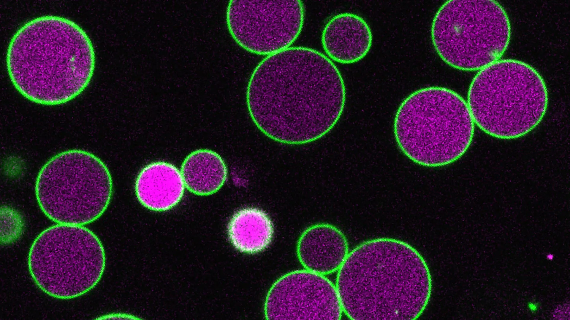
    These mutant cells also lacked vesicles, small membrane-bound compartments that typically form inside the peroxisome during fatty acid processing. Under normal conditions, these vesicles develop as the peroxisome grows and appear to remove portions of its outer membrane.

    “The vesicles taking pieces of membrane as they form may help control the peroxisome’s growth,” Tharp said. “In our PEX11 mutants, these vesicles either don’t form or are abnormally small and rare, and so we see these massive peroxisomes, way larger than normal.”

    Findings Extend Beyond Plants to Other Species

    Although the research focused on plants, Tharp wanted to know whether the same mechanism might exist in other organisms. To test this idea, he introduced the yeast version of the protein, called Pex11, into the mutant plant cells.

    “We put yeast Pex11 into our mutant plant cells to see if it could return the peroxisomes back to normal,” Tharp said. “And it did.”

    This result suggests that Pex11 serves a similar function in yeast as it does in plants, despite the vast evolutionary distance between them. Because of this, the protein may also play a comparable role in other types of cells, including human cells.

    “Finding that this protein fills the same role in yeast and plant cells suggests that it may be a highly conserved protein,” Bartel said. “Our findings in plants, in this relatively easy-to-study model, may thus be applicable to human cells and cells used for bioengineering.”

  • Scientists discover AI can make humans more creative

    Scientists discover AI can make humans more creative

    Artificial intelligence (AI) is commonly viewed as a technology designed to automate work and potentially replace human labor. However, new research from Swansea University offers a different perspective. The findings suggest that AI can also function as a creative collaborator that encourages exploration, engagement, and inspiration.

    Researchers from the University’s Computer Science Department carried out one of the largest studies so far examining how people work alongside AI during creative design tasks. More than 800 participants joined an online experiment where they used an AI-supported system to design virtual cars.

    How AI Generated Diverse Design Ideas

    Rather than quietly optimizing designs behind the scenes, the system used a method called MAP-Elites to produce visual galleries filled with many different design possibilities. These galleries showed a wide spectrum of car concepts, including highly effective designs, unusual ideas, and even some intentionally flawed options.

    Turing Fellow Dr. Sean Walton, Associate Professor of Computer Science and the study’s lead author, explained: “People often think of AI as something that speeds up tasks or improves efficiency, but our findings suggest something far more interesting. When people were shown AI-generated design suggestions, they spent more time on the task, produced better designs and felt more involved. It was not just about efficiency. It was about creativity and collaboration.”

    Why Traditional AI Evaluation May Be Too Limited

    The study, published in the ACM journal Transactions on Interactive Intelligent Systems, also highlights a problem with how AI design tools are typically assessed. Standard metrics often focus on simple behaviors, such as how frequently users click on or copy AI suggestions. According to the researchers, these measures overlook important aspects of the experience, including how the technology influences people’s thoughts, emotions, and willingness to explore new ideas.

    The Swansea researchers argue that AI systems should be evaluated using broader methods that capture these deeper effects. Understanding how AI shapes human thinking and engagement could provide a more complete picture of its impact.

    Why Imperfect Ideas Can Boost Creativity

    Dr. Walton emphasized that variety in AI-generated output played a crucial role in the experiment. “Our study highlights the importance of diversity in AI output. Participants responded most positively to galleries that included a wide variety of ideas, including bad ones! These helped them move beyond their initial assumptions and explore a broader design space. This structured diversity prevented early fixation and encouraged creative risk-taking.

    “As AI becomes increasingly embedded in creative fields, from engineering and architecture to music and game design, understanding how humans and intelligent systems work together is essential. As the technology evolves, the question is not only what AI can do but how it can help us think, create and collaborate more effectively.”

  • Scientists built the hardest AI test ever and the results are surprising

    Scientists built the hardest AI test ever and the results are surprising

    As artificial intelligence systems began scoring extremely high on long used academic benchmarks, researchers noticed a growing issue. The tests that once challenged machines were no longer difficult enough. Well known evaluations such as the Massive Multitask Language Understanding (MMLU) exam, which had previously been seen as demanding, now fail to properly measure the capabilities of today’s advanced AI models.

    To solve this problem, a worldwide group of nearly 1,000 researchers, including a professor from Texas A&M University, developed a new type of test. Their goal was to build an exam that is broad, difficult, and grounded in expert human knowledge in ways that current AI systems still struggle to handle.

    The result is “Humanity’s Last Exam” (HLE), a 2,500 question assessment covering mathematics, humanities, natural sciences, ancient languages, and a wide range of highly specialized academic fields. Details of the project appear in a paper published in Nature, and additional information about the exam is available at lastexam.ai.

    Among the many contributors is Dr. Tung Nguyen, instructional associate professor in the Department of Computer Science and Engineering at Texas A&M. Nguyen helped write and refine many of the exam questions.

    “When AI systems start performing extremely well on human benchmarks, it’s tempting to think they’re approaching human-level understanding,” Nguyen said. “But HLE reminds us that intelligence isn’t just about pattern recognition — it’s about depth, context and specialized expertise.”

    The purpose of the exam was not to trick or defeat human test takers. Instead, the goal was to carefully identify areas where AI systems still fall short.

    A Global Effort to Measure AI’s Limits

    Specialists from around the world wrote and reviewed the questions included in Humanity’s Last Exam. Each problem was carefully designed so it has one clear, verifiable answer. The questions were also crafted to prevent quick solutions through simple internet searches.

    The topics come from advanced academic challenges. Some tasks involve translating ancient Palmyrene inscriptions, while others require identifying tiny anatomical structures in birds or analyzing detailed features of Biblical Hebrew pronunciation.

    Researchers tested every question against leading AI systems. If any model was able to answer a question correctly, that question was removed from the final exam. This process ensured the test remained just beyond what current AI systems can reliably solve.

    Early testing confirmed that the strategy worked. Even powerful AI models struggled with the exam. GPT-4o achieved a score of 2.7 percent, while Claude 3.5 Sonnet reached 4.1 percent. OpenAI’s o1 model performed somewhat better with 8 percent. The most capable systems so far, including Gemini 3.1 Pro and Claude Opus 4.6, have reached accuracy levels between about 40 percent and 50 percent.

    Why New AI Benchmarks Are Needed

    Nguyen explained that the issue of AI surpassing older tests is more than a technical concern. He contributed 73 of the 2,500 publicly available questions in HLE, the second highest number among contributors, and wrote the most questions related to mathematics and computer science.

    “Without accurate assessment tools, policymakers, developers and users risk misinterpreting what AI systems can actually do,” he said. “Benchmarks provide the foundation for measuring progress and identifying risks.”

    According to the research team, high scores on tests originally designed for humans do not necessarily indicate genuine intelligence. Those benchmarks mainly measure how well AI can complete specific tasks created for human learners, rather than capturing deeper understanding.

    Not a Threat, but a Tool

    Despite the dramatic name, Humanity’s Last Exam is not meant to suggest that humans are becoming obsolete. Instead, it highlights the large amount of knowledge and expertise that still remains uniquely human.

    “This isn’t a race against AI,” Nguyen said. “It’s a method for understanding where these systems are strong and where they struggle. That understanding helps us build safer, more reliable technologies. And, importantly, it reminds us why human expertise still matters.”

    Building a Long Term AI Benchmark

    Humanity’s Last Exam is designed to serve as a durable and transparent benchmark for future AI systems. To support that goal, the researchers have released some questions publicly while keeping the majority hidden so that AI models cannot simply memorize the answers.

    “For now, Humanity’s Last Exam stands as one of the clearest assessments of the gap between AI and human intelligence,” Nguyen said, “and despite rapid technological advances, it remains wide.”

    A Massive International Research Effort

    Nguyen emphasized that the scale of the project demonstrates the value of collaboration across disciplines and countries.

    “What made this project extraordinary was the scale,” he said. “Experts from nearly every discipline contributed. It wasn’t just computer scientists; it was historians, physicists, linguists, medical researchers. That diversity is exactly what exposes the gaps in today’s AI systems — perhaps ironically, it’s humans working together.”

  • Scientists create smart synthetic skin that can hide images and change shape

    Scientists create smart synthetic skin that can hide images and change shape

    Synthetic materials are widely used across science, engineering, and industry, but most are designed to perform only a narrow range of tasks. A research team at Penn State set out to change that. Led by Hongtao Sun, assistant professor of industrial and manufacturing engineering (IME), the group developed a new fabrication technique that can produce multifunctional “smart synthetic skin.” These adaptable materials can be programmed to perform a wide variety of tasks, including hiding or revealing information, enabling adaptive camouflage, and supporting soft robotic systems.

    Using this new approach, the researchers created a programmable smart skin made from hydrogel, a soft, water-rich material. Unlike conventional synthetic materials with fixed behaviors, this smart skin can be tuned to respond in multiple ways. Its appearance, mechanical behavior, surface texture, and ability to change shape can all be adjusted when the material is exposed to external triggers such as heat, solvents, or physical stress.

    The findings were published in Nature Communications, where the study was also selected for Editors’ Highlights.

    Inspired by Octopus Skin and Living Systems

    Sun, the project’s principal investigator, said the concept was inspired by cephalopods such as octopuses, which can rapidly alter the look and texture of their skin. These animals use such changes to blend into their surroundings or communicate with one another.

    “Cephalopods use a complex system of muscles and nerves to exhibit dynamic control over the appearance and texture of their skin,” Sun said. “Inspired by these soft organisms, we developed a 4D-printing system to capture that idea in a synthetic, soft material.”

    Sun also holds affiliations in biomedical engineering, material science and engineering, and the Materials Research Institute at Penn State. He described the process as 4D printing because the printed objects are not static. Instead, they can actively change in response to environmental conditions.

    Printing Digital Instructions Into Material

    To achieve this adaptability, the team used a method called halftone-encoded printing. This technique converts image or texture data into binary ones and zeros and embeds that information directly into the material. The approach is similar to how dot patterns are used in newspapers or photographs to create images.

    By encoding these digital patterns within the hydrogel, the researchers can program how the smart skin reacts to different stimuli. The printed patterns determine how various regions of the material respond. Some areas may swell, shrink, or soften more than others when exposed to temperature changes, liquids, or mechanical forces. By carefully designing these patterns, the team can control the material’s overall behavior.

    “In simple terms, we’re printing instructions into the material,” Sun explained. “Those instructions tell the skin how to react when something changes around it.”

    Hiding and Revealing Images on Demand

    One of the most eye-catching demonstrations involved the material’s ability to conceal and reveal visual information. Haoqing Yang, a doctoral candidate in IME and the paper’s first author, said this capability highlights the potential of the smart skin.

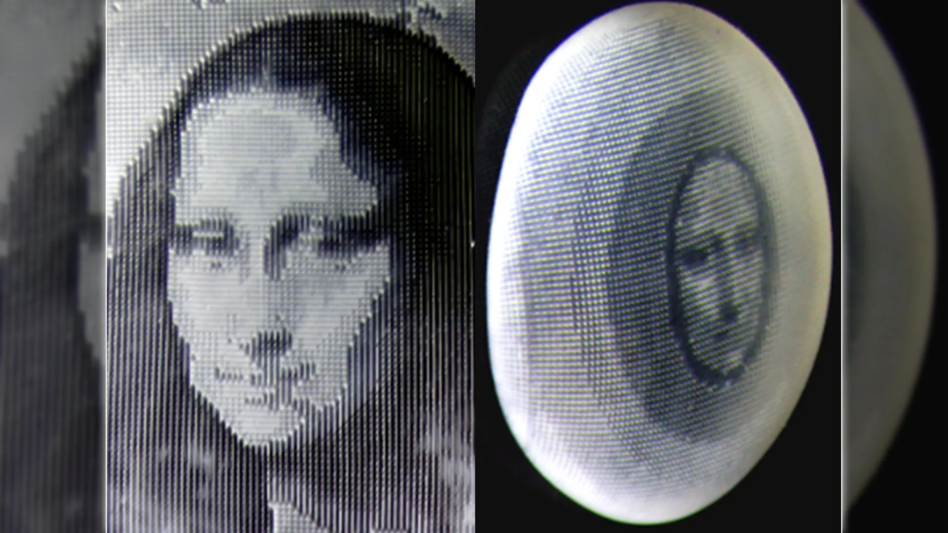
    To demonstrate the effect, the team encoded an image of the Mona Lisa into the hydrogel film. When the material was washed with ethanol, it appeared transparent and showed no visible image. The hidden image became clear only after the film was placed in ice water or gradually heated.

    Yang noted that the Mona Lisa was used only as an example. The printing technique allows virtually any image to be encoded into the hydrogel.

    “This behavior could be used for camouflage, where a surface blends into its environment, or for information encryption, where messages are hidden and only revealed under specific conditions,” Yang said.

    The researchers also showed that concealed patterns could be detected by gently stretching the material and analyzing how it deforms using digital image correlation analysis. This means information can be revealed not only visually, but also through mechanical interaction, adding an extra level of security.

    Shape Shifting Without Multiple Layers

    The smart skin also demonstrated remarkable flexibility. According to Sun, the material can easily shift from a flat sheet into complex, bio-inspired shapes with detailed surface textures. Unlike many other shape-changing materials, this transformation does not require multiple layers or different substances.

    Instead, the changes in shape and texture are controlled entirely by the digitally printed halftone patterns within a single sheet. This allows the material to replicate effects similar to those seen in cephalopod skin.

    Building on this capability, the team showed that multiple functions can be programmed to work together. By carefully designing the halftone patterns, they encoded the Mona Lisa image into flat films that later transformed into three-dimensional forms. As the sheets curved into dome-like shapes, the hidden image slowly appeared, showing that changes in shape and visual appearance can be coordinated within one material.

    “Similar to how cephalopods coordinate body shape and skin patterning, the synthetic smart skin can simultaneously control what it looks like and how it deforms, all within a single, soft material,” Sun said.

    Expanding the Potential of 4D-Printed Hydrogels

    Sun said the new work builds on earlier research by the team on 4D-printed smart hydrogels, which was also published in Nature Communications. That earlier study focused on combining mechanical properties with programmable transitions from flat to three-dimensional forms. In the current research, the team expanded the approach by using halftone-encoded 4D printing to integrate even more functions into a single hydrogel film.

    Looking ahead, the researchers aim to create a scalable and versatile platform that allows precise digital encoding of multiple functions within one adaptive material.

    “This interdisciplinary research at the intersection of advanced manufacturing, intelligent materials and mechanics opens new opportunities with broad implications for stimulus-responsive systems, biomimetic engineering, advanced encryption technologies, biomedical devices and more,” Sun said.

    The study also included Penn State co-authors Haotian Li and Juchen Zhang, both doctoral candidates in IME, and Tengxiao Liu, a lecturer in biomedical engineering. H. Jerry Qi, professor of mechanical engineering at Georgia Institute of Technology, also collaborated on the project.

  • Scientists found a way to cool quantum computers using noise

    Scientists found a way to cool quantum computers using noise

    Quantum computers only work when they are kept extremely cold. The problem is that today’s cooling systems also create noise, which can interfere with the fragile quantum information they are supposed to protect. Researchers at Chalmers University of Technology in Sweden have now introduced a new type of minimal quantum “refrigerator” that turns this challenge into an advantage. Instead of fighting noise, the device partially relies on it to operate. The result is highly precise control over heat and energy flow, which could help make large scale quantum technology possible.

    Quantum technology is widely expected to reshape major areas of society. Potential applications include drug discovery, artificial intelligence, logistics optimization, and secure communications. Despite this promise, serious technical barriers still stand in the way of real world use. One of the most difficult challenges is maintaining and controlling the delicate quantum states that make these systems work.

    Why Quantum Computers Must Be Near Absolute Zero

    Quantum computers built with superconducting circuits must be cooled to temperatures very close to absolute zero (around — 273 °C). At these temperatures, materials become superconducting, allowing electrons to move without resistance. Only under these extreme conditions can stable quantum states form inside qubits, the basic units of quantum information.

    These quantum states are extremely sensitive. Small changes in temperature, electromagnetic interference, or background noise can quickly erase stored information. This sensitivity makes quantum systems difficult to operate and even harder to expand.

    As researchers attempt to scale up quantum computers to solve practical problems, heat and noise become harder to control. Larger and more complex systems create more opportunities for unwanted energy to spread and disrupt fragile quantum states.

    “Many quantum devices are ultimately limited by how energy is transported and dissipated. Understanding these pathways and being able to measure them allows us to design quantum devices in which heat flows are predictable, controllable and even useful,” says Simon Sundelin, doctoral student of quantum technology at Chalmers University of Technology and the study’s lead author.

    Using Noise as a Cooling Tool

    In a study published in Nature Communications, the Chalmers team describes a fundamentally different kind of quantum refrigerator. Instead of trying to eliminate noise, the system uses it as the driving force behind cooling.

    “Physicists have long speculated about a phenomenon called Brownian refrigeration; the idea that random thermal fluctuations could be harnessed to produce a cooling effect. Our work represents the closest realisation of this concept to date,” says Simone Gasparinetti, associate professor at Chalmers and senior author of the study.

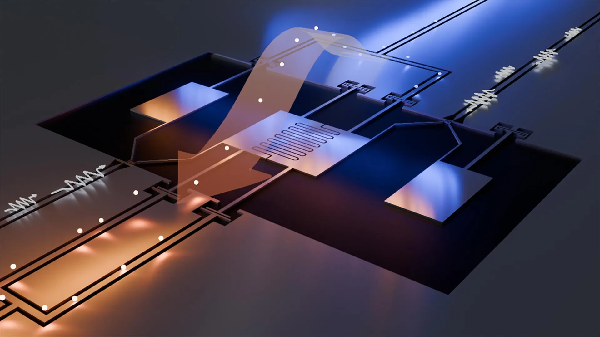
    At the core of the refrigerator is a superconducting artificial molecule created in Chalmers’ nanofabrication laboratory. It behaves much like a natural molecule, but instead of atoms, it is built from tiny superconducting electrical circuits.

    The artificial molecule is connected to multiple microwave channels. By adding carefully controlled microwave noise in the form of random signal fluctuations within a narrow frequency range, the researchers can guide how heat and energy move through the system with remarkable precision.

    “The two microwave channels serve as hot and cold reservoirs, but the key point is that they are only effectively connected when we inject controlled noise through a third port. This injected noise enables and drives heat transport between the reservoirs via the artificial molecule. We were able to measure extremely small heat currents, down to powers in the order of attowatts, or 10-18 watt. If such a small heat flow were used to warm a drop of water, it would take the age of the universe to see its temperature rise one degree Celsius,” explains Sundelin.

    New Paths Toward Scalable Quantum Technology

    By carefully adjusting reservoir temperatures and tracking minuscule heat flows, the quantum refrigerator can operate in multiple ways. Depending on conditions, it can function as a refrigerator, act as a heat engine, or amplify thermal transport.

    This level of control is especially important in larger quantum systems, where heat is produced locally during qubit operation and measurement. Managing that heat directly inside quantum circuits could improve stability and performance in ways conventional cooling systems cannot.

    “We see this as an important step towards controlling heat directly inside quantum circuits, at a scale that conventional cooling systems can’t reach. Being able to remove or redirect heat at this tiny scale opens the door to more reliable and robust quantum technologies,” says Aamir Ali, a researcher in quantum technology at Chalmers and co-author of the study.

    More Information

    The study Quantum refrigeration powered by noise in a superconducting circuit was published in the scientific journal Nature Communications. The authors are Simon Sundelin, Mohammed Ali Aamir, Vyom Manish Kulkarni, Claudia Castillo-Moreno, and Simone Gasparinetti from the Department of Microtechnology and Nanoscience at Chalmers University of Technology.

    The quantum refrigerator was fabricated at the Nanofabrication Laboratory, Myfab, at Chalmers University of Technology.

    Funding for the research was provided by the Swedish Research Council, the Knut and Alice Wallenberg Foundation through the Wallenberg Centre for Quantum Technology (WACQT), the European Research Council, and the European Union.

  • Scientists teach bacteria the octopus’s secret to camouflage

    Scientists teach bacteria the octopus’s secret to camouflage

    Scientists at the University of California, San Diego have come closer to understanding one of nature’s most fascinating abilities: the art of disguise.

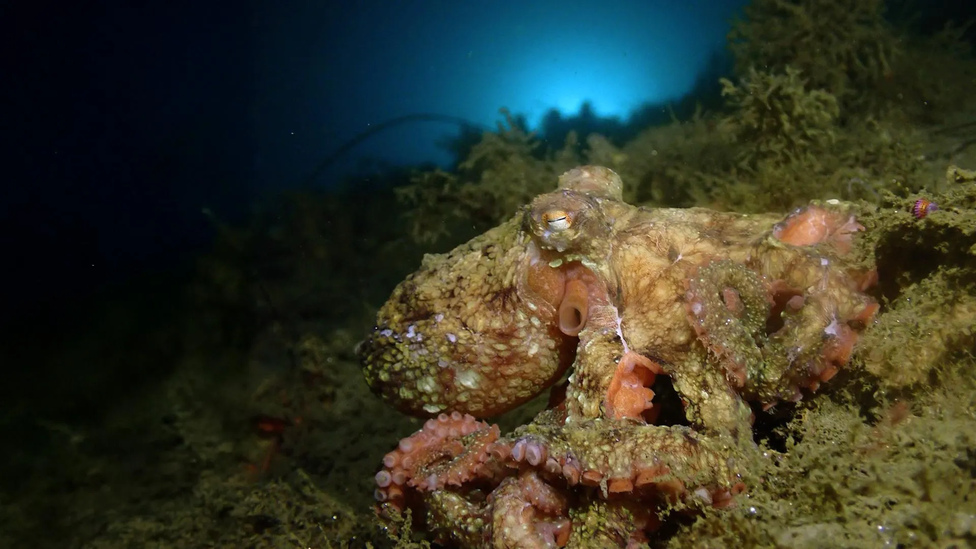
    Octopuses, squids, cuttlefish, and their cephalopod relatives are masters of camouflage, able to instantly shift their skin color to blend into their surroundings. This extraordinary transformation is driven by a natural pigment called xanthommatin, which plays a key role in their color-changing skin.

    For years, researchers and even defense organizations have been captivated by xanthommatin’s light-responsive qualities. Yet replicating and studying this pigment in the lab has been extremely challenging — until now.

    In a new breakthrough from UC San Diego’s Scripps Institution of Oceanography, scientists successfully created a method to produce large quantities of xanthommatin. This marks a major step forward in decoding how animals achieve their remarkable camouflage.

    Bacteria Turned Into Natural Pigment Factories

    Using a biologically inspired approach, the research team was able to generate the pigment inside bacteria, achieving production levels up to 1,000 times greater than previous methods. This innovation could pave the way for sustainable new uses in materials and cosmetics, including applications in photoelectronics, thermal coatings, dyes, and UV-protective products.

    “We’ve developed a new technique that has sped up our capabilities to make a material, in this case xanthommatin, in a bacterium for the first time,” said Bradley Moore, senior author of the study and a marine chemist with appointments at Scripps Oceanography and the UC San Diego Skaggs School of Pharmacy and Pharmaceutical Sciences. “This natural pigment is what gives an octopus or a squid its ability to camouflage — a fantastic superpower — and our achievement to advance production of this material is just the tip of the iceberg.”

    Published today (Nov. 3) in Nature Biotechnology, the study received support from the National Institutes of Health, the Office of Naval Research, the Swiss National Science Foundation, and the Novo Nordisk Foundation.

    According to the researchers, this achievement not only deepens our understanding of the biological and chemical foundations of animal coloration, but also highlights a powerful new biotechnology. The same technique could be used to create other valuable compounds, helping industries transition away from petroleum-based products toward more sustainable, nature-inspired materials.

    A Promising Pigment

    Beyond cephalopods, xanthommatin is also found in insects within the arthropod group, contributing to the brilliant orange and yellow hues of monarch butterfly wings and the bright reds seen in dragonfly bodies and fly eyes.

    Despite xanthommatin’s fantastic color properties, it is poorly understood due to a persistent supply challenge. Harvesting the pigment from animals isn’t scalable or efficient, and traditional lab methods are labor intensive, reliant on chemical synthesis that is low yielding.

    Researchers in the Moore Lab at Scripps Oceanography sought to change that, working with colleagues across UC San Diego and at the Novo Nordisk Foundation Center for Biosustainability in Denmark to design a solution, a sort of growth feedback loop they call “growth coupled biosynthesis.”

    The way in which they bioengineered the octopus pigment, a chemical, in a bacterium represents a novel departure from typical biotechnological approaches. Their approach intimately connected the production of the pigment with the survival of the bacterium that made it.

    “We needed a whole new approach to address this problem,” said Leah Bushin, lead author of the study, now a faculty member at Stanford University and formerly a postdoctoral researcher in the Moore Lab at Scripps Oceanography, where her work was conducted. “Essentially, we came up with a way to trick the bacteria into making more of the material that we needed.”

    Typically, when researchers try to get a microbe to produce a foreign compound, it creates a major metabolic burden. Without significant genetic manipulation, the microbe resists diverting its essential resources to produce something unfamiliar.

    By linking the cell’s survival to the production of their target compound, the team was able to trick the microbe into creating xanthommatin. To do this, they started with a genetically engineered “sick” cell, one that could only survive if it produced both the desired pigment, along with a second chemical called formic acid. For every molecule of pigment generated, the cell also produced one molecule of formic acid. The formic acid, in turn, provides fuel for the cell’s growth, creating a self-sustaining loop that drives pigment production.

    “We made it such that activity through this pathway, of making the compound of interest, is absolutely essential for life. If the organism doesn’t make xanthommatin, it won’t grow,” said Bushin.

    To push the bacteria to make even more pigment, the researchers turned to robotics and automation. They used robotic systems to guide the microbes through two rounds of high-throughput adaptive laboratory evolution, a process designed to help the cells gradually improve their performance. This advanced method was developed by the lab of study co-author Adam Feist, a professor in the Shu Chien-Gene Lay Department of Bioengineering at the UC San Diego Jacobs School of Engineering and a senior scientist at the Novo Nordisk Foundation Center for Biosustainability.

    The researchers also used specialized bioinformatics software from the Feist Lab to pinpoint genetic changes that increased the microbes’ productivity. These key mutations allowed the engineered bacteria to produce the pigment efficiently using only a single nutrient source.

    “This project gives a glimpse into a future where biology enables the sustainable production of valuable compounds and materials through advanced automation, data integration and computationally driven design,” said Feist. “Here, we show how we can accelerate innovation in biomanufacturing by bringing together engineers, biologists and chemists using some of the most advanced strain-engineering techniques to develop and optimize a novel product in a relatively short time.”

    Traditional approaches yield around five milligrams of pigment per liter “if you’re lucky,” said Bushin, while the new method yields between one to three grams per liter.

    Getting from the planning stages to the actual experimentation in the lab took several years of dedicated work, but once the plan was put into motion, the results were almost immediate.

    “It was one of my best days in the lab,” Bushin recalled of the first successful experiment. “I’d set up the experiment and left it overnight. When I came in the next morning and realized it worked and it was producing a lot of pigment, I was thrilled. Moments like that are why I do science.”

    Next Steps

    Moore anticipates that this new biotech methodology, which is fully nature-inspired and non-invasive, will transform the way in which biochemicals are produced.

    “We’ve really disrupted the way that people think about how you engineer a cell,” he said. “Our innovative technological approach sparked a huge leap in production capability. This new method solves a supply challenge and could now make this biomaterial much more broadly available.”

    While some applications for this material are far-out, the authors noted active interest from the U.S. Department of Defense and cosmetics companies. According to the researchers, collaborators are interested in exploring the material’s natural camouflage capabilities, while skincare companies are interested in using it in natural sunscreens. Other industries see potential uses ranging from color-changing household paints to environmental sensors.

    “As we look to the future, humans will want to rethink how we make materials to support our synthetic lifestyle of 8 billion people on Earth,” said Moore. “Thanks to federal funding, we’ve unlocked a promising new pathway for designing nature-inspired materials that are better for people and the planet.”

    Additional study authors are Tobias Alter, María Alván-Vargas, Daniel Volke, Òscar Puiggené and Pablo Nikel from the Novo Nordisk Foundation Center for Biosustainability; Elina Olson from UC San Diego’s Shu Chien-Gene Lay Department of Bioengineering; Lara Dürr and Mariah Avila from Scripps Institution of Oceanography at UC San Diego; and Taehwan Kim and Leila Deravi from Northeastern University.