Author: admin

  • The State of AI in Drug Discovery

    The State of AI in Drug Discovery

    Panelists:

    Image of Aliza Apple, PhD

    Aliza Apple, PhD

    Vice President
    Catalyze360 AI/ML, Eli Lilly

    Panelist

    Image of Aliza Apple, PhD

    Aliza Apple, PhD

    Aliza Apple, PhD, is a vice president of Catalyze360 AI/ML at Eli Lilly, where she leads the strategy, build and launch of Lilly’s external-facing AI/ML efforts for drug discovery. She previously served as the COO and head of Lilly Gateway Labs West Coast, where she supported the local biotech ecosystem through early engagement and providing tailored offerings to meet their needs. Prior to Lilly, Aliza served as a co-founder at Santa Ana Bio, a venture-backed precision biologics company focused on autoimmune disease, and as an advisor to the founders of Firefly Biologics. Prior to biotech, Aliza was a partner in McKinsey’s life sciences practice. During her nine years at McKinsey in New York and San Francisco, she collaborated with leaders from more than 20 biopharma companies to develop their corporate and portfolio strategies, evaluate business development opportunities, and improve their organizational effectiveness.

    Aliza completed her PhD in bioengineering at UC Berkeley and UCSF and holds a BS in physics from Georgetown University. She completed a postdoctoral fellowship at Cornell Medical College, supported by the NIH Kirschstein National Research Service Award.
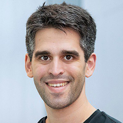
    Image of Sam Rodriques, PhD

    Sam Rodriques, PhD

    Co-Founder, Director, and CEO
    Edison Scientific and FutureHouse

    Panelist

    Image of Sam Rodriques, PhD

    Sam Rodriques, PhD

    Physicist and bioengineer Sam Rodriques, PhD, is the co-founder, director, and CEO of Edison Scientific and FutureHouse. Sam has invented technologies for spatial and temporal transcriptomics, brain mapping, gene therapy, and nanofabrication, and briefly ran an academic lab at the Francis Crick Institute in London prior to launching FutureHouse in San Francisco in 2023. FutureHouse is a new research lab focused on building an AI Scientist to accelerate research in biology and other complex sciences. In 2025, Sam launched Edison Scientific to commercialize the agents being built at FutureHouse, with the goal of wanting “science to move faster.”

    Sam graduated summa cum laude with highest honors in physics from Haverford College, where he worked on new methods for calculating quantum entanglement in multipartite quantum systems. He holds a PhD in physics from MIT, having worked between the MIT Media Lab, the MIT Department of Brain and Cognitive Sciences, and the Broad Institute. Among his awards and fellowships are the Hertz Foundation Graduate Fellowship, an NSF Graduate Research Fellowship, and a Churchill Scholarship.
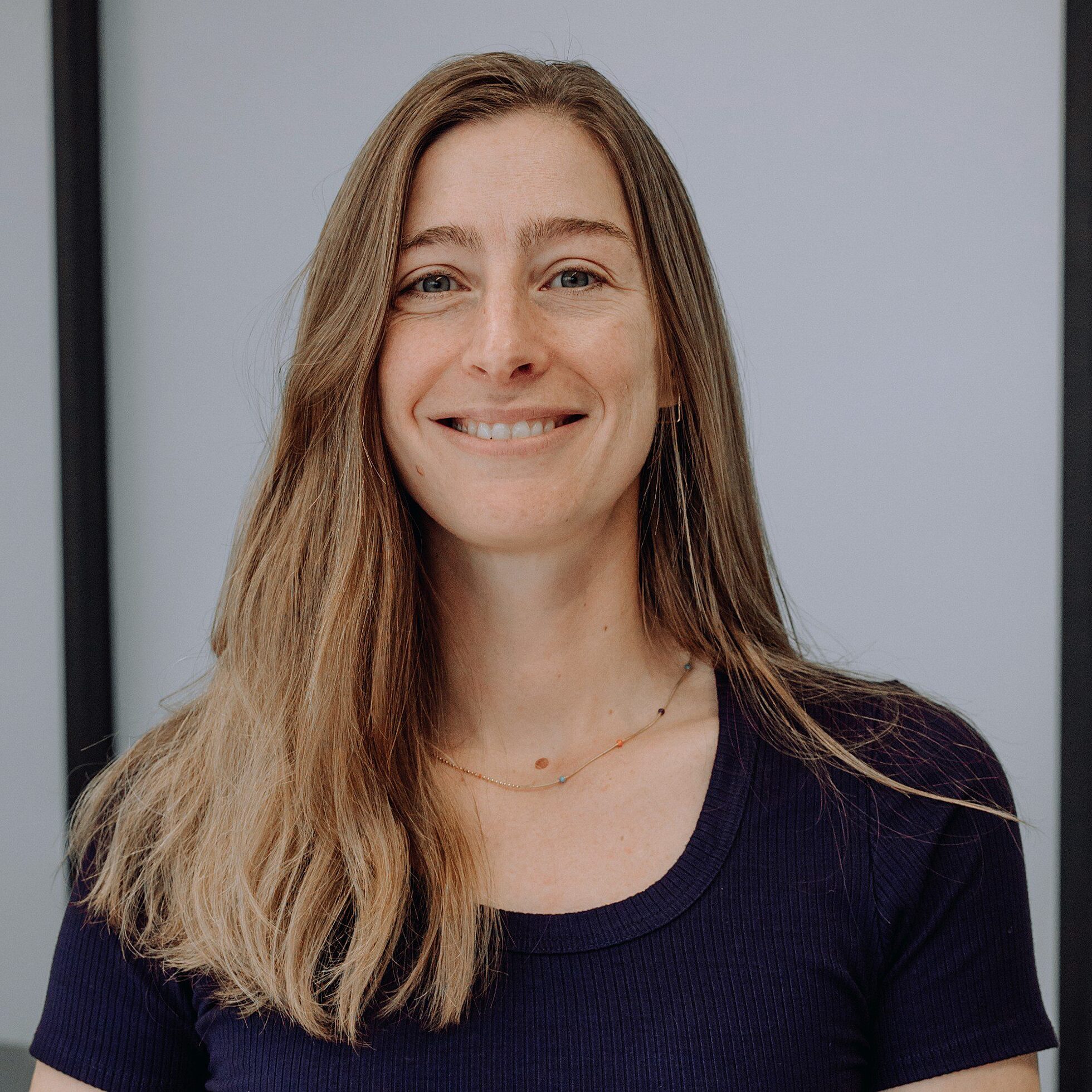
    Image of Peyton Greenside, PhD

    Peyton Greenside, PhD

    Co-Founder and CEO
    BigHat Biosciences

    Panelist

    Image of Peyton Greenside, PhD

    Peyton Greenside, PhD

    Peyton Greenside, PhD, is the co-founder and CEO of BigHat Biosciences, a Bay Area biotechnology company pioneering an ML-driven platform to accelerate the design of next-generation antibodies and other therapeutic proteins.

    Peyton holds a BA in applied mathematics from Harvard University, a PhD from Stanford University, and an MPhil in computational biology from the University of Cambridge, U.K.

    Image of Marc Tessier-Lavigne, PhD

    Marc Tessier-Lavigne, PhD

    Co-Founder, Chairman, and CEO
    Xaira Therapeutics

    Panelist

    Image of Marc Tessier-Lavigne, PhD

    Marc Tessier-Lavigne, PhD

    Marc Tessier-Lavigne, PhD, is the co-founder, chairman, and CEO of Xaira Therapeutics. Previously, he held executive positions in the private sector at Genentech, where he was executive vice president for research and chief scientific officer, and in the academic sector, where he served as president of The Rockefeller University and then for seven years at Stanford University, where he remains a professor of biology (on leave). He also served previously on the faculty of the University of California, San Francisco, and was an investigator with the Howard Hughes Medical Institute. A Rhodes Scholar, Marc has been a leader in understanding mechanisms that direct the wiring of the brain and has helped elucidate mechanisms of neurodegeneration. He is the recipient of numerous scientific awards, including the 2020 Gruber Neuroscience Prize, and has been elected to multiple scientific societies, including the U.S. National Academy of Sciences and U.S. National Academy of Medicine; he is also an officer of the Order of Canada, one of that country’s highest honors. Marc is the co-founder of two neuroscience biotech companies, Renovis and Denali, and has served on the boards of Agios, Regeneron, Pfizer, Juno, and Denali.

    Marc holds a BS degree in physics from McGill University in his native Canada. He earned a BA in philosophy and physiology from the University of Oxford as a Rhodes Scholar, and a PhD in physiology from the University of London.

    Broadcast Date: 

    • Time: 

    From billion-dollar funding rounds and waves of IPOs to the first AI-guided molecules entering the clinic, the industry is signaling a clear inflection point: AI is set to fundamentally reshape drug discovery. At the center of this shift are maturing foundation models for molecules and cellular systems that unveil insights into complex biology.

    Yet the challenge is no longer prediction alone. Building the infrastructure that connects accelerated computing, generative design, and experimental feedback will be critical for transforming AI into a fully integrated engine for therapeutic innovation.

    GEN’s virtual summit, The State of AI in Drug Discovery, returns on November 4 to examine how AI is reshaping R&D timelines and real-world patient translation. From big pharma to AI-natives, this year’s program features the leaders of many of today’s outstanding companies and organizations powering the AI revolution, including Xaira Therapeutics, Edison Scientific, Eli Lilly, Chai Discovery, Lila Sciences, BigHat Biosciences, and more!

    Among the program highlights during this 4-hour event are:

    • Building the AI scientist with Sam Rodriques, PhD (Edison Scientific)  
    • A fireside chat with Xaira Therapeutics CEO Marc Tessier-Lavigne, PhD 
    • Powering AI adoption through strategic enterprise partnerships with Aliza Apple, PhD (Lilly TuneLab), Simon Barnett (Dimension), and Elliot Hershberg, PhD (Amplify Ventures)  
    • The role of agentic workflows in accelerating drug discovery with Le Cong, PhD (Stanford and Phylo) and Andrew Beam, PhD (Lila Sciences) 
    • The new era of programmable therapeutics with Josh Meier (Chai Discovery) and Peyton Greenside, PhD (BigHat Biosciences) 
    • And breakout sessions from our four event sponsors: A-AlphaBio, Benchling, LabVantage, and Revvity Signals.

    Don’t miss this exceptional virtual summit, featuring many of the visionaries shaping the future of AI in drug discovery and biotech. Registration, as always, is free.

    Guest Speakers Include
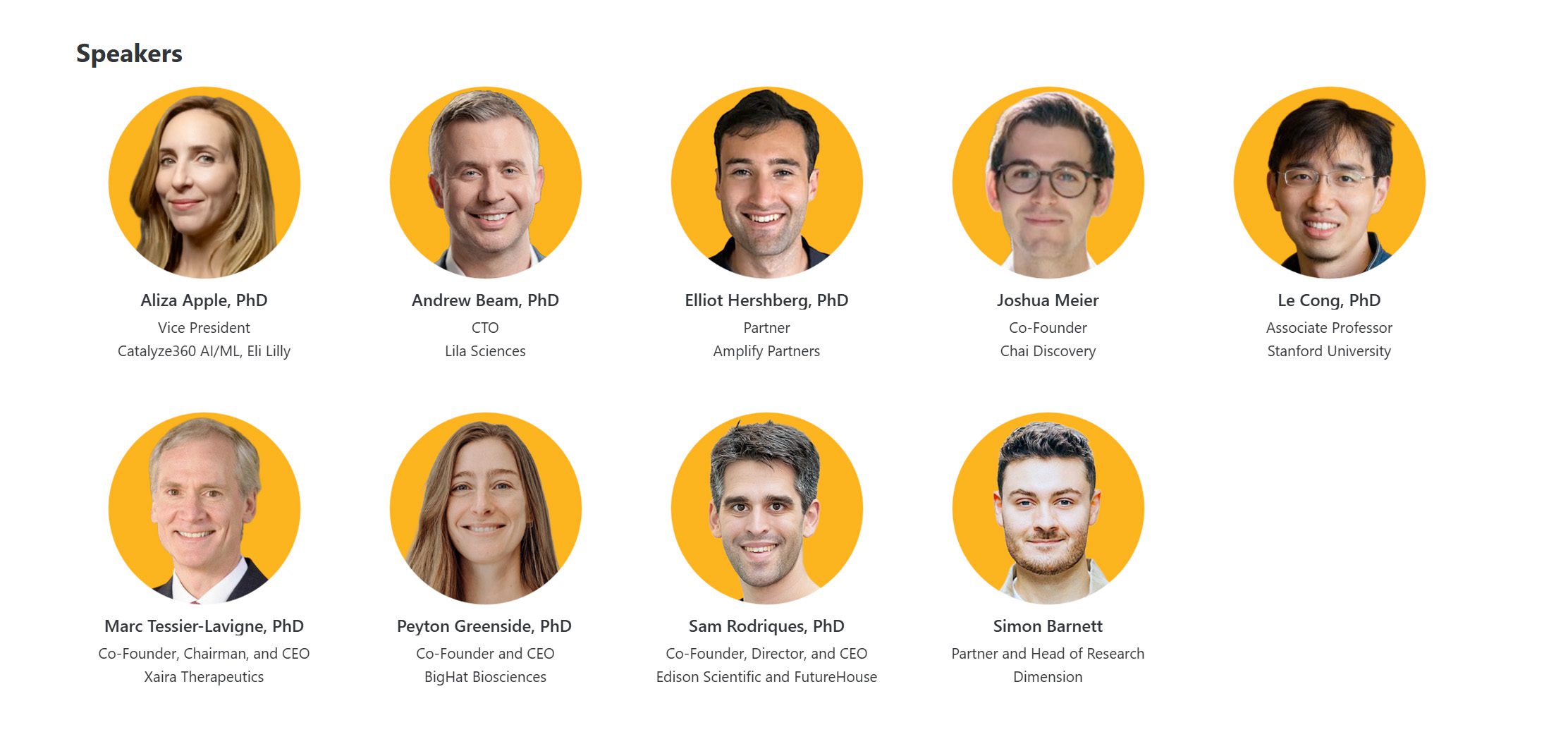
    The State of AI in Drug Discovery speakers

  • FDA-approved epilepsy drug may help reverse osteoarthritis damage

    FDA-approved epilepsy drug may help reverse osteoarthritis damage

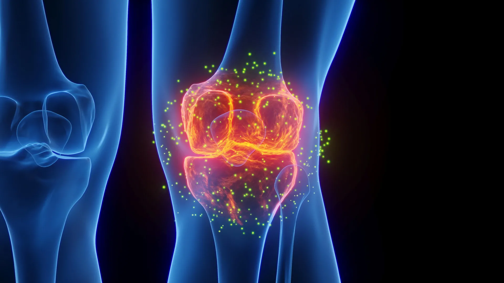
    For millions of people with osteoarthritis, pain and stiffness can make everyday activities increasingly difficult. Common treatments, including over-the-counter medications and steroid injections, may reduce symptoms for a time, but they do not prevent the underlying joint damage from progressing.

    New research from Yale suggests a different approach. In a study published in Bioactive Materials, scientists found that the medication lacosamide can serve two purposes in osteoarthritis, reducing joint pain while also reversing cartilage damage. The effects were particularly strong when the drug was delivered directly into the joint using a specialized hydrogel.

    Why osteoarthritis damages cartilage

    Osteoarthritis is often called a condition caused by wear and tear, but that description leaves out much of the biology involved.

    Within a healthy joint, cells known as chondrocytes help maintain cartilage by continuously balancing the creation of new tissue with the removal of old material. Osteoarthritis disrupts that balance. Cartilage begins to break down faster than it can be replaced, eventually allowing bones to rub against one another. As the disease advances, some patients may ultimately require joint reconstruction procedures such as a total knee replacement.

    “There is a major unmet need in osteoarthritis,” says the study’s principal investigator and Charles W. Ohse Professor of Orthopaedics & Rehabilitation, Chuan-Ju Liu, PhD. “We need therapies that don’t just mask pain but actually change how the disease progresses.”

    At present, no medicine approved by the U.S. Food and Drug Administration can both stop osteoarthritis pain and prevent the structural breakdown of cartilage. The work led by Liu suggests that it may be possible to address both problems with a single therapeutic strategy.

    The researchers repurposed an existing medication and paired it with an advanced gel designed to keep the drug inside the joint. The approach could potentially preserve joint tissue while delivering sustained pain relief without relying on addictive opioids.

    A protein linked to pain and cartilage loss

    The research focuses on Nav1.7, a protein that functions as a sodium channel. These channels act as microscopic gates in cell membranes and play an important role in electrical signaling.

    Nav1.7 was long thought to operate primarily in specialized nerve cells that transmit pain signals to the brain. More recent work from Liu and his team, however, revealed that the protein is also highly active in chondrocytes, the cells responsible for maintaining cartilage.

    Nav1.7 is relatively quiet in healthy joints. In osteoarthritis, however, its activity increases substantially. The researchers found that this heightened activity can intensify pain signaling while also pushing chondrocytes toward breaking down the cartilage they would normally help preserve.

    That makes Nav1.7 an unusual therapeutic target because it appears to influence both the sensation of pain and the physical deterioration of joint tissue.

    “When Nav1.7 becomes dysregulated, it contributes to both joint degeneration and pain,” Liu says. “Our findings suggest that Nav1.7 is a dual-acting target. By blocking this single protein, we can potentially quiet the pain nerves and tell the cartilage cells to not only stop breaking down but start repairing as well.”

    Epilepsy drug shows potential for cartilage repair

    Instead of creating an entirely new medication, Liu’s team tested drugs that already inhibit sodium channels. Among them, lacosamide produced strong biological effects at considerably lower concentrations and offered a better safety profile than older drugs in the same class.

    Lacosamide is already used as a treatment for epilepsy, but the researchers found that its effects on cartilage depended heavily on the dose.

    More was not necessarily better. At an ideal low concentration, lacosamide encouraged cells to produce proteins involved in building cartilage while also suppressing processes that break tissue down. When the concentration was either too high or too low, those benefits began to fade.

    “This tells us the system is finely tuned,” Liu notes. “There is an optimal range where the drug helps restore balance without overcorrecting. What stood out was not just its effectiveness, but how little of a dose was needed.”

    Further investigation revealed that lacosamide also changes the way cells communicate. The drug stimulated the release of two beneficial signaling proteins, HSP70 and midkine.

    HSP70 helps cells respond to stress and supports tissue repair, while midkine helps regulate inflammation and protect joint tissue from degeneration. Together, the proteins appear to create conditions that are more favorable for maintaining cartilage.

    “These proteins create a supportive environment for cartilage maintenance,” Liu explains. “They allow the effects of the drug to extend beyond individual cells and influence the entire tissue.”

    Smart hydrogel keeps the drug inside the joint

    Oral treatment with lacosamide was effective in preclinical testing, but drugs taken by mouth circulate throughout the body, increasing the potential for unwanted effects elsewhere.

    The researchers therefore investigated intra-articular injection, which allows the medication to be placed directly inside the affected joint.

    There was still a significant problem to overcome. “The knee joint, which is also the most common location for osteoarthritis, naturally acts like a leaky bucket,” Liu says. “The body’s drainage system can clear out liquids injected into the knee within hours.”

    To keep the medication in place longer, the team created a specialized hydrogel made from Collagen II. The material responds to temperature. It remains liquid while inside a cool syringe, then becomes a firm, jelly-like material after reaching body temperature.

    Once inside the joint, the gel acts as a storage site for lacosamide, keeping the medication concentrated in the affected area while gradually releasing it over several weeks.

    “The hydrogel acts as a local reservoir,” Liu says. “It holds the drug in place in the location it is needed most and releases it slowly over time. It transforms a daily pill into a long-lasting, local treatment that stays active for a month or longer.”

    In the same preclinical studies, one injection of the lacosamide-containing gel every four weeks prevented cartilage loss more effectively than taking an oral dose every day.

    Existing drug could speed clinical testing

    One important advantage of lacosamide is that the medication is already approved for use in people. That could make it possible to move toward clinical studies in osteoarthritis patients more quickly than would be possible with a completely new drug.

    Lacosamide has also been tested in humans with certain nerve-related pain conditions caused by Nav1.7 mutations. Those results give the researchers greater confidence that the effects seen in laboratory studies could eventually translate into meaningful pain relief for patients.

    The work also reflects a broader direction in medicine that combines drugs with advanced biomaterials to control exactly where and how treatments are delivered. If the approach proves successful in people, it could eventually reduce the number of procedures patients need, limit unwanted side effects, and provide longer-lasting protection against structural joint damage.

    “We are not just developing a treatment,” Liu concludes. “We are developing a system that allows the medicine to work more effectively where it matters most. Our goal is to move beyond symptom control and towards true disease modification. This effort brings us closer to that reality.”

  • Taking antibiotics? Sugar may be making the damage worse

    Taking antibiotics? Sugar may be making the damage worse

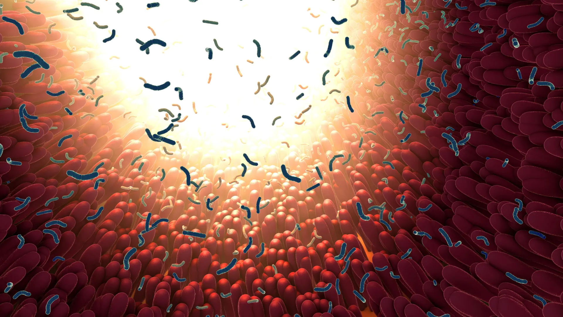
    Eating more sugar while taking antibiotics may intensify the disruption those drugs cause to beneficial gut bacteria, according to new research.

    The study focused on the intestinal microbiome, the vast community of bacteria that has evolved alongside humans and helps with digestion, immune function, and other essential processes. Antibiotics are among the most commonly prescribed medicines worldwide, but they can kill beneficial bacteria as well as harmful ones. In some cases, that disruption allows potentially dangerous species to gain an advantage.

    A diverse microbiome is generally better able to resist the expansion of harmful pathogens. Antibiotic treatment can reduce that diversity, leaving the gut ecosystem more vulnerable to disease, according to the researchers.

    Sugar Linked to Greater Microbiome Damage

    Researchers from NYU Langone Health, City of Hope, and Memorial Sloan Kettering closely monitored 9,419 meals consumed by 173 hospitalized cancer patients and tracked changes in their gut microbiomes.

    For every additional 100 grams of sugar eaten during the 48 hours before treatment, the researchers observed an additional 21% decline in alpha diversity, a measure of the variety of beneficial bacterial species found in the gut.

    The findings, published online Sept. 30 in the journal Nature, also showed that sugary diets combined with antibiotic treatment were associated with increases in Enterococcus faecium among patients. This bacterial species has been linked to infections that can be difficult to treat.

    Mouse experiments produced a similar result. Animals exposed to the antibiotic biapenem while consuming dietary sucrose experienced a 16.3-fold increase in the gut Enterococcus population by day 3 of the study. By day 6, the bacterial burden was 33.4-fold higher.

    “As we are learning how collateral damage of antibiotics to the microbiome are linked with long-term health problems, it’s important to identify factors that limit these unintended health risks,” said the study’s co-senior author Jonas Schluter, PhD, assistant professor in the Department of Microbiology at NYU Langone. “Our study, which used more than 9,000 recorded meals, suggests that modifying diet during and after antibiotics, specifically by reducing sweets intake, could help with that.”

    Why Researchers Studied Blood Cancer Patients

    The team focused on people with blood cancers because these patients frequently receive antibiotics and their food intake can be monitored closely during hospitalization. Researchers recorded what patients ate at every meal and collected microbiome samples each day.

    That combination of detailed diet records and daily biological sampling made the project one of the largest and most detailed studies of its kind to date.

    “The microbiome is emerging as an important factor in cancer care,” said co-senior study author Marcel van den Brink, MD, PhD, chief physician executive at City of Hope. “For patients undergoing intensive treatments such as bone marrow transplantation, the healthy diversity of the gut microbiome can have important implications for outcomes. Studies like this help us better understand how everyday factors, including diet, may affect a delicate balance that influences how patients respond to treatment, recover, and experience side effects.”

    Patients with blood cancers who receive stem cell transplants are often hospitalized for several weeks while undergoing chemotherapy and antibiotic treatment. During that period, both their diets and their gut microbiomes can change dramatically.

    In these patients, potentially harmful species such as Enterococcus faecium can flourish after much of the surrounding microbial ecosystem has been “destroyed.”

    Gut Microbiome Damage and Cancer Outcomes

    “Especially in blood cancer patients, microbiome injury is associated with worse outcomes, including higher overall mortality and infections,” said Jonathan Peled, co-senior author on the study and associate director of the Center for Hematological Malignancies at Memorial Sloan Kettering Cancer Center. “An important next step will be to design clinical trials to test whether short-term dietary changes can improve clinical outcomes in patients.”

    The researchers say dietary sugar may magnify the microbiome disruption caused by antibiotics because potentially harmful bacteria that survive treatment may be able to use those sugars to expand.

    “The new work identifies dietary sugars as an amplifier of antibiotic-induced microbiome disruption, probably because the potentially harmful bacteria that survive the antibiotics are benefiting from the dietary sugars that they can use to expand,” said study second author William Jogia, a PhD student in Dr. Schluter’s lab in the Institute for Systems Genetics. “While the findings suggest that reducing simple sugars may limit microbiome injury, whether sugar directly supports Enterococcus, weakens competing bacteria, or acts through another mechanism in patients remains to be seen.”

    The findings therefore suggest that cutting back on simple sugars during and after antibiotic treatment could potentially reduce microbiome damage. However, the researchers emphasize that more work is needed to determine exactly how sugar produces the effect and whether dietary changes will improve outcomes in patients.

    Study Authors and Collaborators

    Along with Drs. Schluter and Jogia, study authors from the Schluter lab at the Department of Microbiology and Institute for Systems Genetics at NYU Langone Health were Caichen Duan, Chenzhen Zhang, Fanny Matheis, and Alexis Sullivan.

    Authors from the Adult Bone Marrow Transplantation Service in the Department of Medicine at Memorial Sloan Kettering Cancer Center were co-senior author Jonathan Peled, co-first authors Anqi Dai and Annamaria Ballweg, along with Madhumitha Rangesa, Peter Adintori, Mirae Baichoo, Nicholas Waters, Abrielle Swartz, Sukanya Sahu, Brianna Gipson, Sandeep Raj, Zoe Pierce, Kenton Wu, Justin Neuberger, Adam Warren, Marissa Lubin Buchan, Ana Gradissimo, Luigi Amoretti, Annelie Clurman, Daniel Brereton, Paul Giardina, Miguel-Angel Perales, and Sergio Giralt. Also authors from Memorial Sloan Kettering Cancer Center were Teng Fei in the Department of Epidemiology and Biostatistics, Oriana Miltiadous in the Department of Pediatrics, Tatanisha Peets in Food and Nutrition Services, and Marina Burgos da Silva in the Human Oncology & Pathogenesis Program.

    Other authors were Tyler Funnell, Natalie Smith, John Slingerland, Antonio Gomes, and Robert Jenq in the Department of Hematology and Hematopoietic Cell Transplantation at City of Hope National Medical Center in Los Angeles; Eiko Hayase in the Departments of Genomic Medicine and Stem Cell Transplantation Cellular Therapy in the Division of Cancer Medicine at University of Texas MD Anderson Cancer Center; Corrado Zuanelli Brambilla in the Hematology Unit, Department of Cellular Therapy, Hematology and Laboratory Medicine at Azienda Ospedaliera Universitaria Senese in Siena, Italy; Abigail Johnson in the Division of Epidemiology and Community Health in the School of Public Health at the University of Minnesota, Minneapolis; Dan Knights in Department of Computer Science and Engineering at the University of Minnesota; and Kate Markey in the Translational Science and Therapeutics Division at Fred Hutchinson Cancer Center in Seattle.

    Funding and Disclosures

    Funding for the study was by National Institutes of Health grants K08HL143189, P30 CA008748, P01CA023766, R35CA284024, DP2AI164318, R01CA269617, P30CA0160087, R01-CA228358, R01-CA228308, F31-CA261086, P30 CA008748, P01-CA023766; R01-HL123340, R01-HL147584, and P01-AG052359. Other support came from the Gabrielle’s Angel Foundation, a Helen Hay Whitney postdoctoral fellowship, the Starr Cancer Consortium, The Tri-Institutional Stem Cell Initiative, the Lymphoma Foundation, the Susan and Peter Solomon Divisional Genomics Program, Cycle for Survival, the Parker Institute for Cancer Immunotherapy, the American Society of Clinical Oncology Young Investigator Award, a Hyundai Hope on Wheels Young Investigator Award, a Tow Center for Developmental Oncology Career Development Award, and an AIRC fellowship for Abroad.

    Dr. Schluter has filed intellectual property applications related to the microbiome (reference numbers #63/299,607), serves on an advisory board and holds equity in Jona Health, and is cofounder of Postbiotics Plus Research. These relationships are being managed in keeping with the policies of NYU Langone Health.

  • A new way to fight gum disease without wiping out good bacteria

    A new way to fight gum disease without wiping out good bacteria

    Bacteria are remarkably good at adapting. That ability helps them survive changing conditions, but it can also create problems when harmful species become resistant to antibiotics and disinfectants.

    At the same time, bacteria are not simply enemies to eliminate. Many species play important roles in human health, including the hundreds of different microbes that live in the mouth. That raises an intriguing possibility: instead of trying to kill bacteria, could scientists influence how they behave and encourage healthier microbial communities?

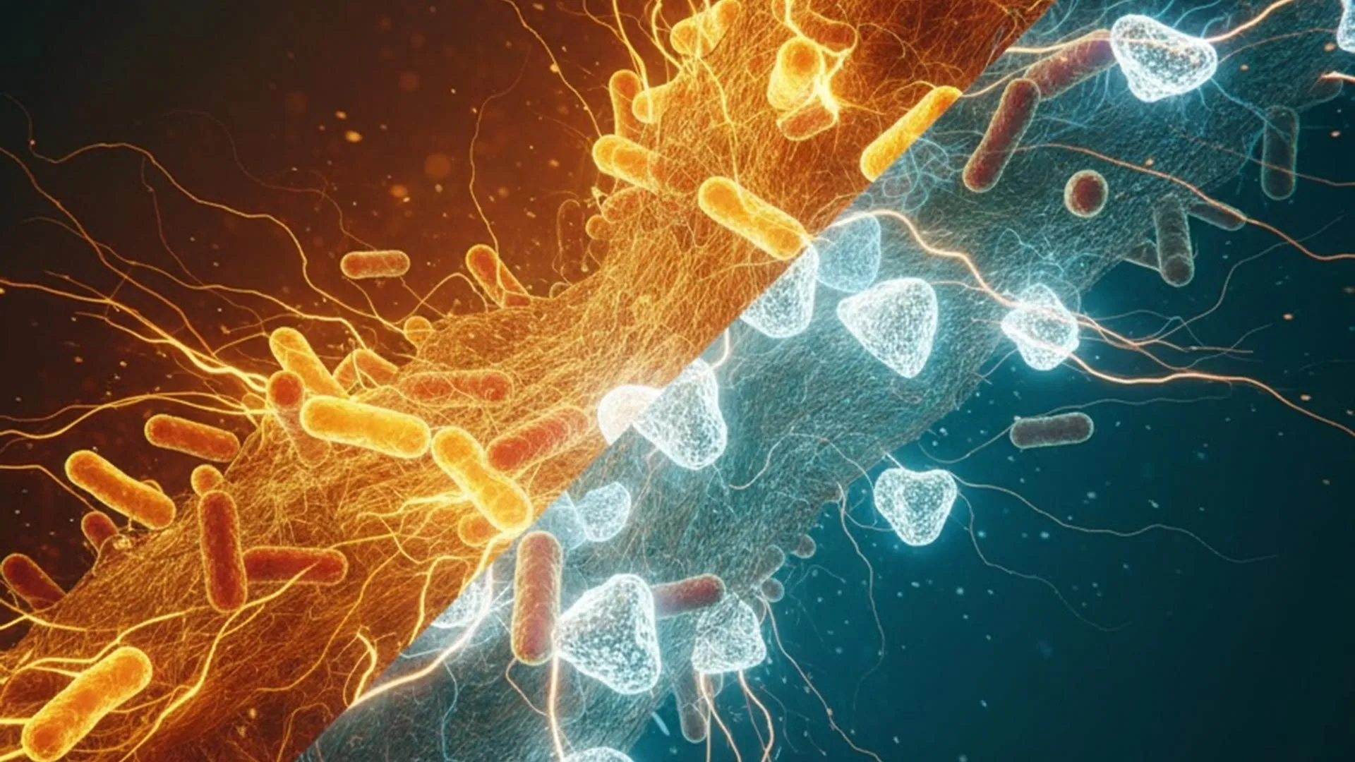
    Research published in 2025 in npj Biofilms and Microbiomes suggests that may be possible. Scientists studying dental plaque found that interfering with the chemical signals bacteria use to communicate can change which species thrive, potentially shifting the community toward bacteria associated with better oral health.

    How Bacteria Communicate in the Mouth

    The human mouth is home to roughly 700 species of bacteria. These microbes do not simply grow alongside one another. Many exchange chemical signals through a process called quorum sensing.

    Quorum sensing allows bacteria to detect how many neighboring cells are present and coordinate group behaviors. Some oral bacteria communicate using molecules known as N-acyl homoserine lactones (AHLs).

    Researchers from the College of Biological Sciences and the School of Dentistry examined how these signals shape dental plaque. Their goal was to determine whether bacterial communication could be manipulated to help maintain a healthier oral microbiome.

    The oral microbiome refers to the community of microorganisms living in the mouth. When that community becomes unbalanced, potentially harmful bacteria can become more dominant and contribute to conditions such as periodontal disease.

    Chemical Signals Can Cross the Gumline

    The researchers found that bacteria within dental plaque produced AHL signals in aerobic environments (such as above the gumline), where oxygen is available.

    Those chemical messages could also be detected by bacteria living in anaerobic environments (beneath the gumline), where oxygen levels are much lower.

    This connection was important because conditions below the gumline can favor bacteria associated with periodontal disease.

    The team also used specialized enzymes called lactonases to remove AHL signals. Lactonases break down these communication molecules, effectively interfering with the bacterial conversation.

    When researchers disrupted AHL signaling, the dental plaque community shifted toward species more strongly associated with oral health.

    Their findings suggest that carefully chosen enzymes might someday be used to alter the makeup of dental plaque and help maintain a healthier balance of microorganisms.

    Dental Plaque Behaves Like an Ecosystem

    “Dental plaque develops in a sequence, much like a forest ecosystem,” said Mikael Elias, associate professor in the College of Biological Sciences and senior author of the study. “Pioneer species like Streptococcus and Actinomyces are the initial settlers in simple communities — they’re generally harmless and associated with good oral health. Increasingly diverse late colonizers include the ‘red complex’ bacteria like Porphyromonas gingivalis, which are strongly linked to periodontal disease. By disrupting the chemical signals bacteria use to communicate, one could manipulate the plaque community to remain or return to its health-associated stage.”

    In other words, dental plaque changes over time rather than appearing all at once. Early communities may contain relatively harmless bacteria, while more complex communities can eventually include species strongly associated with gum disease.

    The researchers hope that interfering with bacterial communication could make it possible to guide plaque toward an earlier, healthier state rather than attempting to eliminate the entire microbial population.

    Oxygen Changes How Bacterial Signals Behave

    One of the most important findings was that the effects of bacterial communication depended heavily on oxygen levels.

    “What’s particularly striking is how oxygen availability changes everything,” said lead author Rakesh Sikdar. “When we blocked AHL signaling in aerobic conditions, we saw more health-associated bacteria. But when we added AHLs under anaerobic conditions, we promoted the growth of disease-associated late colonizers. Quorum sensing may play very different roles above and below the gumline, which has major implications for how we approach treatment of periodontal diseases.”

    That difference suggests that the same chemical signals can influence microbial communities in very different ways depending on where they occur in the mouth.

    Above the gumline, where oxygen is more plentiful, disrupting AHL signaling favored bacteria linked with better oral health. Beneath the gumline, adding those signals encouraged later colonizing species associated with disease.

    A Different Strategy for Preventing Gum Disease

    The researchers now want to investigate how bacterial communication varies throughout the mouth and among people at different stages of periodontal disease.

    Rather than broadly attacking oral bacteria with antimicrobial treatments, the long term goal is to find ways to influence the balance of the microbial community itself.

    “Understanding how bacterial communities communicate and organize themselves may ultimately give us new tools to prevent periodontal disease — not by waging war on all oral bacteria, but by strategically maintaining a healthy microbial balance,” said Elias.

    The approach could eventually have implications beyond dentistry. Similar microbial imbalances, often called microbiome dysbiosis, occur elsewhere in the body and have been associated with a range of health problems, including certain types of cancer.

    Researchers hope that learning how to manipulate bacterial communication could eventually provide a foundation for therapies designed to steer microbial communities toward healthier states rather than simply destroying them.

    The research was funded by the National Institutes of Health.

  • Goodbye joint replacements? Stanford scientists found a way to regrow cartilage and stop arthritis

    Goodbye joint replacements? Stanford scientists found a way to regrow cartilage and stop arthritis

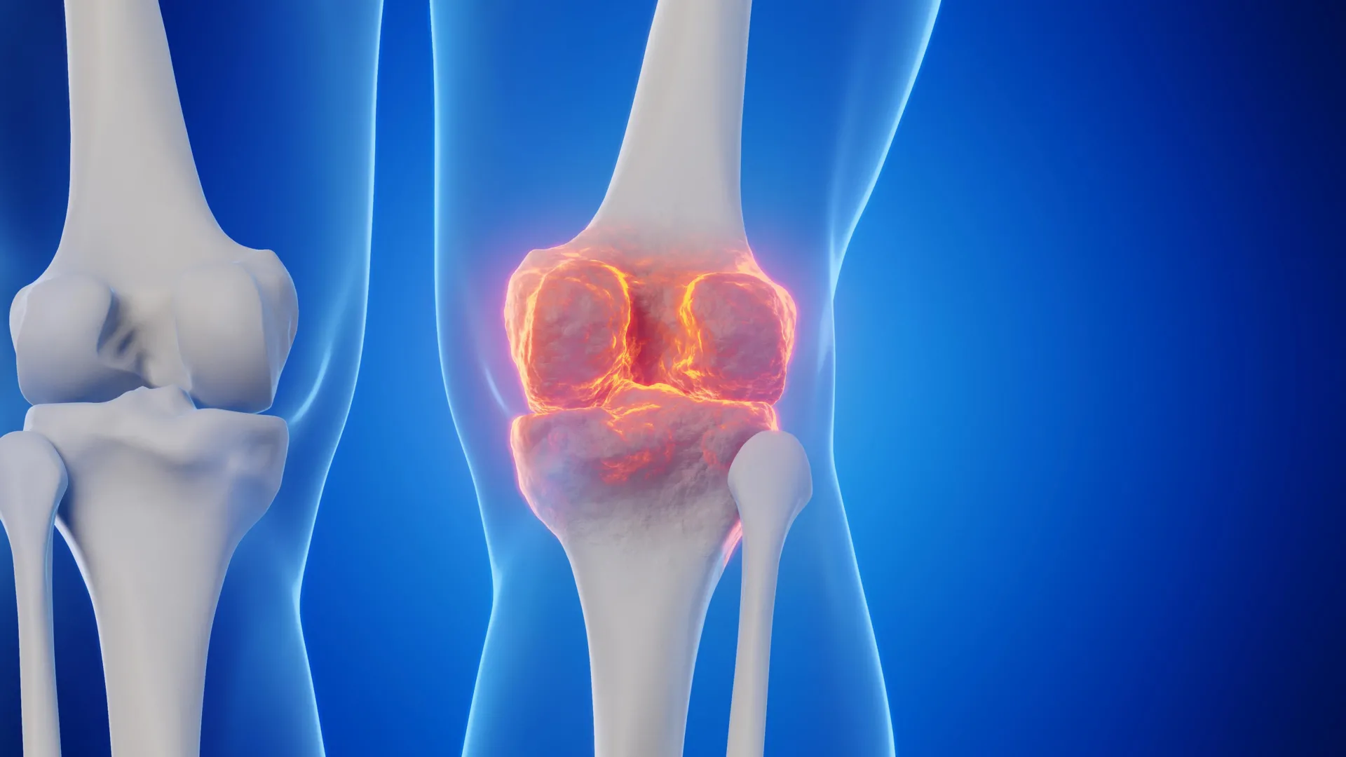
    Scientists have identified a way to restore cartilage in aging knee joints, at least in mice, by blocking a protein whose levels rise with age. The approach not only reversed naturally occurring cartilage loss in older animals, but also protected mice from developing arthritis after knee injuries similar to ACL tears in people.

    The findings also showed promise in human tissue. Cartilage samples collected during knee replacement surgeries responded to the treatment by beginning to produce new, functional cartilage.

    Together, the results suggest that cartilage damaged by aging or arthritis may be more capable of repair than previously thought. If the strategy ultimately works in people, researchers believe it could potentially lead to an oral medicine or injection that regenerates cartilage and reduces the need for knee or hip replacement surgery.

    Targeting the damage behind osteoarthritis

    Osteoarthritis is a degenerative joint disease in which cartilage gradually breaks down, leaving joints painful, swollen and increasingly difficult to move. It affects about one in five adults in the United States and is estimated to account for roughly $65 billion in direct health care costs each year.

    Current treatments mainly focus on controlling pain and other symptoms. Once the disease becomes severe, replacing the damaged joint surgically may be the only remaining option. There is currently no drug that can reliably slow or reverse osteoarthritis itself.

    The Stanford Medicine-led study instead focused on a protein called 15-PGDH. The researchers have described it as a gerozyme, a term for enzymes that become more abundant with age and contribute to the gradual loss of tissue function.

    Previous work by the same group showed that 15-PGDH acts as an important regulator of aging in multiple tissues. Blocking the protein with a small molecule increased muscle mass and endurance in old mice. Doing the opposite, increasing 15-PGDH in young animals, caused their muscles to shrink and weaken. The protein has also been linked to the regeneration of bone, nerve and blood cells.

    In many of those tissues, healing depends on tissue-specific stem cells multiplying and developing into specialized cells. Cartilage turned out to behave differently. Instead of relying on stem cells, existing cartilage cells called chondrocytes changed their patterns of gene activity and shifted toward a more youthful state.

    “This is a new way of regenerating adult tissue, and it has significant clinical promise for treating arthritis due to aging or injury,” said Helen Blau, PhD, professor of microbiology and immunology. “We were looking for stem cells, but they are clearly not involved. It’s very exciting.”

    Blau, who directs the Baxter Laboratory for Stem Cell Biology and is the Donald E. and Delia B. Baxter Foundation Professor, and Nidhi Bhutani, PhD, associate professor of orthopaedic surgery, are the senior authors of the research, which was published in Science. Instructor of orthopedic surgery Mamta Singla, PhD, and former postdoctoral scholar Yu Xin (Will) Wang, PhD, are the lead authors of the study. Wang is now an assistant professor at the Sanford Burnham Institute in San Diego.

    “Dramatic regeneration” of cartilage

    “Millions of people suffer from joint pain and swelling as they age,” Bhutani said. “It is a huge unmet medical need. Until now, there has been no drug that directly treats the cause of cartilage loss. But this gerozyme inhibitor causes a dramatic regeneration of cartilage beyond that reported in response to any other drug or intervention.”

    Cartilage is not all the same. The human body contains three major types, each designed for a different job.

    Elastic cartilage is soft and flexible and helps form structures such as the outer ear. Fibrocartilage is tougher and better suited to absorbing force, including in areas between the vertebrae of the spine. Hyaline cartilage is smooth and slippery, allowing bones to move against one another with very little friction in joints such as the ankles, hips, shoulders and parts of the knee.

    In joints, hyaline cartilage is also known as articular cartilage. It is the type most commonly damaged by osteoarthritis.

    Osteoarthritis can develop as joints experience stress from aging, injury or obesity. Chondrocytes begin producing inflammatory molecules while also breaking down collagen, a structural protein that gives cartilage much of its strength.

    As collagen disappears, cartilage becomes thinner and softer. Inflammation adds swelling and pain, creating the familiar symptoms of osteoarthritis.

    The problem is that articular cartilage normally has very little ability to repair itself. Researchers have identified possible stem or progenitor cells capable of forming cartilage in bone, but efforts to find similar cell populations within articular cartilage itself have not been successful.

    An aging protein becomes a target

    Earlier research from Blau’s laboratory found that a molecule called prostaglandin E2 is important for the function of muscle stem cells. The protein 15-PGDH breaks down prostaglandin E2.

    Blocking 15-PGDH, or otherwise raising levels of prostaglandin E2, has been shown to support regeneration in damaged muscle, nerve, bone, colon, liver and blood cells in young mice.

    That raised an important question for Blau, Bhutani and their colleagues: Could the same biological pathway contribute to the deterioration of cartilage as animals age or recover from injury?

    The researchers compared 15-PGDH levels in knee cartilage from young and old mice. They found that the amount of the gerozyme roughly doubled with age.

    They then gave older animals a small molecule drug designed to inhibit 15-PGDH. In one experiment, the drug was injected into the abdomen so it could have effects throughout the body. In another, it was delivered directly into the knee joint.

    Both approaches produced striking results.

    Cartilage in the knees of older animals had been noticeably thinner and less functional than cartilage in young mice. After treatment, however, it became thicker across the surface of the joint.

    Importantly, the cells were making hyaline cartilage, the smooth articular cartilage needed for healthy joint movement, rather than the less suitable fibrocartilage.

    “Cartilage regeneration to such an extent in aged mice took us by surprise,” Bhutani said. “The effect was remarkable.”

    Protecting knees after ACL injuries

    The researchers also tested whether the treatment could help after traumatic knee injuries.

    ACL tears are particularly common in sports such as soccer, basketball and skiing, where athletes frequently pivot, stop abruptly or jump. Surgery can repair the torn ligament, but fixing the immediate injury does not necessarily prevent long-term joint damage.

    About 50% of people who experience these injuries go on to develop osteoarthritis in the affected joint within approximately 15 years.

    In the mouse experiments, researchers administered the gerozyme inhibitor twice a week for four weeks after injury. The treatment dramatically reduced the likelihood that the animals would develop osteoarthritis.

    Mice that received a control drug had 15-PGDH levels twice those of animals whose knees had not been injured, and they developed osteoarthritis within four weeks.

    The treated mice also moved more normally and placed more weight on the paw attached to the injured leg than untreated animals did.

    “Interestingly, prostaglandin E2 has been implicated in inflammation and pain,” Blau said. “But this research shows that, at normal biological levels, small increases in prostaglandin E2 can promote regeneration.”

    Making old cartilage cells act younger

    To understand what was happening inside the joint, the researchers took a closer look at chondrocytes from young and old mice.

    Older cartilage cells showed more activity in genes associated with inflammation and with the unwanted conversion of hyaline cartilage into bone. At the same time, genes involved in normal cartilage development were less active.

    Treatment shifted that balance.

    One population of old chondrocytes that produced 15-PGDH and expressed genes associated with cartilage degradation dropped from 8% of cells to 3%.

    A second population, which did not produce 15-PGDH but expressed genes linked to fibrocartilage formation, fell from 16% to 8%.

    Meanwhile, a third group moved sharply in the opposite direction. These cells did not produce 15-PGDH and expressed genes involved in forming hyaline cartilage and maintaining the extracellular matrix needed for healthy cartilage function. Their share increased from 22% to 42%.

    The extracellular matrix is the network of proteins and other molecules that surrounds cells and gives tissues their structure. In cartilage, it is especially important because it helps the tissue withstand pressure while maintaining the smooth surface joints need for movement.

    Overall, the treatment appeared to push cartilage toward a younger biological state without recruiting stem or progenitor cells.

    Human cartilage also responded

    The researchers then examined cartilage taken from people with osteoarthritis who were undergoing total knee replacement surgery.

    After one week of treatment with the 15-PGDH inhibitor, the human tissue contained fewer chondrocytes expressing 15-PGDH. Activity in genes associated with cartilage degradation and fibrocartilage also declined compared with untreated tissue.

    Most notably, the samples began regenerating articular cartilage.

    “The mechanism is quite striking and really shifted our perspective about how tissue regeneration can occur,” Bhutani said. “It’s clear that a large pool of already existing cells in cartilage are changing their gene expression patterns. And by targeting these cells for regeneration, we may have an opportunity to have a bigger overall impact clinically.”

    The findings do not yet establish that the treatment can regrow cartilage or prevent osteoarthritis in people. The mouse results and experiments on human tissue represent important early evidence, but clinical trials specifically testing cartilage regeneration will be needed to determine whether the approach is safe and effective for patients.

    An oral 15-PGDH inhibitor has already reached clinical testing for a different age related problem: muscle weakness.

    Blau added, “Phase 1 clinical trials of a 15-PGDH inhibitor for muscle weakness have shown that it is safe and active in healthy volunteers. Our hope is that a similar trial will be launched soon to test its effect in cartilage regeneration. We are very excited about this potential breakthrough. Imagine regrowing existing cartilage and avoiding joint replacement.”

    Researchers from the Sanford Burnham Prebys Medical Discovery Institute contributed to the work.

    The study was funded by the National Institutes of Health (grants R01AR070864, R01AR077530, R01AG069858 and R00NS120278), the Baxter Foundation for Stem Cell Biology, the Li Ka Shing Foundation, the Stanford Cardiovascular Institute, the Milky Way Research Foundation, the Canadian Institutes of Health Research, a Stanford Translational Research and Applied Medicine Pilot grant, a GlaxoSmithKline Sir James Black Postdoctoral Fellowship, and a Stanford Dean’s Postdoctoral Fellowship.

    Blau, Bhutani and other coauthors are inventors on patent applications held by Stanford University involving 15-PGDH inhibition for cartilage and tissue rejuvenation. Those applications are licensed to Epirium Bio. Blau is a cofounder of Myoforte/Epirium and holds equity and stock options in the company.

  • High-throughput metabolic engineering in mammalian cells

    High-throughput metabolic engineering in mammalian cells

    • Research Briefing
    • Published:

    Nature Biotechnology (2026) Cite this article

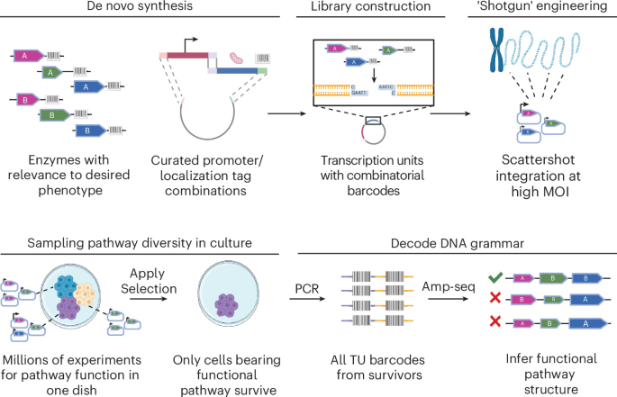
    Engineering complex phenotypes into mammalian cells holds profound potential but remains a largely intractable engineering challenge. We developed shotgun genetic engineering (SGE), a multiplexed strategy that screens millions of combinatorial solutions in a single experiment. This approach uses selection strategies that reveal effective designs, facilitating rapid engineering of complex biological functions.

    This is a preview of subscription content, access via your institution

    Access options

    Access through your institution

    Access Nature and 54 other Nature Portfolio journals

    Get Nature+, our best-value online-access subscription

    $32.99 / 30 days

    cancel any time

    Subscribe to this journal

    Receive 12 print issues and online access

    $259.00 per year

    only $21.58 per issue

    Buy this article

    • Purchase on SpringerLink
    • Instant access to the full article PDF.

    USD 39.95

    Prices may be subject to local taxes which are calculated during checkout

    Fig. 1: Highly multiplexed exploration of combinatorial design spaces with SGE.

    Subjects

    References

    1. Lim, W. A. The emerging era of cell engineering: harnessing the modularity of cells to program complex biological function. Science 378, 848–852 (2022). This review article outlines potential therapeutic applications of cellular engineering.

      Article  CAS  PubMed  Google Scholar 

    2. Nielsen, J. & Kiesling, J. Engineering cellular metabolism. Cell 164, 1185–1197 (2016). This review article outlines to our knowledge of cellular metabolism, how they impede metabolic engineering efforts, and how they are addressed using the DBT framework.

      Article  CAS  PubMed  Google Scholar 

    3. Srinivasan, P. & Smolke, C. D. Biosynthesis of medicinal tropane alkaloids in yeast. Nature 585, 614–619 https://doi.org/10.1038/s41586-020-2650-9 (2020). This article describes metabolic engineering feat in yeast: the successful biosynthesis of medicinal tropane alkaloids by overexpression of 26 genes across six subcellular locations.

      Article  CAS  PubMed  PubMed Central  Google Scholar 

    4. Rai, K. et al. Ultra-high-throughput mapping of genetic design space. Nature 650, 1035–1044 (2026). This paper describes the CLASSIC method for optimizing gene circuit designs.

      Article  CAS  PubMed  Google Scholar 

    5. Trolle, J. et al. Resurrecting essential amino acid biosynthesis in mammalian cells. eLife 11, e72847 (2022). This paper features our previous work to engineer valine biosynthesis in CHO cells.

      Article  CAS  PubMed  PubMed Central  Google Scholar 

    Download references

    Additional information

    Publisher’s note Springer Nature remains neutral with regard to jurisdictional claims in published maps and institutional affiliations.

    Trolle, J. et al. Highly multiplexed mammalian metabolic engineering with a shotgun approach. Nat. Biotechnol. https://doi.org/10.1038/s41587-026-03318-7 (2026).

    About this article

    Check for updates. Verify currency and authenticity via CrossMark

    Cite this article

    High-throughput metabolic engineering in mammalian cells. Nat Biotechnol (2026). https://doi.org/10.1038/s41587-026-03317-8

    Download citation

    • Published:

    • Version of record:

    • DOI: https://doi.org/10.1038/s41587-026-03317-8

  • Cancer-fighting t cells burn out. Scientists may have found a fix

    Cancer-fighting t cells burn out. Scientists may have found a fix

    Cancer immunotherapy can unleash T cells, the immune system’s specialized cancer killers, against tumors. But there is a major limitation: These cells can become worn down before the cancer is eliminated. This condition, known as T cell exhaustion, leaves the cells unable to maintain a strong attack or keep tumor growth under control.

    That problem has been particularly important for checkpoint inhibitors, immunotherapy drugs designed to remove biological restraints that normally limit T cell activity.

    “A tragic part of T cell exhaustion is that the immunotherapy seems to be working for patients, and then it fades,” says Santosha Vardhana, MD, PhD, a physician-scientist at Memorial Sloan Kettering Cancer Center (MSK) who treats people with lymphoma. “Many of them experience a brief wisp of promise only to have it taken away.”

    Researchers in Dr. Vardhana’s laboratory have now identified a signaling molecule called MEK as an important driver of this exhaustion process. Based on animal studies published in Immunity, the findings suggest that blocking MEK might slow T cell exhaustion and potentially make immunotherapy more effective.

    “We’re excited about applying this finding to enhance multiple forms of immunotherapy,” Dr. Vardhana says. “FDA-approved MEK inhibitors are already available, so this approach could be tested in humans without much delay.”

    How MEK Pushes T Cells Toward Exhaustion

    Scientists have only recently begun to understand the processes that cause T cells to become exhausted. In 2020, Dr. Vardhana’s laboratory identified an important piece of the puzzle: the cells’ metabolism, meaning the chemical processes they use to turn nutrients into energy.

    When T cells are continuously exposed to tumor antigens (the cancer proteins the immune system sees as foreign), the mitochondria inside the cells can become overburdened. Mitochondria are responsible for transforming nutrients into energy that cells can use.

    “There is a large metabolic demand being imposed as T cells encounter cancer cells and try to produce cancer-killing, or cytotoxic, proteins,” Dr. Vardhana says. “It turns out that the decision to make high levels of these proteins is regulated by MEK.”

    If MEK becomes excessively active, it can eventually drive T cells into terminal exhaustion, a severely depleted state in which immunotherapy can no longer reactivate them.

    “We realized T cell exhaustion isn’t simply a loss of function — it reflects an imbalance between what these cells are being asked to do and the energy they have available,” says Tanmana Mitra, PhD, a student in the Vardhana lab and the study’s first author.

    Surprisingly, the team discovered that exhausted T cells were not metabolically sluggish. They were actually highly active. When researchers treated the cells with MEK inhibitors, the T cells multiplied more while consuming less energy.

    “That paradox made us ask where all that energy was going, and we discovered that these cells were investing enormous resources into making proteins,” Dr. Mitra explains. “It changed how we think about T cell exhaustion — from a problem of too little energy to one of excessive energy demand.”

    Helping T Cells Pace Themselves

    The findings suggested that reducing MEK signaling could lower the pressure on T cells to continuously manufacture cytotoxic proteins. That, in turn, allowed some of the cells to stay active and capable of renewing themselves for longer, potentially extending the effectiveness of immunotherapy.

    The concept is similar to pacing yourself during a long road trip instead of driving at full speed from beginning to end. Reducing the intensity can help preserve enough fuel to keep going.

    In laboratory models, blocking MEK signaling allowed T cells to persist even in the difficult conditions surrounding a tumor.

    But the researchers caution that suppressing MEK is unlikely to be the right approach for every cancer patient.

    T Cell Exhaustion Can Also Be Protective

    Scientists now understand that T cell exhaustion is more complicated than simply having immune cells lose their strength. MSK immunologist Andrea Schietinger, PhD, previously found that T cells can enter an exhausted state as a survival mechanism. By reducing their activity, they avoid becoming overstimulated and dying.

    “As we’ve learned more about T cell exhaustion,” Dr. Vardhana says, “we’ve increasingly understood that it’s not the case that exhausted T cells are bad, so let’s try to reverse the process with a drug. Instead, exhaustion is more of an equilibrium state that lets the cells survive and keep going — almost like a ‘safe mode’ for T cells.”

    When a T cell is actively attacking cancer and producing cytoxic proteins, its mitochondria must turn nutrients from food into adenosine triphosphate (ATP). ATP is the molecule cells primarily use to store and transfer energy.

    “Think of ATP as the currency in a fund that the cell spends down,” Dr. Vardhana says. “If you spend ATP on one thing, you don’t have enough to do something else. The exhaustion program is a sign that the cell’s bank account is getting close to zero. MEK tells exhausted cells whether to conserve fuel or go for broke. What we found is that inhibiting MEK makes the cells more conservative — helping them live longer while reducing the rate at which they produce the proteins that actually kill cancer cells.”

    That makes MEK both part of the problem and part of the solution. Strong MEK activity helps T cells attack at maximum intensity, but it can also push them toward complete burnout. Blocking MEK therefore involves a tradeoff: The immune attack may become less intense, but the cancer-fighting cells can survive longer.

    Whether a short, powerful immune response or a slower, longer-lasting one is preferable may depend on the characteristics of an individual patient’s cancer.

    When T Cells May Need To Go Full Speed

    According to Dr. Vardhana, MEK inhibitors would need to be used selectively. Two factors can indicate that a patient is likely to respond well to immunotherapy:

    1. The tumors are small.
    2. The patients have a high number of immune cells attacking the tumor — usually because the tumor has many mutations that make it recognizable.

    “In these patients, conservation of T cells is not that important,” he says. “It’s like being in a car with 1/8 of a tank left, but you can see the finish line. In these patients, you would just let the car keep burning the gas — in other words, take the traditional immunotherapy approach. These are the patients in whom MEK inhibition is probably not needed.”

    The situation may be different for patients with large tumors or relatively few tumor-fighting immune cells. Their immune response may not be powerful enough to eliminate the cancer quickly.

    In those cases, creating a slower, sustained response with a MEK inhibitor could help T cells remain present for longer, even if they are partly exhausted. That persistence may be especially important when either the task at hand (the tumor) is large or the workforce (the number of T cells) is small.

    Potential Uses Across Cancer Immunotherapy

    Dr. Vardhana says carefully applied MEK inhibition could potentially improve several types of immunotherapy.

    Checkpoint inhibitors: MEK inhibition has already shown effectiveness in melanoma when combined with checkpoint inhibitors and a targeted treatment known as a BRAF inhibitor.

    Chimeric antigen receptor (CAR) T cell therapy: “We think this approach could dramatically boost T cell persistence, which has been a big problem with CAR T cell therapy,” Dr. Vardhana says.

    Tumor infiltrating lymphocyte (TIL) therapy: This treatment takes advantage of immune cells that have already been attacking a patient’s cancer and expands their numbers. Using MEK inhibition before or after TIL therapy could potentially help the most effective tumor-fighting TILs survive longer.

    Bispecific antibodies: These laboratory-made proteins are designed to attach to two different targets at the same time. They can strongly activate T cells, but that intense stimulation may also contribute to exhaustion.

    “This study shows the importance of understanding core principles of T cell biology — what sets the balance between conservation of energy and strong, cancer-fighting activity,” Dr. Vardhana says. “Once we know the answer to that, the therapeutic possibilities really start to fan out.”

    Key Takeaways

    • T cells can become exhausted as they continuously recognize and attack cancer, reducing their ability to keep tumors under control.
    • The signaling molecule MEK appears to play an important role in pushing T cells toward exhaustion.
    • Blocking MEK may slow that process by reducing the cells’ energy demands, potentially helping them persist for longer during cancer treatment.
    • The researchers suggest this strategy could be particularly useful for patients who respond poorly to traditional immunotherapy, including those with large tumors or relatively few immune cells capable of recognizing the cancer.

    Additional authors on the study include Jahan Rahman, Madeline Hwee, Yan-Ting Chen, Ruben Jose Jesus Faustino Ramos, Hui Liu, Travis Hartman, Justin Cross, Miguel de Jesus, Morgan Huse, Valerie Longo, and Pat Zanzonico.

  • The “glue” holding your cells together has a surprising second job

    The “glue” holding your cells together has a surprising second job

    A protein best known for helping cells and tissues stay connected has another unexpected role. Researchers have found that it also helps epithelial cells, which form continuous sealed layers throughout the body, engulf nearby dead cells.

    The discovery could have implications for chronic inflammation. Debris from dying cells is a major contributor to inflammatory responses, so understanding how tissues remove that material may reveal new clues about what happens when the cleanup process fails.

    The study, published in Nature Communications, focuses on the E-cadherin complex. This molecular system includes E-cadherin along with three additional proteins. Together, they connect epithelial cells lining areas such as the skin, gut and airways, giving tissues the structural strength they need to remain intact. In these tissues, each cell connects to E-cadherin molecules on neighboring cells.

    Cellular Glue Takes on a Cleanup Role

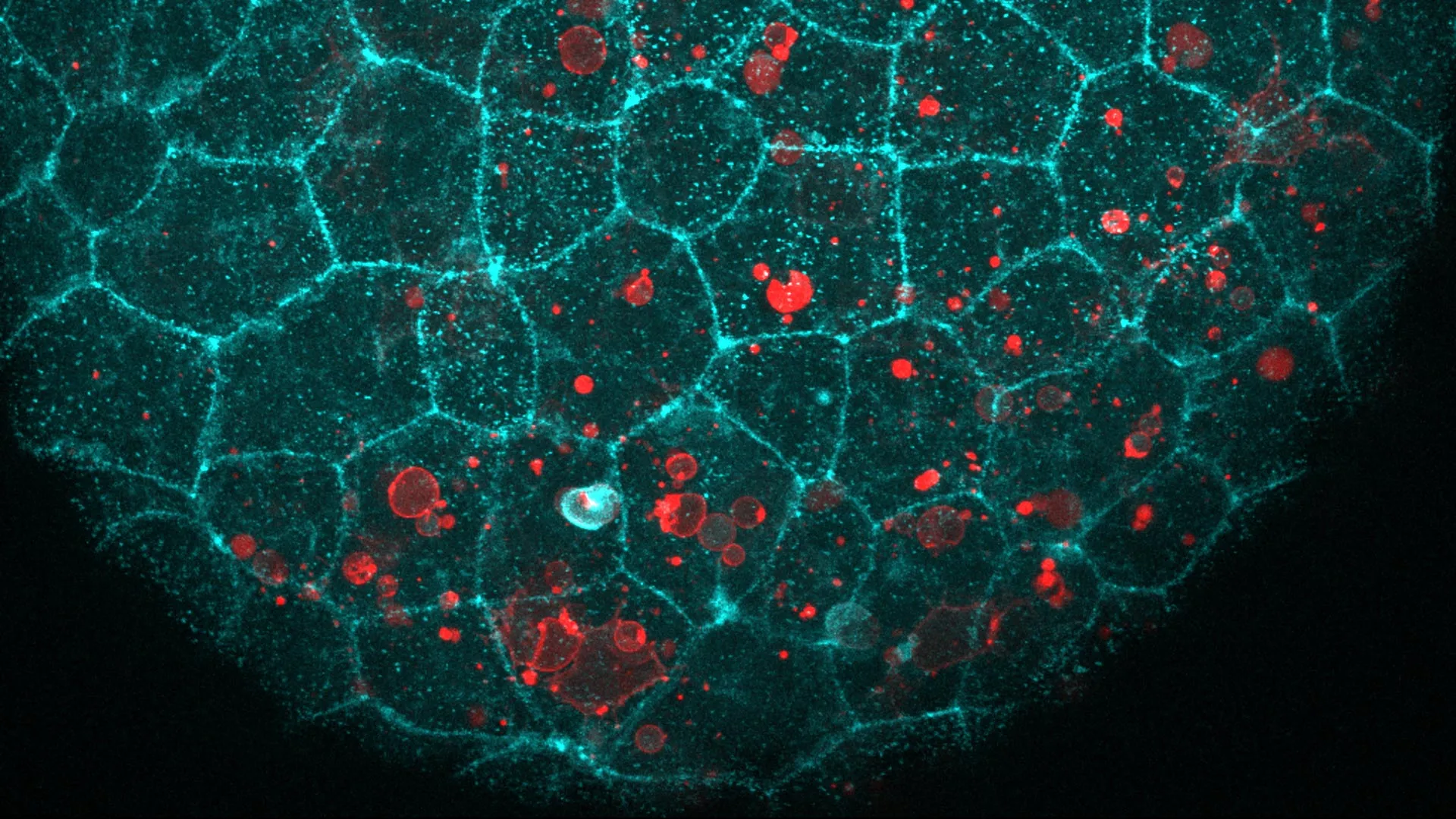
    A team led by Verena Ruprecht examined epithelial tissues in living zebrafish and mouse embryos. They found that the same molecular machinery also gathers at the exact location where a dying cell comes into contact with the tissue.

    The researchers wanted to know whether E-cadherin and its partners were attaching to the dying cell in the same way they normally attach neighboring epithelial cells. To test that possibility, they carried out two experiments.

    First, they presented the tissue with dying cells that had been stripped of E-cadherin. The epithelial tissue removed those cells just as effectively as it removed normal dying cells. Next, the team introduced fat droplets that contained no protein at all but carried a signal normally displayed on the surface of dying cells. The epithelial cells engulfed those droplets as well.

    “We were intrigued to find out that epithelial cells repurpose their molecular adhesion machinery — the ‘glue’ that normally holds them together — to engulf dying cells,” says ICREA Research Professor Verena Ruprecht, senior author of the study.

    How Cells Eat Without Breaking the Barrier

    Engulfing something roughly the size of another cell presents a difficult mechanical problem. Epithelial cells are tightly packed into barriers that often need to remain sealed, even while individual cells reshape themselves to remove debris.

    Live imaging revealed how they accomplish this. The upper and lower surfaces of the same epithelial cell can behave differently from one another. The lower surface stretches and bends around the dead cell, while the opposite side remains relatively unchanged.

    That upper surface, which may face the outside environment or an open space such as the inside of a lumen, continues maintaining the tissue barrier. Measurements taken before, during and after engulfment showed that the area of the upper surface changed very little. By contrast, the lower surface underwent substantial deformation during the ‘eating’ process.

    Ruprecht compares the behavior to a row of dancers standing with their arms linked. Their upper bodies remain steady while their feet perform increasingly complicated movements when a dying cell appears. “It’s the same dancer with a different choreography,” she says.

    A Molecular Rope and Brake

    The researchers also examined the mechanics that allow the cells to perform this cleanup.

    One protein in the E-cadherin complex acted much like a rope. It connected the molecular assembly to the cell’s internal skeleton, allowing force to be transmitted across the surface of the material being engulfed. When cells lacked this tethering protein, or lacked the specific region that attaches it to the skeleton, they could no longer swallow dead cells.

    Another component behaved more like a brake on the cell’s contractile machinery. Surprisingly, removing this brake did not make the cleanup process more effective. Instead, the cell became too stiff and lost its ability to properly remove dying cells.

    The Same Mechanism Appears in Mammals

    The team next investigated whether the process extends beyond zebrafish.

    In early mouse embryos, blocking E-cadherin caused dying cells to remain uncleared. This matched the results seen in zebrafish and suggests that the mechanism is shared among vertebrates.

    The new work builds on earlier research from Ruprecht showing that embryos can use epithelial tissues to cooperatively remove dying cells. That behavior represents a form of early innate immune defense.

    Embryos are particularly useful for studying these events because they are transparent. Researchers can observe living cells and tissues directly at a level of detail that cannot currently be achieved inside the human body.

    Could the Same Process Work in Adult Tissues?

    An important question remains unanswered. Researchers do not yet know whether this same E-cadherin dependent mechanism operates in adult zebrafish or mice, or in any type of human tissue.

    There are reasons to think it could. Epithelial tissues in adults are already known to remove dying cells in places including the retina, colon, airways and mammary gland. E-cadherin is also found throughout epithelial tissues in the body, and its structure has remained remarkably similar across species. Those characteristics make it a strong candidate for a more broadly used cleanup mechanism.

    The potential medical importance comes from what can happen when dead cells are not removed efficiently. Dying cells that remain in tissues can eventually rupture and release their contents, contributing to chronic inflammation.

    The findings suggest that successful cleanup depends on more than receiving the correct chemical signal to engulf apoptotic debris. Cells must also be physically capable of changing shape, applying force and wrapping around dead material without compromising themselves or the surrounding tissue.

    “Studying the mechanisms of how dying cells can be removed efficiently from tissues is of very high relevance to human health,” concludes Ruprecht.

    The work was led by joint first authors Hanna-Maria Häkkinen, Marta Batet Palau and Laura F. Bianchi and supervised by Verena Ruprecht. It was funded by the Spanish Ministry of Science and Innovation, the Human Frontier Science Program, the European Union’s Horizon Europe program, and the “la Caixa” Foundation, with additional support from the European Social Fund. It made use of the CRG Core Facilities for Advanced Light Microscopy, Tissue Engineering and Protein Technologies.

  • Behind (cell) walls

    While mammalian delivery technologies are advancing to translation at an exceptional rate, delivery to plant tissues faces challenges, limiting their practical applications.

    Crops genetically engineered for higher environmental stress and herbicide tolerance, enriched nutrition, disease resistance, and carbon sequestration for carbon management are important to meet global food demand. Gene editing technologies appear to promise faster and easier adaptation than traditional breeding approaches. However, to generate a desired genetic edit that is also transmitted across generations, the gene-editing machinery must be delivered to the appropriate plant tissue. In mammals, delivery systems have become increasingly sophisticated, with ongoing improvements in immunogenicity, targeting precision and efficiency1. Plants, however, have distinct physical barriers, have different transport systems (xylem and phloem) and are under continuous environmental stress. Mammalian delivery systems such as lipid nanoparticles (LNPs) are optimized for tissue-specific barriers2 and cannot traverse the plant cell wall. In addition, gene editing of human tissues is restricted to somatic cells to minimize safety concerns. In plants, the opposite is desired: for heritability, delivery systems must be capable of reaching reproductive tissues, or the editing reagents must be applied directly to these cells.

    Growing plants for transformation takes time. The most common method is Agrobacterium-mediated transformation, whereby a natural pathogen transfers DNA containing an editing construct into a mass of undifferentiated plant cells called a callus. The callus can be screened for cells that have successfully been edited, and these cells can regenerate into a whole plant in appropriate culture conditions. Occasionally, other tissues such as the growing meristem or immature embryos can be transformed instead of a callus. As an alternative to Agrobacterium-mediated transformation, DNA has been attached to tiny gold particles that are physically propelled into plant cells through the cell wall and membrane, but DNA integrations using this method can be random, and many cells are damaged or die during the process3. Other approaches, such as viral delivery or regeneration of edited protoplasts that lack cell walls, rely heavily on tissue culture-based or transgenic intermediates, which are often time-consuming, labor-intensive, dependent on genotype and species, and bottlenecked by difficulties in plant regeneration or viral host range4.

    The vast species variety of plants is another limitation. As in mammalian tissue delivery, what works well in one plant may not work in another. For plants, germline or meristematic tissues must be targeted, ideally in a species-agnostic way. Also, as in mammalian cells, edits need to be done efficiently, fast, and at scale.

    Recent cell culture- and transgene-free “in planta” approaches avoid the time-consuming step of tissue culture for regeneration and have produced widespread interest in accelerating heritable editing. In these methods, developmental regulators are co-delivered with gene-editing reagents to induce new shoot meristems and enable heritable genome editing in whole plants5. Another recently published method grafts transgenic rootstocks to wild-type shoots to generate transgene-free offspring in one step6. While applications to tobacco, tomato, potato and grape (and in 2025 to soybean) have been successful, this crop-engineering workflow is not yet routine5.

    Another recent in planta approach, virus-induced genome editing (VIGE), uses viral vectors to transiently deliver gene editing CRISPR components into plant cells directly, with high editing efficiencies and without the need for tissue culture intermediates7,8,9. Common viruses used for VIGE will not work for all plants, and currently the technology is not scalable. Whereas expanding the host range of mammalian viruses often requires altering cell entry viral receptor binding (tropism), broadening the host range of plant viruses may instead depend on overcoming constraints related to replication, systemic spread and host–virus molecular compatibility. VIGE approaches have worked well in dicot species, but their application in major monocot crops such as rice and wheat remains more challenging as a result of a more limited viral vector toolkit8. In addition, the virus must access the plant germline and deliver its payload to the meristem. It will be difficult to find a broadly applicable virus that can act on a wide variety of crops and cultured plants. Adding RNA mobility sequences can help to move the cargo, but not the virus10. Impairing plant immunity gives viruses better access to plant cells, and thus higher delivery efficiencies11, but plant-specific mechanistic and physical barriers are not easy to overcome.

    Working with viruses as delivery technologies in plants also creates regulatory challenges. The use of infectious agents in the food supply requires rigorous demonstration that engineered viruses do not acquire unintended properties, harm non-target species or create new ecological risks. Addressing these regulatory concerns will be essential for broader adoption of such approaches. As an alternative, existing viral vectors with known safety risks could be engineered, with recognized engineering principles potentially adapted to broaden host tropism in plants.

    Lipid nanoparticles (LNPs) have transformed the field of therapeutic genome editing in humans, but their application in plants remains far less developed. LNPs and virus-like particles are already known to carry larger editing machineries in mammalian cells, like those required for prime and base editing, and could be functionalized in plants. LNPs would need to navigate several barriers, including transport to the target tissue, passage through the cell wall, cellular uptake and intracellular cargo release. Nanosheets functionalized with lysozymes have been used to loosen the cell wall in target plants and deliver RNA cargo and plasmid DNA, demonstrating delivery to reproductive tissues (pollen) in tomato. Yet this was limited to GFP nucleic acid delivery; no editing machinery was delivered to enable heritable genome editing12.

    Plant biotechnology companies seem to be striking a balance between investing in advanced transgene- and cell culture-free methods and more traditional routes of transformation. Terrana Biosciences is introducing engineered self-replicating, native plant RNAs into the plant that could move throughout the plant and deliver information or desired traits, avoiding the regeneration bottleneck of traditional methods. Synthetic latent RNAs can travel through the seed of corn, bell peppers and almond. Qi Biodesign is also looking at RNA-based grafting methods and alternative approaches including direct and transient protein delivery to the meristem in plants such as tomato. Vireo modifies plants directly in the greenhouse using shoot generation, also eliminating the need for tissue culture while still editing cells that can produce new shoots. Yet these technologies each need to be optimized with individual adaptions for every species and are not general solutions.

    Considering the magnitude of the challenge and the benefits that time-saving advances could bring, the level of activity in plant delivery systems compared to that in mammalian ones appears modest. Of course, plant transformation approaches that perform efficiently in laboratory genotypes often show substantially reduced efficiency, or may fail altogether, when applied to elite commercial cultivars because of strong genotype dependence in transformation and regeneration processes. This could limit their wider adoption. But this is true for mammalian gene editing technology as well. Today, regulatory requirements for transgene-free approaches in plants vary between countries and are continually being modified to keep up with the pace of advances.

    Gene editing in plants seems no longer primarily limited by the editing machinery itself, but by the ability to deliver it. If the field continues to improve the editor while leaving delivery largely unchanged, the practical reach of gene editing will remain limited by the biology of transformation. But if delivery becomes an equally deliberate target of innovation, the set of plants that can be engineered — and the traits that can be addressed — could expand dramatically.

  • This new qubit could be 100 times less error-prone in superfluid quantum computer breakthrough

    This new qubit could be 100 times less error-prone in superfluid quantum computer breakthrough

    Researchers at the University of Surrey have proposed a new kind of qubit that could help address one of the biggest obstacles in quantum computing: keeping error rates low as systems become larger.

    The concept uses superfluid helium, a form of helium with unusual quantum properties, to create a qubit that may be less sensitive to some of the disturbances that affect today’s leading quantum computers.

    Why Quantum Computers Struggle With Errors

    Quantum computers rely on qubits to store and process information in ways that conventional computers cannot. Many current systems use superconducting circuits, but these are highly sensitive to electromagnetic noise and stray electrical charges – similar to the static electricity that can make hair cling to a balloon.

    Even very small disturbances can disrupt the fragile quantum information stored in a qubit. As more qubits are added, controlling those errors becomes increasingly difficult, making scalability one of the central challenges in quantum computing.

    In a study published in npj Quantum Information, researchers from Surrey’s Quantum Sciences Group describe a different approach built around superfluid helium-3 – an unusual form of liquid helium that can flow without friction.

    Their proposed device, called the Superfluid Helium Oscillator Quantum (SHOQ) device, would use charge-neutral superfluid helium. Because the helium carries no electric charge, the design could be naturally protected from certain types of electromagnetic noise.

    A Qubit With Much Lower Predicted Error Rates

    According to the researchers, this is the first reported design for a qubit based on a superfluid. Their calculations suggest the SHOQ device could have error rates about 100 times lower than conventional superconducting qubits.

    Dr. Priya Sharma, Daphne Jackson Fellow in Hybrid Quantum Systems at the University of Surrey’s School of Mathematics and Physics, and lead author of the study, said:

    “We are not the first to think about the individual components behind this idea, but what we have done for the first time is bring them together in a microfluidic device and work out the specific details that could enable the device to function as a qubit.

    “The maths tells us that it should work. We have taken what we already know about superfluid helium and quantum technologies and turned that into an educated design, with the parameters and specifications needed to build one. The next step is to make a prototype and put those predictions to the test.”

    Rather than replacing existing quantum computing hardware, the researchers say the SHOQ device could potentially be integrated with superconducting quantum technology.

    That could allow different types of qubits to perform different jobs within the same system, depending on their strengths.

    A Possible Role as Quantum Memory

    One longer-term possibility is to use the SHOQ device as a form of quantum memory. In that setup, a superfluid based qubit could store quantum information while other hardware carries out calculations.

    Dr. Eran Ginossar, Associate Professor at the University of Surrey’s Department of Physics and Advanced Technology Institute, and co-author of the study, said:

    “We don’t necessarily need one type of qubit to do everything. Combining different quantum technologies could allow us to take advantage of the strengths of each.

    “Superfluid helium gives us a fundamentally different type of quantum hardware to explore. If the predicted performance can be demonstrated experimentally, it could eventually work alongside existing superconducting technologies as part of a larger quantum system.”

    The Next Step Is Building a Prototype

    The team now plans to build a prototype to find out whether the predicted performance can be reproduced in a real device. That effort is being supported by an IAA Commercialisation Fellowship awarded to Dr. Priya Sharma.

    The SHOQ device would need to operate at extremely low temperatures, but researchers have already reached the necessary conditions in previous experiments involving superfluid helium-3.

    The project was led by the University of Surrey in collaboration with Professor Jens Koch at Northwestern University in the United States. Koch was one of the researchers involved in developing the transmon – a superconducting qubit design that is now widely used in quantum computing.