Author: admin

  • How COVID, Characterization, and Commerce Are Driving an RNA Revolution

    How COVID, Characterization, and Commerce Are Driving an RNA Revolution

    The technology underpinning RNA vaccines1 and therapeutics2 has been around for decades. However, until recently, only a handful of products were on the market.

    Everything changed in 2020 when mRNA vaccines proved3 to be effective against SARS-CoV-2, the virus that causes COVID-19. In the years since, many more RNA products entered the clinic as developers recognized both their therapeutic and commercial potential.

    The pandemic also kick-started efforts to optimize RNA manufacturing that will likely shape the sector for decades to come, according to David Sokolowski, global workflow manager for nucleic acid therapeutics at Cytiva.

    “The COVID vaccines heralded the rapid industrialization of RNA and LNP [lipid nanoparticle] technologies, and due to the nature of a pandemic, large amounts of vaccine safety data were generated. However, it remains a nascent industry with limited applications commercially available, and this space is where developers are rushing to build.

    “Manufacturing good quality RNA and LNP became easier with the proliferation of high-quality reports from successful studies, trials, and campaigns occurring all over the world.

    “Now, we’re supporting the development of therapeutics with even grander aspirations and increasingly exotic biology—whether they are encoding and producing mAb via RNA, or enabling next-generation in vivo cell therapy, LNP-mediated therapeutics have no shortage of potential advances ahead,” he continued.

    Manufacturability

    The key to developing any RNA-based product—be it a vaccine or a therapeutic—is to ensure it can be produced in bulk in a cost-effective and efficient manner, says Luis Avila, PhD, a scientist at services firm Novoprotein Scientific.

    “RNA design fundamentally determines both manufacturability and biological performance, but optimizing for one often comes at the expense of the other.” The best approach is to design for manufacturability from the start,” he says, adding “the right template architecture can contribute with optimized promoter design, transcription initiation sequences, encoded polyA tails, transcript boundaries, and sequence composition to improve IVT [in vitro transcription] performance while minimizing problematic secondary structures.”

    “Combined with other factors, such as engineered RNA polymerases, optimized reaction chemistries and advanced capping technologies, these design strategies will improve RNA yield, integrity, and translational performance all while reducing the generation of dsRNA,” he tells GEN.

    The type of RNA being made should be the key consideration in any assessment of manufacturability, according to Avila, who cites the differences between messenger RNA and self-amplifying RNA to illustrate the point.

    “For mRNA, coding-sequence composition, transcript length, GC content, and secondary structure strongly influence IVT efficiency. Whereas optimized UTRs [untranslated regions], cap structures, and poly(A) tails primarily govern RNA stability and translational efficiency.

    “For saRNA, the incorporation of the large alphavirus replicase cassette substantially increases transcript length and structural complexity, thus making IVT more challenging while enabling intracellular RNA amplification that dramatically lowers the therapeutic dose required,” he says.

    Producing circular RNA—a covalently closed-loop structure that is stable and resistant to exonuclease degradation—can be even harder, according to Avila.

    “circRNA presents an even greater manufacturing challenge because linear IVT is followed by an additional circularization step whose efficiency remains a major bottleneck. Following circularization, translation relies on efficient cap-independent initiation through optimized IRES [internal ribosome entry site] elements and RNA architecture.”

    Characterization

    So, designing an RNA therapeutic or vaccine with manufacturing in mind is critical. However, the ability to realize that optimized blueprint on the factory floor is entirely dependent on being able to characterize the product in real-time, as it is being made.

    And, unfortunately for the industry, RNA characterization is a complex challenge, says Matthew Ranaghan, PhD, applications scientist at analysis technology firm Refeyn.
    TwoMP with model
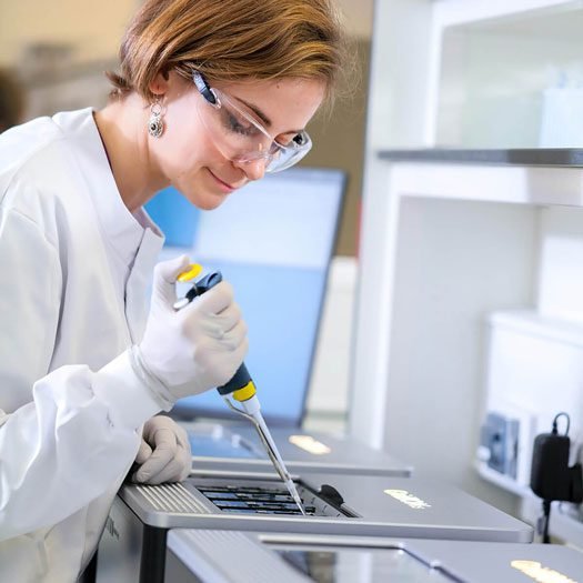
    Analyzing multiple RNA molecules in a single pass reduces the experimentation burden on RNA developers. Being able to visualize characteristics within minutes—as shown here—would benefit any developer, says Refeyn. [Refeyn]

    “Thorough Characterization of RNA is critical to ensuring the safety, stability, and efficacy of next-generation RNA therapeutics, yet it remains a significant analytical bottleneck.”

    “Current methods are limited to various types of chromatography, which struggle with poor resolution for large RNAs, or mass spectrometry, which requires digesting samples into smaller oligos with RNases. While both have advantages, they demand extensive and time-consuming method development.”

    Traditional RNA analysis usually consists of separate assays looking at specific characteristics. For example, size exclusion chromatography is used to assess aggregation while enzyme-linked immunosorbent assays are used to check for trace amounts of double-stranded RNA.

    One promising alternative is mass photometry (MP), a technique that can analyze RNA molecules within minutes based on only nanograms of material within minutes in a single experiment. There are multiple potential advantages according to Ranaghan.

    “A single readout replaces the need for multiple instruments, drastically reducing hands-on time, reagent costs, and the need for extensive method development across different platforms. A single readout also enables at-line process monitoring, allowing for immediate go/no-go decisions during bioprocessing and purification.”

    “We envision the faster speed and higher resolution of MP to enable its use as a multi-attribute tool for at-line assessment of the various critical quality attributes (CQAs) of RNA therapeutics,” he says.

    “We have already demonstrated how MP can rapidly assess integrity, fragments, multimer, and purity in a single measurement. We are also actively building out more applications,” Ranaghan adds.

    Commercial concerns

    Manufacturers of RNA products also need to develop commercialization plans as early as possible—in parallel with process development—to ensure they reach the patients who need them and garner the revenues needed to fund future research.

    Rajiv Gangurde
    Rajiv Gangurde, PhD
    Vice President
    Parexel International

    And, for some firms, developing such plans can be a steep learning curve, says Rajiv Gangurde, PhD, vice president of chemistry, manufacturing, and controls (CMC) for cell and gene therapies at contract research organization Parexel International.

    “Like many newer modalities, RNA-based therapies typically emerge out of academia or start-up biotech, and transitioning from academia to industry can be challenging due to differences in objectives, differences in expertise, fundraising, etc.”

    “In addition, newer modalities require heavier than usual investment in CMC activities to establish proof-of-concept as well as manufacturability. There is heavy reliance on external partners, creating capability and capacity constraints.

    “Most importantly,” Gangurde continued, “a phase-appropriate CMC strategy aimed at global approvals can significantly derisk development and help minimize costs and timelines.”

    Yet preparing a CMC strategy that will win approval in multiple markets is no small undertaking, particularly, as Gangurde points out, because regulatory guidelines for RNA-based therapies are still evolving.

    Another challenge is the lack of consistency. For instance, the U.S. Food and Drug Administration (FDA) and EU EMA use different definitions and guidelines for CMC.

    Gangurde told us, “Harmonization of guidelines and expectations across the two agencies will be hugely beneficial to developers of RNA therapies. For example, while both agencies recognize potency/efficacy assessment as a critical quality attribute (CQA), there are some differences in method validation requirements, timing, and regulatory expectations across clinical phases.”

    “While the FDA accepts real-world experience and surrogate endpoints, particularly for rare or life-threatening diseases, EMA requires more comprehensive clinical data and demonstration of long-term efficacy for approval. The EMA also mandates an appropriate degree of method validation at each stage of development, whereas the FDA does not expect validation of the potency assays during Phase I,” he added.

    The product component designations used by regulatory agencies can also differ. For example, the lipid nanoparticle for the COVID-19 vaccine, Spikevax, consists of four lipids: SM-102, PEG2000-DMG, DSPC, and cholesterol.

    The FDA designates each of these components as a “drug substance,” whereas the EMA designates them as “novel excipients”—each designation has differing data submission requirements.

    Continuous process

    In the future, RNA manufacturing methods are likely to keep improving, according to Novoprotein’s Avila, who says continuous production processes are likely to have a major impact.

    “One of the most exciting developments is the emergence of continuous or flow-based IVT manufacturing. This has the potential to improve productivity, reduce costs, and enable more consistent large-scale RNA production through continuous enzyme utilization and process integration.”

    “As equally transformative are advances in engineered low-dsRNA T7 RNA polymerases, improved co-transcriptional capping chemistries, and integrated continuous purification. All of which will enable improvements in RNA quality, simplify downstream processing, and accelerate GMP manufacturing,” he added.
    TwoMP- Auto with model
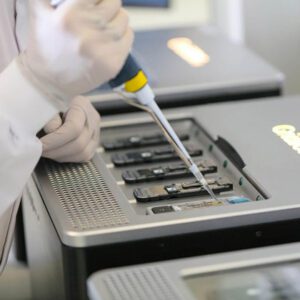
    RNA characterization is a challenge using current technologies. Refeyn claims its mass photometry system—seen here at lab in Waltham, Massachusetts—reduces the number of assays required. [Refeyn]

    This view is shared by Ranaghan from Refeyn, who cites the shift toward continuous flow manufacturing as one of the most exciting advances in the field.

    “Moving away from traditional batch processing to a continuous flow model is highly logical because it minimizes the time that the fragile mRNA product spends exposed to active reaction components that drive fragmentation and aggregation. This shift not only improves overall yield and intactness, especially for massive transcripts like saRNA, but it also creates a mandate for faster, real-time analytics.”

    For Parexel’s Gangurde, efforts to make production more efficient and sustainable will drive innovation, with the WHO’s recently launched mRNA Technology Transfer Programme Phase 2.04 being a case in point.

    “This is a major global initiative aimed at scaling up commercially viable mRNA-based vaccine and therapeutic production in low- and middle-income countries—where the need for these therapies is most critical.”

    Launched in 2021 in collaboration with the Medicines Patent Pool (MPP), the initiative has enabled 15 partners across Latin America, Africa, Eastern Europe, and Asia to receive foundational mRNA technology.

    Phase 2.0 focuses on building local manufacturing capacity so countries can make mRNA vaccines and therapeutics for future outbreaks and diseases like influenza, TB, HIV, malaria, dengue, and leishmaniasis, as well as oncology drugs and mAbs.

    Gangurde says, “By decentralizing production, the program reduces reliance on a few high-income countries for mRNA supply, supporting global access for mRNA-based therapies.”

    References

    1. Beyrer C. The Long History of mRNA Vaccines. Johns Hopkins Bloomberg School of Public Health; 2021. Accessed August 12, 2026.
    2. Chatterjee S, Bhattacharya M, Lee SS, Chakraborty C. An insight of different classes of RNA-based therapeutic, nanodelivery and clinical status: Current landscape. Current Research in Biotechnology 2023;6:100150-100150. doi:10.1016/j.crbiot.2023.100150
    3. Parvin N, Mandal TK, Joo SW. The Impact of COVID-19 on RNA Therapeutics: A Surge in Lipid Nanoparticles and Alternative Delivery Systems, Systems. Pharmaceutics 2024; 16(11): 1366. Doi: 10.3390/pharmaceutics16111366
    4. World Health Organization: WHO. mRNA Technology Transfer Programme’s Phase 2.0 discussed with partners on the sidelines of G20 Summit. Who. June 13, 2025. Accessed August 12, 2026.
  • Spatial Biology Moves Toward Translation

    Spatial Biology Moves Toward Translation

    If “Spatial Biology” conjures images of a science conducted in zero gravity aboard the International Space Station, think again. As one of the most rapidly advancing areas in biomedical research and precision medicine, spatial biology probes the inner landscape of intact cells and tissues down to single-molecule resolution. By preserving location and organization, instead of dissociating tissue into component parts, this discipline maps fine architecture, cellular neighborhoods, and individual molecules.

    Maintaining that context is driving new ways of characterizing disease progression, identifying biomarkers, advancing therapeutics, and monitoring treatment responses. Fueling these advances is an expanding toolbox that encompasses high-plex imaging, spatial transcriptomics and proteomics, multiomic profiling, and increasingly sophisticated computational analyses. We consulted industry leaders about how the field is progressing, barriers that remain, and their vision for what is needed to propel spatial biology into its next phase.

    Scaling spatial discovery

    While genomics and single-cell sequencing profile molecular features and cellular states, spatial biology answers questions that neither can address, such as where those features and cells are located and how they are organized.

    Kamila Belhocine
    Kamila Belhocine, PhD
    Senior Director
    10x Genomics

    “When you dissociate a tissue into individual cells, you lose something fundamental: where those cells were and how they were organized relative to one another,” observes Kamila Belhocine, PhD, senior director of spatial biology at 10x Genomics. She continues: “Spatial biology puts that context back. In cancer, for example, knowing that a particular immune cell population is present within a tumor sample is useful. But knowing whether those cells have infiltrated the tumor core, are sitting at its margin, or are excluded from it altogether can tell you something very different about the biology, and potentially, about response to treatment. The same principle applies to understanding tissue architecture in cases like development and cell differentiation, and disease progression.”

    Several barriers have prevented spatial biology from going mainstream in the biopharma/biotech industry. Belhocine says, “I think about four things: speed, reliability, cost, and interpretability. Spatial needs to be fast enough to fit into routine research workflows, robust enough to generate consistent results at scale, affordable enough to use across large studies and, ultimately, straightforward enough to turn the resulting data into biological insight. The first three are fundamentally technology and engineering challenges. Interpretability is harder because it also requires accumulating evidence and validating which spatial signatures are truly meaningful and actionable.”

    However, according to Belhocine, single-cell technologies had to address similar issues. “A decade ago, single-cell analysis was largely a specialized technique. Continued innovation made it easier, more scalable, more robust, and dramatically more cost-effective, opening it to a much broader community and enabling much larger studies. We think spatial is following a similar trajectory.”

    Gary Latham
    Gary Latham, PhD
    Chief Technology Officer
    Bio-Techne

    Belhocine says the company’s newest spatial instrument, Atera, which will be shipping in the second half of the year, is an important step in that progression. “This technology combines whole-transcriptome spatial profiling with single-cell sensitivity and subcellular resolution at a massive scale designed for much larger studies.”

    Looking ahead, Belhocine predicts, “As spatial technologies become more scalable, robust and cost-effective, researchers can apply them across much larger patient cohorts to identify biomarkers and signatures of treatment response. Additionally, AI will undoubtedly accelerate that trajectory even further.”

    Spatial multiomics

    Gary Latham, PhD, CTO, Bio-Techne, provides another perspective on the role of integrating protein analysis and gene expression in spatial biology. “A fuller understanding of tissue biology requires seeing where proteins and genes are expressed, how they relate, and how they interact within the tissue architecture.”

    Human breast cancer
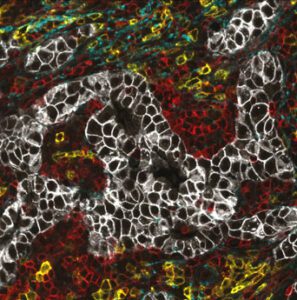
    Automated spatial biology systems enable high-throughput, hyperplexed analysis of tissue samples. Human breast cancer sample stained with 105-plex multiomics panel on COMET. The image shows a HER2+ tumor with HER+ cells and immune infiltrates. Markers shown: HER2 (white), collagen I (teal), CD45 (red), and CD38 (yellow). [Bio-Techne]

    This molecular multiplexing can provide a more complete view of tissue biology. “Spatial biology is unique from other technologies because it takes us from seeing what is there to understanding what is where,” says Latham. He continues, “This is critical because biology can’t be understood as a parts list. Instead, it is a dynamic system of interactions and networks where location defines meaning. While single-cell sequencing can tell you which cell types exist and their molecular states, spatial biology tells you how those cells are organized, how they may be interacting, and how these relationships may influence disease biology and clinical outcomes.”

    Latham indicates that the field is working to overcome several challenges. “There is a need for reproducible, standardized automation for today’s biomarkers and tomorrow’s multiomic precision medicine. Many spatial biology workflows remain exploratory and manual during early discovery phases. As programs move toward translational research, the focus shifts to robustness, reproducibility, and standardized, validated workflows. Ultimately, clinical adoption requires validated panels, automation, and throughput suited to routine testing.”

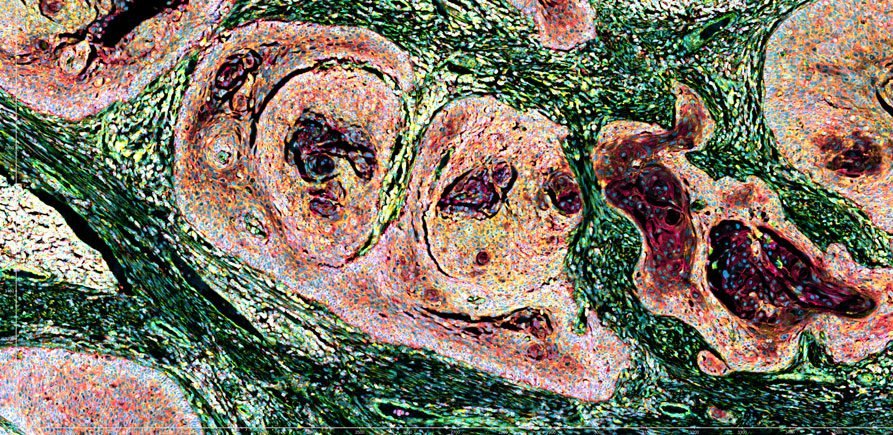
    Human prostate cancer
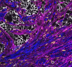
    Automated spatial biology systems enable high-throughput, hyperplexed analysis of tissue samples. Human prostate cancer sample stained with a 105-plex multiomics panel on COMET. The image shows tissue fibrosis associated with cancer-associated fibroblasts (CAFs). Markers shown: COL1A1 RNA (yellow), E-Cadherin (white), alpha-SMA (blue), and FAP (magenta). [Bio-Techne]

    According to Latham, Bio-Techne is helping address these barriers in several ways. Its COMET Suite provides an end-to-end automated platform for hyperplex immunofluorescence and multiomics staining, imaging, and analysis of tissue samples. Additionally, its RNAscope portfolio enables same-section multiomics, with 80,000 RNA probes spanning 450 species and compatibility with multiple automated staining instruments.

    Latham believes the spatial multiomics approach can connect gene and protein expression with tissue architecture within the same cells and microenvironment, revealing biological relationships and linking state to function that no single modality can uncover.

    He says several areas are seeing the fastest adoption of spatial biology. “Oncology and immuno-oncology continue to dominate spatial biology applications, while immunology and neuroscience represent the next-most prominent areas of focus. Biodistribution of gene therapies is an especially high-impact application: spatial multiomics can localize an AAV vector, transgene mRNA, or oligonucleotide therapeutics such as ASOs and siRNAs to specific cells, then measure changes in the target or downstream gene expression and the associated protein phenotype.”

    Integrated discovery & translation

    Spatial biology is increasingly moving beyond simply identifying where cells are located to understanding how cellular neighborhoods, cell–cell interactions, and biological pathways shape disease biology. As Thomas Campbell, PhD, senior director of product management for Bruker Spatial Biology, explains, “Biology is inherently multi-dimensional, and no single spatial technology can or will capture every aspect of that complexity. The field needs multiple fit-for-purpose approaches that allow researchers to ask different questions and connect insights across DNA, RNA, proteins, and cellular function.” This ability to examine biology from complementary perspectives is becoming increasingly important as spatial research moves from discovery toward translation.

    According to Campbell, understanding these context-dependent differences is driving the field. “The next phase of spatial biology will be about using high-plex discovery to develop focused, quantitative assays that can be deployed across large cohorts. One of the biggest challenges in translational biopharma is the discovery-to-translation gap. A study may identify dozens or hundreds of potentially relevant biomarkers, but a translational or clinically deployable assay demands a much smaller, highly reproducible panel.”

    Thus, defining a clinically relevant spatial signature requires insights across the genome, transcriptome, and proteome. Campbell says Bruker Spatial Biology’s portfolio comprises platforms spanning multiple omic layers—including DNA, RNA, proteins, post-translational modifications, miRNA and live cell function—“because we recognize that no single modality can fully resolve the biological complexity that drives disease.”

    Thomas Campbell
    Thomas Campbell, PhD
    Senior Director
    Bruker Spatial

    That integrated approach is reflected in several of the company’s platforms. CosMx® Spatial Molecular Imager (SMI) provides a view of the whole transcriptome at subcellular resolution, while enabling the simultaneous measurement of RNA and protein within the same tissue section. The CellScape™ XR platform provides quantitative spatial proteomics using flexible assay chemistry. Meanwhile, the GeoMx® Digital Spatial Profiler (DSP) profiles the whole transcriptome and more than 1,200 proteins from distinct tissue compartments. Campbell says that, together, these three platforms “provide complementary views of biology and, when used together, enable a more complete understanding of cellular function across multiple omic layers.”

    Campbell sees this transition from discovery to translation as one of the most important opportunities for spatial biology. “Whether it’s in oncology, immunology, or neuroscience applications, the future of spatial biology is not simply just about generating more data; it’s translating increasingly sophisticated spatial discoveries into assays that can answer focused biological and clinical questions across large numbers of samples,” he says. “That requires a portfolio of complementary technologies. High-plex discovery platforms can reveal the complexity and heterogeneity of disease, while more focused and quantitative approaches can translate those findings into reproducible biomarkers and clinically relevant assays. Ultimately, the value of spatial biology will be measured by how effectively we can connect discovery to clinically actionable insights.”

    From plex to precision

    “Historically, spatial biology involved a fundamental tradeoff between plex, sensitivity, and resolution—researchers had to sacrifice one to gain another,” suggests Mike Miller, PhD, CTO and CPO at Quanterix. He says that is now shifting. “With today’s tools, those tradeoffs have narrowed considerably. The current challenge becomes building a platform that delivers reproducibility, rigor, and clinical-grade integrity at scale.”

    The company is addressing those needs in several ways. Miller reports: “Our Akoya PhenoCycler-Fusion platform enables researchers to visualize and quantify 100+ proteins in a single tissue section at single-cell resolution.” The platform uses molecular barcoding chemistry. Miller elaborates: “Molecular barcoding is a way of identifying and measuring multiple proteins in a tissue while avoiding many of the limitations of traditional immunohistochemistry and conventional multiplexing approaches.”

    Mike Miller, PhD
    Mike Miller, PhD
    Chief Technology Officer
    Quanterix

    This approach combines three main components: an antibody that recognizes the target of interest; a unique molecular barcode, consisting of a short oligonucleotide sequence attached to the antibody; and a complementary oligonucleotide carrying a fluorescent reporter that binds to the barcode, enabling target visualization and quantification. “The key advantage is that the fluorescent reporter can be reversibly hybridized and removed without removing the antibody or repeatedly staining and stripping the tissue. This enables a cyclical imaging workflow.”

    The company also recently introduced its PhenoCode2.0 chemistry as part of its new molecular barcoding kit, “which lets researchers reliably barcode virtually any antibody of their choice.” Miller continues: “This means researchers can easily expand and customize their biomarker panels to address new hypotheses, incorporate emerging biomarkers, and scale between unbiased discovery and targeted validation, accelerating the path to new insights.”

    Beyond expanding what can be measured, Miller believes the next phase of spatial biology will focus on translating high-plex discoveries into larger, clinically relevant studies. “That’s ultimately what will separate platforms going forward, bridging the gap between generating a striking image in discovery and generating a measurement that holds up in a clinical trial or diagnostic setting.”

  • Trends in NGS

    Trends in NGS

    Next-generation sequencing (NGS) has matured into an essential part of modern drug development and diagnostic laboratories, and its steadily dropping costs are making it affordable for many facilities that a few years ago couldn’t justify the expense.

    The coming years will build on that foundation by extracting more and different types of data from samples, leveraging AI both upstream and downstream, and streamlining workflow. The entire workflow—not just the sequencing itself—will become more cost effective, industry experts tell GEN.

    The next wave

    “The next major breakthrough in sequencing technology will likely come not from the sequencer alone, but from the ability to extract more meaningful biological insights from every sample,” Karen B. Chapman, PhD, associate VP, R&D Genomics, Agilent Technologies, says.

    Karen B. Chapman
    Karen B. Chapman, PhD
    Associate VP
    Agilent

    She envisions “the convergence of improved long-read sequencing, intelligent assay design, AI-enabled analysis, and integrated automation across sample preparation, target enrichment, quality control, sample tracking, and data interpretation.”

    Multiomics assays will likely be the next big thing.

    Mark Van Oene
    Mark Van Oene
    President and CEO
    PacBio

    “Sequencing has traditionally focused on the four canonical bases, but DNA molecules contain much more information, including methylation, damage, and secondary structure, that can reveal different layers of biology,” Mark Van Oene, president and CEO, PacBio, points out. “We see sequencing moving toward a rapid test that can deliver comprehensive, highly accurate results within hours, enabling scientists and clinicians to make decisions faster.”

    The experts at New England Biolabs (NEB) concur. “That’s one of the next big opportunities in sequencing: to gather different data types and provide them in a way that allows them to be interpreted usefully in the context of the human genome,” Andrew Barry, associate director, product portfolio management, NEB, says.

    Such multiomics assays will “enable richer and more comprehensive biological insights,” Lakmal Jayasinghe, PhD, CSO, Oxford Nanopore Technologies, says. “The next major breakthrough will be the ability to analyze DNA, RNA, and proteins on a common sensing platform along with epigenetics, epitranscriptomics, and post-translational modifications of proteins. The next frontier is understanding how that code is regulated and executed in real biological systems. We see proteomics as a key part of that future.”

    Lakmal Jayasinghe
    Lakmal Jayasinghe
    CSO
    Oxford Nanopore Technologies

    As the industry pushes current boundaries, it is also working to improve sequencing performance through parallel workflows and automation that can accommodate a broad range of samples. Opportunities exist “to increase throughput, improve fidelity, and simplify workflows,” Jayasinghe adds.

    To that end, Keerthana “Kit” Krishnan, director, NGS applications and product development, NEB, points out: “Library prep may need a bit more work.” On the upstream side, she identifies sample extraction and sample prep as areas with opportunities for improvement. Downstream opportunities, she says, are in data analysis.

    For NGS to become a routine clinical tool, however, those analyses must be clinically meaningful and linked to follow-up actions, she says. She envisions NGS assay kits that are tailored to specific clinical applications.

    AI adds value today

    “The real breakthrough,” Chapman of Agilent Technologies points out, “will be…in delivering deeper, more actionable biological insights in a way that is economically viable, operationally practical, and accessible to a broad range of laboratories and users. AI will play a critical role in enabling this vision, unlocking new possibilities that lower computational and labor costs while enhancing accuracy and increasing operational speed.”

    sequencing
    As sequencing costs decline, steadily improving technology makes sequencing affordable for an increasing number of users. Photo provided by Nanopore. [Oxford Nanopore Technologies]

    Across the board, instrument developers are leveraging AI both upstream and down. For example:

    • PacBio uses AI upstream to transform raw sequencing data into HiFi reads and to detect DNA modifications. Downstream applications include tertiary analysis, which Van Oene says can be used “to identify and prioritize genetic variants associated with rare disease.”
    • NEB’s bioinformatics team deploys AI for coding support, while the automation team is exploring its application to streamline multi-instrument automation processes for developers and for field application scientists to anticipate pitfalls customers may face. AI is also becoming an important tool in enzyme discovery.
    • Agilent uses AI to learn from large datasets and develop ways to improve probe placement and target-enrichment design to enhance specificity, coverage and uniformity. Other applications may include “improving base calling, variant detection, structural variant characterization, methylation assessment and clinical interpretations,” Chapman says.
    • Oxford Nanopore uses AI to help build more accurate models and to directly detect DNA and RNA modifications. Looking ahead, “AI will become increasingly important in helping scientists, clinicians, and biopharma companies interpret complex multiomics datasets,” Jayasinghe says.
    • Illumina has embedded AI tools in its secondary analysis pipeline and throughout its software and informatics portfolio. It has also created the peer-reviewed Primate, Splice and Promoter AI genomic models.

    Future AI value

    Algorithms are moving “beyond simple rule-based design and increasingly learn from large datasets to optimize enrichment performance,” Chapman says. “Over time, these approaches could evolve into fully-AI-driven panel design systems that automatically optimize probe selection, predict assay performance, minimize sequencing costs, and continuously improve…as more data becomes available.”

    “Looking ahead, the most transformative role for AI may be guiding researchers toward the right biological signals to measure, rather than merely processing larger volumes of data,” she continues. “AI can help to identify the most relevant genomic regions, uncover intricate molecular patterns, integrate genomic and methylation information, and bridge genomics discoveries with protein, cellular, and metabolic biology.”

    Andrew Barry
    Andrew Barry
    Associate Director
    New England Biolabs

    As AI becomes increasingly ubiquitous, explainable AI will become a requirement, these experts agree. The ability to interrogate, assess, and validate the AI’s reasoning is imperative not only for on-instrument applications such as base-calling, but especially for tertiary analysis where understanding the evidence behind a result is critical.

    As Barry says, “To use AI effectively, we have to know what’s going on underneath the hood.”

    Continuing, overall cost reduction

    The lab cost of genome sequencing broke $100 two years ago, and some labs are advertising 30x whole-genome sequencing and analysis for about $500. And, Van Oene from PacBio says, “There is potential to drive the cost of whole-genome sequencing down by another order of magnitude.”

    For example, Van Oene says that increasing sample throughput offers a major opportunity “to reduce instrument amortization by spreading the cost of the instrument across many more genomes.” Other opportunities are emerging through computing advances that speed processing and analysis as well as through improvements in compute logistics, such as data compression, which reduces storage requirements, and cloud computing, which reduces hardware requirements and expands access to data.

    “The sequencing part of it is becoming a smaller part of the total cost of a workflow,” Fiona Kaper, PhD, VP, head of assay R&D, Illumina, points out.

    So, rather than focusing solely on reducing the cost of sequencing itself, NGS innovators increasingly are targeting efficiencies across the entire workflow, from improving sequencing performance and optimizing library preparation to expanding multiplexing capabilities that generate more insight from every run. “Ultimately,” Chapman, Agilent Technologies, says, “the industry’s focus will shift from the cost of sequencing a genome to the cost of delivering a clinically meaningful answer.”

    “There’s still some meat left on the bone in terms of how you can improve some workflows,” Barry, NEB, observes.

    Scalability is one example. The challenge of sequencing and analyzing a genome varies by size, and solutions need to address that. Therefore, scaling the upstream environment to meet the need is a key consideration for some instrument makers, along with logistical enhancements, such as lyophilization and assay formats, to improve ease of use globally.

    Multiomics and multimodal assays two other examples. “We see a lot more customers starting to layer in additional information like proteomics, transcriptomics, and spatial and single cell analysis,” Kaper says.

    Kit Krishnan
    Kit Krishnan
    Director
    New England Biolabs

    This trend suggests a future, modular, environment in which scientists can select and optimize the assays that best address their questions. “Having a black box [solution]…really doesn’t work for everyone,” Krishnan underscores.

    Further cost reductions, these experts agree, will stem from automating and consolidating workflows, effectively multiplexing assays and developing multimodal assays to generate more actionable information with each run. Such simplification extends to library preparation, which has expanded from DNA and RNA to include proteomics and data from spatial and single-cell analysis.

    Instruments developers also are reducing the quantities of reagents needed. Consequently, Van Oene says, “Reducing reagent volumes in library preparation and sequencing will lower consumable costs as well as shipping and storage requirements—which people don’t usually think about when they think about the cost of a genome.”

    “Time is also a form of cost,” Kaper says. The industry recognizes that by increasing data quality, capabilities, and sequencing speed to help its users do more, better, and faster.

    “The goal,” as Jayasinghe of Oxford Nanopore Technologies says, “is to help customers derive meaningful answers faster, with fewer steps and greater efficiency.”

    Ubiquitous sequencing

    Fiona Kaper
    Fiona Kaper, PhD
    Vice President
    Illumina

    Five to ten years from now, NGS may well be the core technology for research and clinical labs both, Kaper, Illumina, predicts. “Anything that has to do with the genome, like oncology, could really benefit from technologies that interrogate the genome and also the epigenome, proteome, etc.,” she says.

    “Ultimately, the goal is not simply a cheaper genome, but a more informative genome, at a much lower cost,” Van Oene concludes.

  • Science Speeds Up Its Fight Against Prions

    Science Speeds Up Its Fight Against Prions

    Prions have never played by the rules. They aren’t viruses or bacteria. They’re simply proteins folded into the wrong shape, and that structural mistake can cause devastating problems.

    As Alison Armstrong, PhD, global head of scientific & regulatory consultancy, BioReliance Contract Testing Services, Merck KGaA, Darmstadt, Germany, puts it: “Prions are infectious forms of naturally occurring proteins that can alter healthy proteins and lead to neuronal cell death.” As she adds, “They present a potential health risk through animal-to-human transmission, although human-to-human transmission is rare.”

    One prion can trigger a chain reaction, coaxing neighboring proteins to misfold too, and the damage spreads through the brain like a rumor nobody can stop. Prion diseases are called transmissible spongiform encephalopathies (TSEs), and they include bovine spongiform encephalopathy (BSE), better known as mad cow disease, and Creutzfeldt-Jakob disease (CJD). Once symptoms of CJD appear, a person’s decline is often fast, and it is always fatal.

    For scientists, drug developers, and biomanufacturers, that strange biology creates a second, more practical problem: prions are brutally hard to eliminate, detect, and study.

    “For products with a risk of prion contamination, purification processes must be able to eliminate the pathogen,” Armstrong says. “Because methods for directly detecting prions in raw materials are limited by sensitivity, prion-clearance studies have become an industry-standard approach for demonstrating that downstream purification steps can remove or inactivate transmissible spongiform encephalopathy agents.”

    Unique infectious agent

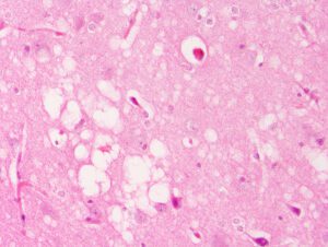
    Prion disease, spongiform encephalopathy
    Credit: UCSF / Getty Images

    Human prion diseases are exceedingly rare. CJD is the most common, and variant CJD (vCJD)—historically linked to eating beef from cattle infected with BSE—is rarer still. Only four vCJD cases have ever been reported in the United States, all in people believed to have been exposed abroad, but rarity doesn’t mean the risk disappears.

    Acquired prion disease can, in unusual circumstances, follow medical procedures involving contaminated equipment or human tissue. That possibility also looms over pharmaceutical manufacturing, particularly wherever human- or animal-derived materials enter the production line.

    “Industry and regulatory authorities address these challenges through risk assessment and controls around animal-derived materials used in biologics manufacturing,” Armstrong says. “A TSE risk assessment typically evaluates factors such as the animal species, tissue source, country of origin, tissue classification, manufacturing controls, and sourcing from countries with negligible or controlled BSE risk. These measures, combined with appropriate manufacturing and quality controls, help mitigate the risk of transmissible prion diseases.”

    For biological products carrying any plausible contamination risk, manufacturers can use prion-clearance studies to test whether individual purification or manufacturing steps actually remove or inactivate prions. Researchers deliberately spike scaled-down versions of a production process with prion material, then measure what’s left afterward.

    Texcell, for instance, describes seeding prion material into crude starting material or mock manufacturing vessels, then tracking removal or inactivation through each subsequent step. Similar studies check whether cleaning procedures between production batches actually destroy prion protein left behind on equipment.

    On paper, it sounds simple: put prions in, run the process, see what comes out. Measuring that convincingly is another matter entirely.

    The measurement problem

    A clearance study is only as trustworthy as the assay behind it.

    ViruSure, whose staff have worked in prion safety since the BSE crisis of the 1990s, relies on the hamster-adapted 263K scrapie strain for many of its studies—a model it describes as accepted by regulatory authorities. The company runs quantitative Western blot assays to check whether manufacturing steps remove or inactivate prion protein, alongside in vivo bioassays for determining prion titer.

    Animal bioassays remain the benchmark, but they are slow, expensive, and nearly impossible to scale. And an assay built on rodent-adapted prions isn’t a stand-in for rapidly experimenting on the real, infectious human thing.

    That distinction matters most in drug discovery. A compound that performs beautifully against an experimental prion model has no guarantee of working against actual human disease.

    A new experimental system might finally close that gap.

    Making human prions grow in cells

    Parmjit Jat, PhD, a professor of molecular biology at University College London, and his colleagues report developing EKV cells: a humanized mouse-cell model capable of supporting robust, indefinite propagation of sporadic CJD (sCJD) prions.

    That solves a problem that has dogged the field for decades. As Jat and his co-authors put it, research into human prion biology and therapeutics “has been severely constrained by the inability to propagate human prions in dividing cells,” leaving scientists dependent on “costly and slow animal bioassays.”

    The team reports that EKV cells reliably generate high-titer infectious sCJD prions. Lysates from those cells triggered lethal prion disease in humanized mice that was clinically and neuropathologically indistinguishable from disease caused by sCJD-infected human brain tissue, which gives a strong signal that the model is capturing the real thing, not a rough approximation of it. Using the cell line, the researchers built the Human Prion Assay (HPA) which quantifies sCJD infectivity with sensitivity they report as comparable to the gold-standard mouse bioassay.

    The crucial difference is time. The HPA compresses the experimental timeline “from years to weeks,” the scientists note.

    A renewable, cell-based model makes experiments more reproducible and far more scalable. Instead of leaning entirely on infected brain tissue or months-long animal studies, scientists can now investigate disease mechanisms and screen candidate therapies directly against infectious human prions.

    It also hands researchers a faster route to arguably the most important question in the field: Can an established prion infection actually be cleared, once it has taken hold?

    To show the platform could actually screen drugs, Jat’s team dosed infected cells with ICSM18, an anti-prion protein monoclonal antibody previously shown to work against nonhuman prions. The result was potent, dose-dependent clearance of human PrPSc in infected EKV cells. An isotype control antibody, used as a check, did nothing.

    These scientists point out that earlier therapeutic screens built on murine prions turned up compounds like quinacrine that later failed in human trials. Infected EKV cells, by contrast, could support high-throughput screening of small-molecule inhibitors against genuine infectious human prions from the start.

    The gap between a years-long assay and one that produces answers in weeks could reshape the scale of that screening. More compounds, more concentrations, more shots on goal become experimentally realistic. Failures get weeded out sooner. Promising candidates move faster.

    Attacking from another direction

    As some researchers build better models, others are widening what counts as a viable drug target in the first place.

    Most therapeutic strategies have focused on blocking the conversion of normal cellular prion protein, PrPC, into its pathological form, PrPSc. Scientists at the laboratory of prion biology at the Scuola Internazionale Superiore di Studi Avanzati in Italy tried something different: reducing prion burden without touching either protein directly.

    Their strategy grew out of an observation that the protease inhibitor SERPINA3, also known as SerpinA3n, is upregulated during prion disease. The researchers identified a small molecule, ARN1468, that inhibits those serpins and reduced prion load in chronically infected cells.

    That compound’s low bioavailability, however, ruled out in vivo studies in prion-infected mice. Even so, the work is proof of concept that a drug can target the biological machinery around prion accumulation rather than the prion protein itself. The researchers call it “a novel and effective approach to the treatment of prion disease.”

    That broader strategy could become especially useful once paired with scalable, human-relevant experimental systems. Researchers could investigate not just molecules that physically bind prion proteins, but the cellular pathways that decide whether misfolded proteins accumulate, persist, or vanish.

    Breaking the bottleneck

    Prion clearance in biomanufacturing and therapeutic drug discovery can look like two unrelated fields. They aren’t. Both are wrestling with the same core problem: scientists need a reliable way to prove that infectious prions have actually been removed.

    For manufacturers, that means stress-testing purification and cleaning processes and quantifying how well they eliminate a remarkably resilient contaminant. For therapeutic researchers, it means building systems where a genuine human prion infection can be established, measured, and cleared, rather than approximated.

    Prions remain exceptionally difficult targets. The distance between clearing PrPSc in a dish and treating a patient with a rapidly progressive, fatal neurodegenerative disease is enormous, and new cell assays will complement rather than replace rigorous animal and validation studies.

    But the tools available to scientists shape the questions they can realistically ask. For decades, prion research has been bottlenecked by exactly that—the sheer time and difficulty of studying the pathogen at all. Human prions resisted scalable propagation, infectivity studies leaned almost entirely on animals, and drug candidates built on nonhuman models kept failing to translate. A system that can propagate, quantify, and experimentally clear genuine human sCJD prions would change that math.

    The most important advance here, then, might not be any single antibody, small molecule, or assay. It’s the possibility of a faster experimental loop—one where scientists can generate human prions, measure them, challenge them with candidate treatments, and get an answer within weeks instead of years.

    For one of biology’s most stubborn infectious agents, simply making the experiments faster might turn out to be one of the most powerful forms of progress available.

  • DNA Typewriter, PEtracer Map Mouse Development Through Organogenesis

    DNA Typewriter, PEtracer Map Mouse Development Through Organogenesis

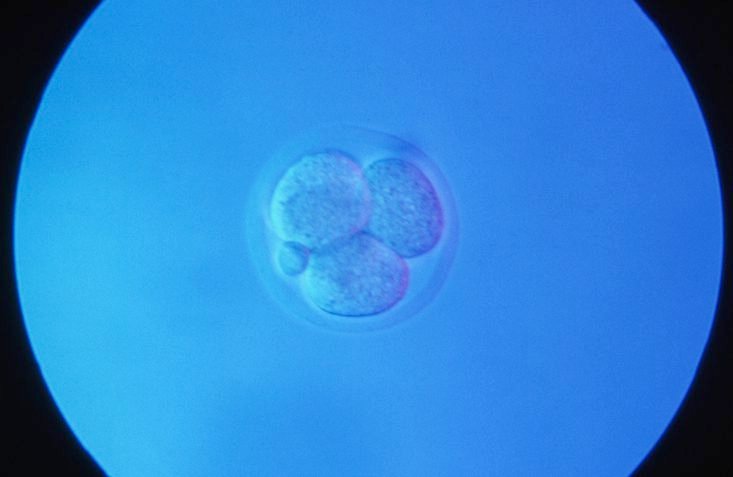
    A mouse embryo at the four cell stage, and polar body. [Clouds Hill Imaging Ltd. / Corbis Documentary / Getty Images]

    Researchers have used two DNA-recording systems to reconstruct cellular family trees across mouse embryonic development. The companion studies—“A DNA typewriter records the cell lineage history of a mouse, from zygote to late organogenesis,” published in Science, and “Comprehensive lineage tracing maps the landscape of cell fate decisions in mouse embryogenesis,” published in Cell—offer high-resolution views of mammalian development, which has been difficult to track because embryos develop inside the uterus and cannot be continuously observed.

    The team from the Science study was led by co-senior authors Jay Shendure, PhD, professor of genome sciences at the University of Washington School of Medicine and a Howard Hughes Medical Institute investigator, and Chengxiang Qiu, PhD, a molecular and systems biologist at Dartmouth College. Their approach centered on DNA Typewriter, a lineage-tracing technology developed by Shendure and Junhong Choi, PhD, that uses a cell’s DNA as a recording medium.

    The researchers redesigned the system’s recording “tape” to make its history easier to recover from individual cells, then inserted it into the genome of a fertilized mouse egg. As cells divided, “a burst of editing unequivocally marks the daughters of the first cleavage, which serve as inline replicates,” the authors wrote in the Science paper. Because daughter cells inherited the marks already written before acquiring new ones, shared patterns revealed common ancestry and the order of developmental branching.

    DNA Typewriter is injected into the genetic material of a fertilized mouse egg. This technology taps into the genetic material in the fertilized egg and resulting embryonic cells as a recording device of the cells' histories. It tracks their relationships as the embryo develops.
    DNA Typewriter is injected into the genetic material of a fertilized mouse egg. This technology taps into the genetic material in the fertilized egg and resulting embryonic cells as a recording device of the cells’ histories. It tracks their relationships as the embryo develops. [Kyle O’Conner/Seattle Hub for Synthetic Biology]

    “Most biological measurements are based on either live imaging, which is limited by the fact that most animal tissues are not transparent, or genomics, which are destructive and only measure a single timepoint,” Shendure said. “Recording techniques like the ones in these studies enable measurements over time including in settings that we can’t directly visualize.”

    The experiment was attempted in 100 fertilized eggs and yielded 10 embryos for examination. One embryo carried the most informative record, allowing the team to trace nearly every profiled cell back to one of the first two cells formed after the fertilized egg divided. Although one founding cell produced more descendants, the two branches generated diverse cell types in nearly equal proportions.

    The ordered recording also allowed the researchers to estimate when cell types diverged from shared developmental paths. Blood and retinal cells committed relatively early, whereas cells forming the skin’s outer layer committed later. The results indicate that cell identity is not fixed in a single coordinated step; instead, different lineages become restricted on distinct schedules.

    Earlier lineage-tracing approaches often relied on DNA-cutting enzymes that could damage cells, erase previous records, exhaust recording capacity, or leave unordered marks whose chronology had to be inferred. “DNA Typewriter avoids all of this,” said co-first author Haedong Kim, PhD, a postdoctoral scientist in genome sciences at UW Medicine. “It writes without fully severing the DNA, keeps recording relatively steadily, and writes everything in strict order, so we can record cell lineages at much higher resolution for a longer time.”

    In the Cell paper, Jonathan Weissman, PhD, and colleagues used a prime-editing system called PEtracer to map lineage dynamics across more than 1.4 million cells from 16 mouse embryos. Together with the DNA Typewriter study in Science, the work demonstrates that mammalian cellular family trees can be reconstructed at unprecedented scale. “It’s really proof of principle that we can do what was done with the roundworm in 1983, but for mammals like you and me,” Weissman said.

    The work provides a framework for investigating how normal organs form and where development can go awry. More complete lineage records could help researchers study congenital malformations, neurodevelopmental and genetic disorders, and cancer, as well as guide stem cell engineering. As Shendure explained, “Recording techniques like the ones in these studies enable measurements over time including in settings that we can’t directly visualize.” Scaling the methods across additional embryos and experimental conditions could support more quantitative—and eventually predictive—models of mammalian development.

  • From Discovery to Development

    From Discovery to Development

    The Era of Modality Convergence: Why Drug Development Is Being Rewritten

    From Discovery to Development: Navigating the New Era of Therapeutic Innovation eBook cover

    The life sciences industry is entering a new era, defined not by a single breakthrough technology but by the convergence of many. Artificial intelligence is accelerating drug discovery, cell and gene therapies are expanding treatment possibilities, and antibody engineering continues to evolve beyond traditional monoclonal antibodies. Meanwhile, mRNA platforms, viral vectors, antibody-drug conjugates (ADCs), multispecific antibodies, and other advanced therapeutic modalities are transforming how developers approach previously intractable diseases. Taken individually, each of these innovations represents a significant advancement. Together, they are reshaping the drug development landscape.

    For decades, therapeutic development followed a familiar path: identify a target, optimize a lead candidate, advance through preclinical studies, and move into clinical evaluation. While the science was challenging, the overall framework remained relatively consistent. Today, that framework is evolving rapidly. Developers are no longer limited to choosing between small molecules and conventional biologics. Instead, they have access to a growing toolkit of therapeutic technologies, each with unique advantages, manufacturing requirements, analytical challenges, and regulatory considerations. At the same time, advances in computational biology and AI-driven discovery are generating more candidates than ever before.

    As a result, the challenge is no longer simply finding promising molecules. Increasingly, success depends on identifying candidates that can be manufactured, scaled, and translated into viable therapies. Decisions once reserved for later stages, including developability, manufacturability, analytical characterization, and clinical strategy, must now be addressed much earlier in the process. Drug development is becoming a continuous, interconnected journey rather than a series of separate steps.

    The boundaries between traditional disciplines are also beginning to blur. Cell therapies may incorporate viral vectors. Gene therapies may leverage lipid nanoparticles. Antibody programs may evolve into multispecific or conjugated formats. The future of drug development is not defined by any single modality, but by the ability to integrate technologies effectively.

    Organizations that combine discovery expertise, modality-specific knowledge, CMC capabilities, and manufacturing execution will be best positioned to navigate this increasingly complex environment. The articles that follow examine key trends driving this transformation, from AI-enabled discovery and experimental validation to advanced cell, gene, and biologic platforms, and explore how the next generation of therapies will move from concept to clinic.

  • How Preclinical Manufacturing Can De-Risk the Path to GMP-Ready Viral Vector Programs

    How Preclinical Manufacturing Can De-Risk the Path to GMP-Ready Viral Vector Programs

    To move viral vector programs from early research toward the clinic, sponsors have to generate the right material and data during development. Non-GMP production of adeno-associated virus (AAV), lentiviral, and adenoviral vectors can support applications ranging from R&D and animal studies to assay development, toxicology planning, and early manufacturability assessments. But to be successful, sponsors have to align production scale, analytical testing, documentation, and material use with the specific questions their studies aim to answer.

    In this GEN webinar, Chris Nieder, an expert in viral vector programs, will examine how strategically designed preclinical manufacturing campaigns provide a practical foundation for GMP production. He will discuss topics such as how to select from among shake flasks, development-scale bioreactors, and pilot-scale approaches; and how to use early process and analytical data to identify yield and scalability challenges. Attendees will also learn how early CDMO engagement can clarify manufacturing scope, timelines, costs, and GMP-readiness without compromising expectations for non-GMP material. Key takeaways from the webinar include:

    • How early-stage sponsors can use non-GMP AAV, lentiviral, and adenoviral production to support R&D, animal studies, toxicology planning, assay development, and manufacturability assessment
    • How process and analytical data generated before GMP can reduce duplicated prep work, lower technical risk, and streamline later scope definition
    • When shake flasks, development-scale bioreactors, or pilot-scale production can provide the most useful insight for the next development step
    • How to align material use, analytical testing, documentation, and expectations without over-positioning preclinical material as clinical-grade supply

    A live Q&A session will follow the presentation offering you a chance to pose questions to our expert panelist.

    Produced with support from:

    skpharmteco logo

  • ARPA-H’s BoSS Program Funds Three Teams Tackling Cold Chain Challenges for Cell Therapies

    ARPA-H’s BoSS Program Funds Three Teams Tackling Cold Chain Challenges for Cell Therapies

    The Advanced Research Projects Agency for Health, (ARPA-H), an agency within the United States’ Department of Health and Human Services (HHS), recently announced awards and teams involved in its BioStabilization Systems (BoSS) program, an initiative that aims to develop new methods for producing, storing, and transporting cell-based biologic medicines at room temperature. ARPA-H has committed to provide up to $87 million across the four-year program. This round of funding will support Phase I of the program.

    The teams have been tasked with pursuing new technologies that confer long-term, shelf-life stability to cell-based therapies and biologics making it possible to store and ship them without refrigeration. To accomplish this goal, BoSS teams will work across two technical areas. They will work on interventions to stabilize cells for storage and then reactivate them on demand as well as on creating novel processing systems that ensure these interventions are efficient and scalable for manufacturing. 

    This week, members of the Reversible Ambient Temperature (RAB) team said that they have been awarded up to $7.3 million initially from ARPA-H. The RAB team includes representatives from Draper, a non-profit research, development, and manufacturing company, which serves as the administrative lead,and Likarda, a biotechnology company that develops hydrogel technologies for delivering and preserving cell therapies and biologics, which serves as the technical lead for the team. Other members of the team include scientists from CaseBioscience, CPSI Biotech, Fresenius Kabi, MiNK Therapeutics, and University of Colorado. 

    For their part, this team will use AI-assisted design of experiments to optimize formulation, quiescence induction parameters, hydrogel composition, and recovery media as well as freeze-assisted droplet drying to achieve a stable product. Specifically, the RAB team’s biostabilization strategy, led by Likarda, will focus on simultaneous control of cell state, physical environment, and the cellular stress responses activated during freezing, dehydration, storage, and recovery. Meanwhile, the bioprocessing engineering team, led by Draper, will convert the manual steps of biostabilization into an automated, commercially viable workflow and will develop custom instruments to scale up the processes.

    Likarda will contribute its proprietary Core-Shell Spherification hydrogel technology, which is designed to encapsulate and protect living cells while maintaining viability and function. CaseBioscience will provide expertise in clinical media formulation, biopreservation, and cellular metabolism to the development and evaluation of novel approaches for long-term ambient stabilization of therapeutic cells.

    “The RAB team brings together deep expertise in cryoprotection, encapsulation, and cell state, with a focus on protecting cell viability throughout the stabilization and reanimation process, and the team has direct experience bringing products to market,” said Jason Fiering, distinguished technical staff at Draper. “Working in parallel, our interdisciplinary engineering team will develop the customized instruments needed to transfer these processes to clinical manufacturing environments.” 

    In his comments, John Baust, PhD, an expert in biopreservation and collaborator on the project with CPSI Biotech noted that “the concept of preserving living cells in a dry state has been pursued for more than two decades, but significant scientific and engineering barriers have yielded limited success.” The BoSS program is an opportunity “to tackle this from multiple scientific and engineering directions simultaneously” that if successful “could represent a paradigm shift in how biologics are preserved, stored, and distributed.”

    Other teams announced by ARPA-H include one led by DesiCorp. This team includes members from the University of Louisville, University of Texas at Austin, Via Therapeutics, VeriSIM Life, and Ossium Health. They will leverage high-throughput screening to identify biocompatible formulations that slow cellular metabolism and AI-assisted process optimization to develop a thin-film freeze-drying (TFFD) system that stabilizes cells at room temperature. 

    DesiCorp will lead integration of the overall workflow by using a high-throughput screening platform to rapidly identify biocompatible formulations that enable cells to survive TFFD and resume function after rehydration. Meanwhile, UT Austin researchers will help optimize the TFFD critical process parameters and composition/material attributes, while also contributing expertise in aseptic processing and scaling up the technology. 

    A third team is led by the University of California, Davis and includes scientists from Mayo Clinic, Case Western Reserve University, University of Georgia, and Sersense. The team will induce a low metabolic state in cells using light-responsive gels and microfluidic processing techniques to produce cells that are stable at room temperature. 

    Recently, that team announced that it had been awarded up to $6 million, initially,  from ARPA-H to fund its project dubbed CYBORGEL. Their efforts will leverage research done in the laboratory of Cheemeng Tan, PhD, a professor of biomedical engineering at UC Davis. Previously, he and his collaborators formed water-rich polymer networks inside living cells to create cell-material hybrids. This process, called intracellular gelation, forms a gel inside the cell and is capable of halting cell division while preserving cellular function. Tan and his team dubbed this cellular product a “cyborg” since the gelation process combines living cells with synthetic materials. 

    Through CYBORGEL, the collaborators will investigate whether this intracellular gelation process can be extended to reduce the cost and simplify the storage, transportation and delivery of cellular therapies. The researchers will also test whether CYBORGEL cells remain therapeutically useful after being returned to their original state. For instance, one study will test the effectiveness of reanimated CAR T cells for existing clinical trials on cancer immunotherapies. Another will test it on placental mesenchymal stem cells used to treat spina bifida, a medical intervention being developed by UC Davis researchers. 

    As part of the initiative, the American Type Culture Collection (ATCC) has been tapped to serve as the IV&V partner. ATCC will provide working cell banks of government-selected cell types that will be used for demonstrations. They will also provide unbiased assessments of both the viability and function of the preserved products and the design features of the preservation systems.  

    The first phase of the BoSS program kicked off in September and is expected to run for 15 months. Over the four-year multi-phase program, the teams’ approaches will be evaluated against increasingly stringent benchmarks for cell viability, production speed, and shelf-life stability. If successful, work under the BoSS program could enable lower costs for cell-based drugs, reduce the risk of therapeutic and pharmaceutical product losses, and enable domestic reserves for public health preparedness. 

  • Epigenetics Underlies Comparatively Accelerated Molecular Aging in Larger and Male Dogs

    Epigenetics Underlies Comparatively Accelerated Molecular Aging in Larger and Male Dogs

    Sophie, a 12-year-old dog, isn’t sure what to make of the much larger Homer after Dog Aging Project co-lead and ASU Professor Noah Snyder-Mackler delivered his lecture to residents at Mirabella at ASU on Thursday, August 6, 2026. Sophie lives at Mirabella with her owner, Bonnie Kasel. Snyder-Mackler and his fellow researchers study various health markers in dogs as they age. [Charlie Leight/ASU News]

    Domestic dogs offer a useful model for understanding the biology of aging because their lifespans vary dramatically with body size. Large dogs tend to be shorter lived than their smaller counterparts, but why? Headed by a team at Arizona State University, a study of nearly 900 dogs now suggests that the answer may be written into the animals’ epigenomes.

    Research professor Noah Snyder-Mackler, PhD, at Arizona State University School of Life Sciences, and colleagues developed an epigenetic clock that predicted mortality in dogs and showed that epigenetic aging is fastest early in life. The study results indicated that male dogs and larger dogs undergo accelerated molecular aging, with male dogs exhibiting pronounced DNA methylation (DNAm) changes on the X chromosome, and larger animals exhibiting methylation changes at transposable elements (TEs), stretches of DNA that can influence genome stability and gene regulation.

    Snyder-Mackler is senior and corresponding author of the researchers’ published paper in Science (“Epigenetic aging and transposon dysregulation reflect size-related lifespan compression in dogs,”) in which they concluded, “Our findings broadly establish companion dogs as a translational model for uncovering molecular mechanisms of lifespan variation, epigenomic instability, and age-related disease.”

    Genome-wide DNA methylation in 894 dogs from the Dog Aging Project reveals accelerated aging in larger dogs and faster loss of repression of transposable elements. [Blaise Mariner & Brianah McCoy]
    Genome-wide DNA methylation in 894 dogs from the Dog Aging Project reveals accelerated aging in larger dogs and faster loss of repression of transposable elements. [Blaise Mariner & Brianah McCoy]

    Aging is a universal process, but its pace and biological mechanisms substantially differ between individuals within even the same species, the authors wrote. “Whether shorter-lived individuals simply die earlier or instead experience accelerated biological aging throughout life remains unclear,” they noted. And while comparative studies across species have identified mechanisms associated with exceptional longevity, they may not be as well suited to identifying the molecular drivers of aging variation. “Domestic dogs provide a model for addressing this challenge because body size strongly predicts lifespan, with larger breeds living substantially shorter lives than smaller breeds despite sharing many physiological and environmental conditions,” the team continued.

    In fact, smaller breeds can live nearly twice as long as larger breeds. And companion dogs live in human environments, consume commercial diets, and receive routine medical care, making them a relevant translational model for studying how genetic background and environmental exposures shape aging, the investigators pointed out.

    Study author with dogs
    Professor and School of Life Sciences Associate Director Noah Snyder-Mackler poses for portraits with his two dogs, 7-year-old Homer, bottom, and 4-year-old Juno, top, on the steps of Old Main on the ASU Tempe campus on Monday morning, September 28, 2026. Snyder-Mackler’s recent research focuses on dog epigenetics. [Deanna Dent/Arizona State University]

    But how intrinsic factors such as size and biological sex influence variation in dog aging and lifespan remains unclear. “It is well established that size and sex influence lifespan in dogs, but it remains unclear whether shorter-lived dogs experience accelerated epigenetic aging,” they stated. Previous research has demonstrated that DNAm is a useful indicator of biological aging, with “epigenetic clocks” offering a way to understand the factors that accelerate or decelerate biological aging. “DNA methylation (DNAm) has emerged as a robust molecular biomarker of age, with ‘epigenetic clocks’ offering a quantitative framework for identifying factors that accelerate or decelerate biological age.”

    For their newly reported study Snyder-Mackler, together with first author Blaise Mariner, PhD, and colleagues generated 1,640 methylomes from a cohort of 894 dogs in the Dog Aging Project. They combined the molecular data with detailed genetic and demographic data. The results indicated that molecular aging occurs most rapidly early in a dog’s life. The study also found that larger and male dogs—both of which have shorter lifespans than their respective counterparts—age more quickly at the molecular level.

    The authors identified different epigenetic patterns underlying these effects. According to the findings, sex-related changes were concentrated on the X chromosome, while body-size-related changes were especially prominent in TEs. “Thus, size and sex, two axes associated with shorter expected lifespan in dogs, appear to shape aging through partially distinct epigenetic architectures,” they stated. “Larger dogs show greater methylation loss at TEs with age, consistent with increased immune remodeling and genomic instability … These data address a major gap in comparative epigenomics by providing evidence that, within a species, demographic groups with shorter expected lifespans show faster epigenetic aging.”

  • The State of AI in Drug Discovery

    The State of AI in Drug Discovery

    Panelists:

    Image of Aliza Apple, PhD

    Aliza Apple, PhD

    Vice President
    Catalyze360 AI/ML, Eli Lilly

    Panelist

    Image of Aliza Apple, PhD

    Aliza Apple, PhD

    Aliza Apple, PhD, is a vice president of Catalyze360 AI/ML at Eli Lilly, where she leads the strategy, build and launch of Lilly’s external-facing AI/ML efforts for drug discovery. She previously served as the COO and head of Lilly Gateway Labs West Coast, where she supported the local biotech ecosystem through early engagement and providing tailored offerings to meet their needs. Prior to Lilly, Aliza served as a co-founder at Santa Ana Bio, a venture-backed precision biologics company focused on autoimmune disease, and as an advisor to the founders of Firefly Biologics. Prior to biotech, Aliza was a partner in McKinsey’s life sciences practice. During her nine years at McKinsey in New York and San Francisco, she collaborated with leaders from more than 20 biopharma companies to develop their corporate and portfolio strategies, evaluate business development opportunities, and improve their organizational effectiveness.

    Aliza completed her PhD in bioengineering at UC Berkeley and UCSF and holds a BS in physics from Georgetown University. She completed a postdoctoral fellowship at Cornell Medical College, supported by the NIH Kirschstein National Research Service Award.

    Image of Sam Rodriques, PhD

    Sam Rodriques, PhD

    Co-Founder, Director, and CEO
    Edison Scientific and FutureHouse

    Panelist

    Image of Sam Rodriques, PhD

    Sam Rodriques, PhD

    Physicist and bioengineer Sam Rodriques, PhD, is the co-founder, director, and CEO of Edison Scientific and FutureHouse. Sam has invented technologies for spatial and temporal transcriptomics, brain mapping, gene therapy, and nanofabrication, and briefly ran an academic lab at the Francis Crick Institute in London prior to launching FutureHouse in San Francisco in 2023. FutureHouse is a new research lab focused on building an AI Scientist to accelerate research in biology and other complex sciences. In 2025, Sam launched Edison Scientific to commercialize the agents being built at FutureHouse, with the goal of wanting “science to move faster.”

    Sam graduated summa cum laude with highest honors in physics from Haverford College, where he worked on new methods for calculating quantum entanglement in multipartite quantum systems. He holds a PhD in physics from MIT, having worked between the MIT Media Lab, the MIT Department of Brain and Cognitive Sciences, and the Broad Institute. Among his awards and fellowships are the Hertz Foundation Graduate Fellowship, an NSF Graduate Research Fellowship, and a Churchill Scholarship.

    Image of Peyton Greenside, PhD

    Peyton Greenside, PhD

    Co-Founder and CEO
    BigHat Biosciences

    Panelist

    Image of Peyton Greenside, PhD

    Peyton Greenside, PhD

    Peyton Greenside, PhD, is the co-founder and CEO of BigHat Biosciences, a Bay Area biotechnology company pioneering an ML-driven platform to accelerate the design of next-generation antibodies and other therapeutic proteins.

    Peyton holds a BA in applied mathematics from Harvard University, a PhD from Stanford University, and an MPhil in computational biology from the University of Cambridge, U.K.

    Image of Marc Tessier-Lavigne, PhD

    Marc Tessier-Lavigne, PhD

    Co-Founder, Chairman, and CEO
    Xaira Therapeutics

    Panelist

    Image of Marc Tessier-Lavigne, PhD

    Marc Tessier-Lavigne, PhD

    Marc Tessier-Lavigne, PhD, is the co-founder, chairman, and CEO of Xaira Therapeutics. Previously, he held executive positions in the private sector at Genentech, where he was executive vice president for research and chief scientific officer, and in the academic sector, where he served as president of The Rockefeller University and then for seven years at Stanford University, where he remains a professor of biology (on leave). He also served previously on the faculty of the University of California, San Francisco, and was an investigator with the Howard Hughes Medical Institute. A Rhodes Scholar, Marc has been a leader in understanding mechanisms that direct the wiring of the brain and has helped elucidate mechanisms of neurodegeneration. He is the recipient of numerous scientific awards, including the 2020 Gruber Neuroscience Prize, and has been elected to multiple scientific societies, including the U.S. National Academy of Sciences and U.S. National Academy of Medicine; he is also an officer of the Order of Canada, one of that country’s highest honors. Marc is the co-founder of two neuroscience biotech companies, Renovis and Denali, and has served on the boards of Agios, Regeneron, Pfizer, Juno, and Denali.

    Marc holds a BS degree in physics from McGill University in his native Canada. He earned a BA in philosophy and physiology from the University of Oxford as a Rhodes Scholar, and a PhD in physiology from the University of London.

    Broadcast Date: 

    • Time: 

    From billion-dollar funding rounds and waves of IPOs to the first AI-guided molecules entering the clinic, the industry is signaling a clear inflection point: AI is set to fundamentally reshape drug discovery. At the center of this shift are maturing foundation models for molecules and cellular systems that unveil insights into complex biology.

    Yet the challenge is no longer prediction alone. Building the infrastructure that connects accelerated computing, generative design, and experimental feedback will be critical for transforming AI into a fully integrated engine for therapeutic innovation.

    GEN’s virtual summit, The State of AI in Drug Discovery, returns on November 4 to examine how AI is reshaping R&D timelines and real-world patient translation. From big pharma to AI-natives, this year’s program features the leaders of many of today’s outstanding companies and organizations powering the AI revolution, including Xaira Therapeutics, Edison Scientific, Eli Lilly, Chai Discovery, Lila Sciences, BigHat Biosciences, and more!

    Among the program highlights during this 4-hour event are:

    • Building the AI scientist with Sam Rodriques, PhD (Edison Scientific)  
    • A fireside chat with Xaira Therapeutics CEO Marc Tessier-Lavigne, PhD 
    • Powering AI adoption through strategic enterprise partnerships with Aliza Apple, PhD (Lilly TuneLab), Simon Barnett (Dimension), and Elliot Hershberg, PhD (Amplify Ventures)  
    • The role of agentic workflows in accelerating drug discovery with Le Cong, PhD (Stanford and Phylo) and Andrew Beam, PhD (Lila Sciences) 
    • The new era of programmable therapeutics with Josh Meier (Chai Discovery) and Peyton Greenside, PhD (BigHat Biosciences) 
    • And breakout sessions from our four event sponsors: A-AlphaBio, Benchling, LabVantage, and Revvity Signals.

    Don’t miss this exceptional virtual summit, featuring many of the visionaries shaping the future of AI in drug discovery and biotech. Registration, as always, is free.

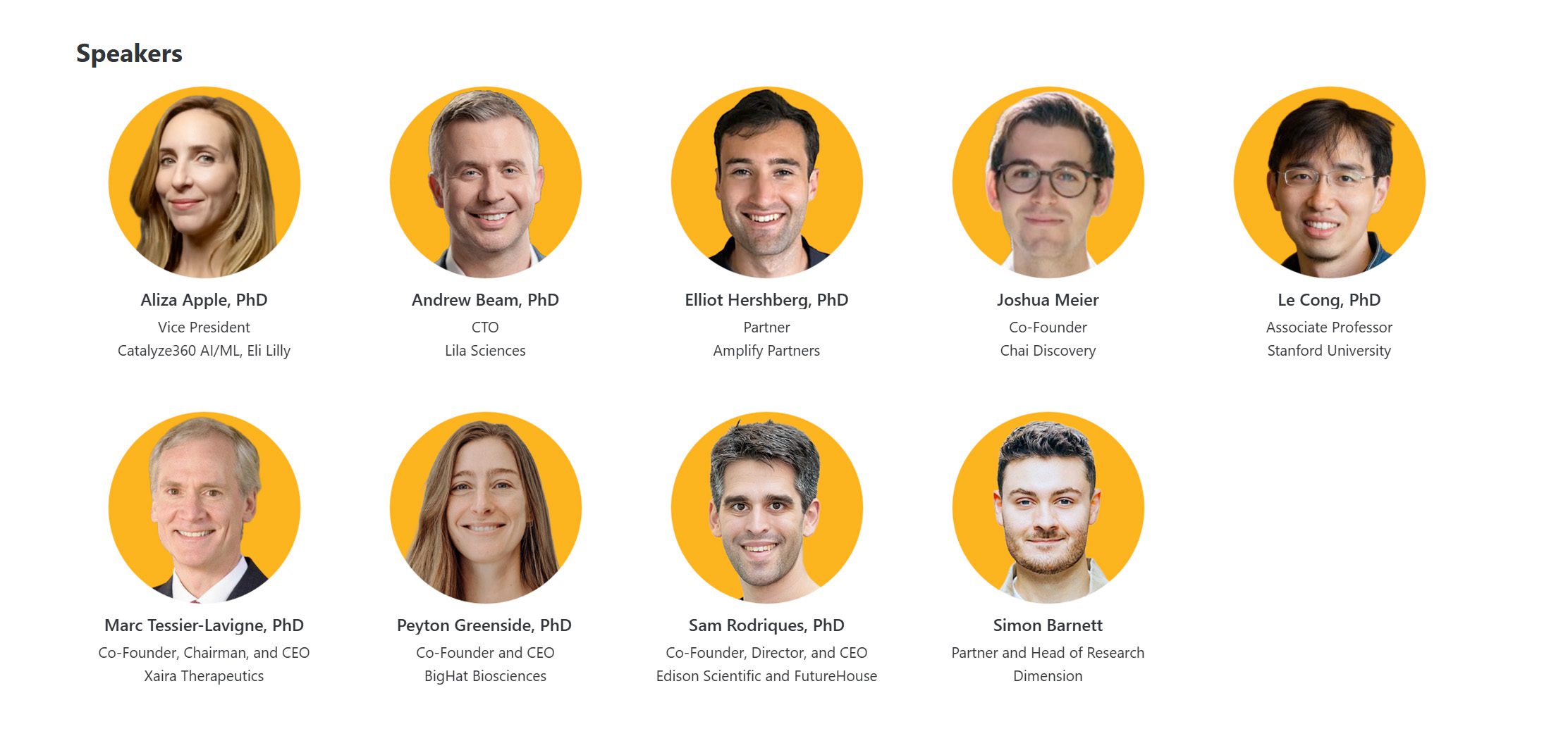
    Guest Speakers Include

    The State of AI in Drug Discovery speakers