Author: admin

  • Scientists discover how gut bacteria “train” the intestine to fight inflammation

    Scientists discover how gut bacteria “train” the intestine to fight inflammation

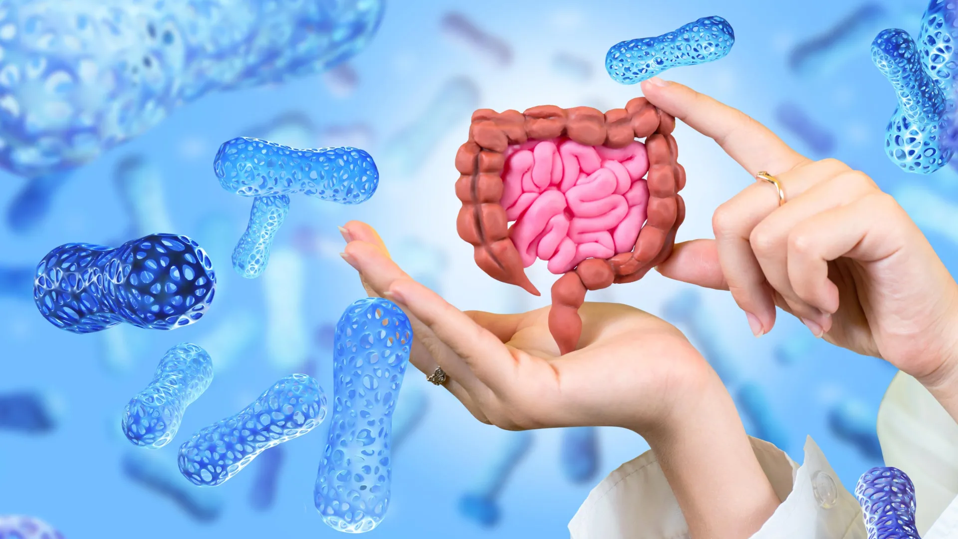
    New Northwestern Medicine research published in Nature Communications suggests that a substance produced when gut bacteria digest dietary fiber may leave a durable molecular mark on cells lining the intestine. That imprint appears to promote immune tolerance and may help protect against inflammatory bowel disease-like conditions even after exposure to the metabolite has ended.

    Yingzi Cong, PhD, the Stanley Gradowski Professor of Gastroenterology and a professor of Microbiology-Immunology and of Pathology, was senior and co-corresponding author of the study.

    The researchers say the results support the idea that beneficial compounds made by gut microbes can effectively “train” the intestinal lining, helping it preserve immune tolerance over time.

    “Our laboratory has long been interested in how the gut microbiota regulates immune responses at intestinal mucosal surfaces,” said first and co-corresponding author of the study Tianming Yu, PhD, research assistant professor of Medicine in the Division of Gastroenterology and Hepatology. “Short-chain fatty acids (SCFAs), which are produced by gut bacteria during the fermentation of dietary fiber, are known to have anti-inflammatory effects in the intestine.”

    How Butyrate May Influence Gut Immunity

    One major question, however, has remained unresolved, Yu said.

    “A large proportion of butyrate in the gut is rapidly absorbed and metabolized by intestinal epithelial cells (IECs), which limits the amount of free butyrate that can directly reach underlying immune cells,” Yu said. “This led us to ask whether butyrate might act through IECs to regulate intestinal immunity.”

    To investigate, the researchers supplied mice with butyrate in their drinking water for a set period and then ended the treatment. Two weeks after the butyrate had been withdrawn, CD4+ T-cells were still producing elevated levels of IL-10, an important anti-inflammatory cytokine that helps control intestinal inflammation.

    The treated mice were also more resistant to chemically induced colitis. Compared with untreated animals, they lost less weight, had lower levels of inflammatory markers, and developed less severe tissue damage. According to the study, this protection depended on IL-10 signaling.

    The researchers also determined that the lasting effect did not result from changes to the gut microbiome itself. In experiments involving germ-free mice, which lack all microbes, butyrate still created a persistent immune-regulating environment.

    “We found that oral butyrate treatment induces a sustained immunoregulatory response in the intestine, characterized by increased IL-10 production in CD4+ T-cells and protection from intestinal inflammation even after butyrate treatment is stopped,” Yu said. “This effect was also observed in germ-free mice, suggesting that butyrate can establish a lasting intestinal environment that does not depend on continuous microbial stimulation.”

    Intestinal Cells May Store a Lasting Biological Signal

    Yu and his colleagues next focused on intestinal epithelial cells (IECs), which form the physical boundary between the body and the microbiome. In laboratory experiments, epithelial cells exposed to butyrate caused a strong increase in IL-10 production in both mouse and human T-cells.

    “We further found that conditioned medium from butyrate-treated IECs strongly induced IL-10-producing CD4+ T-cells in both mouse and human T-cell culture systems,” Yu said. “These findings suggest that IECs secrete some immunoregulatory factors after butyrate treatment, and these factors can act on T-cells.”

    The team then used metabolomic analysis to search for molecules that might be carrying this signal. One likely candidate was N1-acetylspermidine. The compound increased IL-10 production in T-cells and appeared to account for part of the immune-regulating activity generated by epithelial cells exposed to butyrate, Yu said.

    “Mechanistically, we found that butyrate acts on IECs and induces sustained transcriptional and epigenetic activation of Sat1, an acetylpolyamine biosynthetic enzyme,” Yu said. “This promotes production of the metabolite N1-acetylspermidine, which contributes to the ability of butyrate-treated IEC-conditioned medium to induce IL-10 production in CD4⁺ T-cells.”

    The results challenge the traditional idea that intestinal epithelial cells are primarily temporary responders whose main role is to serve as a barrier. Instead, the findings suggest that these cells may retain a longer-lasting molecular record of beneficial signals from the microbiome.

    “The significance of this work is that it identifies a mechanism by which a microbiota-derived metabolite can create durable epithelial T-cell crosstalk,” Yu said. “The intestinal epithelium is often viewed as a short-lived barrier that responds rapidly to luminal stimuli. Our findings suggest that it can also retain a lasting imprint of a microbial metabolite signal. The study also introduces the idea that beneficial microbial metabolites may ‘train’ the IECs to maintain immune tolerance over time.”

    Potential Implications for Inflammatory Bowel Disease

    More research will be necessary to confirm the findings and determine whether the same mechanism operates in people, but Yu said the work could eventually have implications for inflammatory bowel disease.

    “One important next step is to determine how this epithelial metabolic pathway operates in human intestinal disease, especially in patients with inflammatory bowel disease,” Yu said. “We are interested in testing whether the butyrate-Sat1-N1-acetylspermidine pathway is altered in human IECs and whether it correlates with immune regulation or disease activity.”

    The researchers also intend to investigate other metabolites that may contribute to the effect. N1-acetylspermidine by itself did not fully account for all of the immune-regulating activity observed in the experiments.

    Over the longer term, Yu said the findings may expand scientists’ understanding of the relationship among diet, metabolites produced by gut microbes and the intestinal lining.

    “Many studies have focused on how inflammation can leave harmful memory in epithelial cells, but our findings suggest that beneficial microbial metabolites may also establish protective epithelial programs,” he said. “Understanding how diet, microbiota-derived metabolites and inflammation shape intestinal epithelial memory could open new directions for restoring intestinal immune tolerance in inflammatory bowel disease.”

    Wenjing Yang, MD, PhD, research assistant professor of Medicine in the Division of Gastroenterology and Hepatology, was co-first author of the study.

    Additional Feinberg co-authors include Suxia Yao, MD, research associate professor of Medicine in the Division of Gastroenterology and Hepatology; and Parambir Dulai, MD, associate professor of Medicine in the Division of Gastroenterology and Hepatology.

    Cong, Yu, Yang, Yao, and Dulai are members of the Center for Human Immunobiology.

    The study was supported by National Institutes of Health grants DK135193, DK124132 and DK145439.

  • Trump’s FDA pick Overton good for psychedelics, new pain meds but bad for vaccines: Analysts

    Trump’s FDA pick Overton good for psychedelics, new pain meds but bad for vaccines: Analysts

    It’s official. President Donald Trump will nominate Heidi Overton as the next FDA commissioner. Trump announced the news himself on his Truth Social platform early Wednesday afternoon.

    In the post, which refers to Overton as “Dr. Heidi,” Trump called the current White House policy advisor a smart and respected “ROCKSTAR” who would “rapidly” deliver on his priorities of faster cures, innovation, lower drug prices and more wins for Health Secretary Robert F. Kennedy Jr.’s Make America Healthy Again (MAHA) movement. “We need her leadership at the FDA now to ensure that the U.S. remains the WORLD LEADER for Scientific Discovery and CURES,” Trump continued in the post.

    Analysts reacting to reports of Overton’s nomination on Wednesday appear to agree that the Trump administration isn’t looking to rock an already unsteady boat. The general physician, if approved by the Senate, could provide strong support for the administration’s psychedelics and non-opioid pain medicine initiatives—while possibly kowtowing to would-be boss Robert F. Kennedy Jr. on vaccines.

    Overton, who currently serves as the deputy assistant to the president for domestic policy, was reportedly involved in the drafting of an executive order (EO) to accelerate the development of and facilitate broader access to psychedelic therapies.

    “Her visible role in the April 2026 executive order prioritizing FDA review of ibogaine and MDMA-based therapies, highlights the administration’s approach to use of psychedelics as medicine,” BMO Capital Markets wrote in a note to investors on Wednesday.

    Truist Securities in a Tuesday evening note also pointed to Overton’s role in the EO. “She has drawn MAHA attention for the inter-agency process she ran on the psychedelics/ibogaine EO,” the analysts wrote.

    The agency’s finalized recommendations for clinical testing of psychedelic compounds for mental health could pave the way for companies like Compass Pathways, whose psilocybin-based therapy for treatment-resistant depression could win approval as early as this year.

    July 14, 2026

     · 

    3 min read

     · 

    Overton is also likely to support continued progress in the non-opioid pain arena. Since the January 2025 approval of Vertex Pharmaceuticals’ Journavx as the first new mechanism for acute pain in two decades, drug development in the space has taken off. Most recently, AbbVie ventured into the field with an up to $715 million deal with China’s Haisco Pharmaceutical Group in April. Overton’s nomination could be a boon for Vertex, AbbVie and several other companies committed to offering alternatives to the controversial pain drugs at the root of the opioid epidemic.

    “Journavx momentum should continue under Overton,” BMO wrote on Wednesday, additionally noting that “her 2017-2018 opioid prescribing work” with former FDA Commissioner Marty Makary “lines up with the non-opioid pain agenda behind Vertex’s Journavx approval.” Overton and Makary penned a peer-reviewed paper titled “Overprescribing is major contributor to opioid crisis,” in October 2017.

    Overton’s support for non-opioid options should be “an incremental positive as VRTX pushes into broader indications,” BMO said.

    In one hot-button drug development space, however, analysts appear wary of Overton’s influence.

    “Vaccine makers shouldn’t expect a friendlier FDA, given Overton’s involvement in this year’s executive orders reshaping the childhood vaccine schedule,” BMO wrote, “keeping potential regulatory risk alive for Merck, Pfizer, and BioNTech.”

    Melanoma was slower to recur in patients receiving Merck and Moderna’s mRNA vaccine with Keytruda, supporting use of the combination in adjuvant treatment. The partners plan to take the data to the FDA.

    August 19, 2026

     · 

    3 min read

     · 

    Indeed, Overton was in attendance last week when Trump signed an executive order recommending that the measles, mumps and rubella (MMR) vaccine should be split into three separate shots. During the press conference announcing the EO, the president falsely suggested the number or timing of vaccines could play a role in rising rates of autism spectrum disorder, per the Associated Press.

    Regulatory expert Steven Grossman expressed reservations about Overton’s nomination. “I am concerned about: her lack of management experience, her apparent willingness to undercut vaccines, and—especially given her close White House ties—her understanding of science-based decisionmaking,” said Grossman, president of policy and regulatory consulting firm HPS Group, prior to Trump’s announcement.

    Truist was more positive. “Our sense is that Dr. Overton could drive healthy debate internally, though ultimately cede to the administration, which is what we & investors ultimately may want from the seat from our conversations,” the analysts wrote on Tuesday. “[She is] science-driven though diplomatic to the administration, which to us could strike the perfect balance.”

    Moreover, Truist added, “She also appears as the more conventional, pharma-friendly profile on evidence standards.”

    Despite his concerns, Grossman encouraged an open mind to Trump’s FDA pick. “Nonetheless, stakeholders should hear her out before locking in their position.”

    If confirmed, Overton would be the first woman since 2015—and just the third woman ever—to lead the FDA.

  • Scientists turn probiotic bacteria into tiny drug factories for pancreatic cancer

    Scientists turn probiotic bacteria into tiny drug factories for pancreatic cancer

    Cancer immunotherapy has dramatically changed how doctors treat many forms of cancer, yet pancreatic cancer has remained particularly resistant to these advances. A major obstacle is the environment that develops around pancreatic tumors. These tumors often create a “cold” tumor microenvironment that blocks immune cells from launching an effective attack.

    Researchers at the University of Chicago have now developed a new approach that could help overcome this problem. In a study published in Science Advances, the team used BifidoSumIL-2, an engineered strain of Bifidobacterium longum, a probiotic bacterium naturally found in the gut, to carry an immune-stimulating treatment directly into tumors.

    In animal models, the therapy slowed the growth of pancreatic tumors by selectively activating T cells that fight cancer. Its effects became even stronger when researchers combined it with chemotherapy, radiotherapy or immunotherapy. The findings suggest that BifidoSumIL-2 could eventually provide a new way to improve how pancreatic tumors respond to treatment.

    Using Bacteria to Deliver Cancer Therapy

    “A big unmet medical need has been pancreatic cancer, and so that was going to be our mountain to climb,” said Ralph Weichselbaum, MD, the Daniel K. Ludwig Distinguished Service Professor and Chair of Radiation and Cellular Oncology at the University of Chicago.

    BifidoSumIL-2 was created to release a modified version of interleukin-2 (IL-2) once it reaches a tumor. IL-2 is a potent immune signaling molecule that activates T cells involved in attacking cancer. Conventional IL-2 treatment, however, can produce serious side effects and can also stimulate immune cells that actually weaken the antitumor response.

    The researchers sought to avoid these problems by using SumIL-2, a modified form of IL-2 engineered to more precisely activate cancer-fighting T cells while reducing stimulation of regulatory T cells. They then placed SumIL-2 inside Bifidobacterium longum so that the therapeutic molecule could be concentrated within tumors instead of throughout the body.

    Developing the treatment required scientists from several disciplines to work together, including specialists in microbiology, synthetic biology, oncology, and immunology.

    “This was a highly interdisciplinary effort,” said Mark Mimee, PhD, Assistant Professor of Microbiology at the University of Chicago. “We had to bring together people who understand bacteria, people who understand tumors, and people who understand the immune system to make something like this possible.”

    Why Bifidobacterium Can Target Tumors

    Bifidobacterium offered the researchers an unusual advantage as a delivery system. The bacterium grows in anaerobic environments, meaning places with very little oxygen. Low oxygen levels are common inside many solid tumors, including pancreatic tumors, while healthy tissues generally contain more oxygen and are therefore less suitable for the bacteria.

    “Bifidobacterium is an obligate anaerobe, so it doesn’t grow in the presence of oxygen,” Mimee said. When the bacteria are injected systemically, they are cleared from healthy tissues with abundant oxygen. Inside the low-oxygen regions of tumors, however, they can become active.

    That preference allows the engineered bacteria to function as microscopic drug factories inside tumors. Once there, they produce SumIL-2 where the treatment is needed rather than broadly throughout the body. Researchers also noted that Bifidobacterium has shown a favorable safety profile in preclinical models and is already well known as a probiotic organism. It is commonly present in yogurt and is generally recognized as a safe, off-the-shelf probiotic.

    Engineering the organism was not simple.

    “Bifidobacterium is not the easiest organism to work with,” Mimee said. “It’s anaerobic, it grows slowly, and the genetic tools for manipulating it are much more limited compared to model bacteria like E. coli. A lot of the work was just figuring out how to reliably engineer it.”

    Stronger Results With Combination Treatments

    Tests in animal models showed that BifidoSumIL-2 preferentially gathered inside tumors, stimulated immune activity, and slowed the growth of pancreatic cancer. It also changed the tumor microenvironment in a potentially beneficial way by increasing the activity of cancer-fighting CD8+ T cells.

    The results improved further when BifidoSumIL-2 was paired with established cancer treatments. Combining the bacterial therapy with chemotherapy, radiation therapy, or anti-PD-L1 immunotherapy led to better tumor control and longer survival than the individual treatments achieved on their own.

    “This combination potential is one of the study’s most important findings; BifidoSumIL-2 not only works by itself — it works with radiotherapy, chemotherapy, and immunotherapy,” Weichselbaum said.

    Despite the encouraging findings, BifidoSumIL-2 has not been tested in humans. Future research will need to examine its long-term safety, the possibility of effects outside the intended tumor, how long the immune response lasts, and whether the bacteria might eventually be given orally instead of through injection. The researchers also want to investigate whether the strategy can be combined with newer pancreatic cancer treatments, including KRAS inhibitors.

    The Growing “Bugs as Drugs” Approach

    The research adds to growing interest in a strategy known as “bugs as drugs.” By engineering probiotic bacteria to seek out tumors and produce therapies directly inside them, scientists may be able to concentrate powerful immune treatments where they are most useful while reducing unwanted effects elsewhere in the body.

    The study, “Engineered probiotic Bifidobacterium for tumor-targeted pancreatic cancer therapy” was supported by funds from the Ludwig Foundation and the National Institutes of Health.

    Additional authors include Jaehyun Lee, Kaiting Yang, Christina Nowicki, Wei Liu, Emile Naccasha, and Hua Liang from the University of Chicago; Zhichen Sun from the University of Texas Southwestern, Dallas; and Yang-Xin Fu from Tsinghua University, Beijing, China.

    UChicago Medicine and the Biological Sciences Division continue to be at the forefront of cancer care and research. In April 2027, UChicago Medicine will open the AbbVie Foundation Cancer Pavilion, Chicago’s first freestanding cancer pavilion, to bring advanced diagnostics, innovative treatments, translational discoveries, and comprehensive support to patients and the community.

  • Tiny robots powered by light can hunt down and collect bacteria

    Tiny robots powered by light can hunt down and collect bacteria

    Robots small enough to operate in the microbial world could give scientists a new way to directly handle objects that are impossible to manipulate by hand. These tiny machines are around 50 times smaller than the diameter of a human hair, bringing researchers closer to the long-pursued goal of interacting directly with the microscopic world.

    That capability could be especially useful for biological materials in water, including individual cells and bacteria. Precisely controlling and moving objects at this scale has been a persistent challenge. The newly developed nanorobots show that it is already possible to collect bacteria, transport them, and release them at selected locations.

    Light Powers and Steers the Tiny Robots

    One of the biggest obstacles in developing machines this small is finding an effective way to propel and control them. At Julius-Maximilians-Universität Würzburg (JMU), a research team led by Professor Bert Hecht has been developing a solution that uses the recoil produced by individual photons to move microscopic devices known as microdrones.

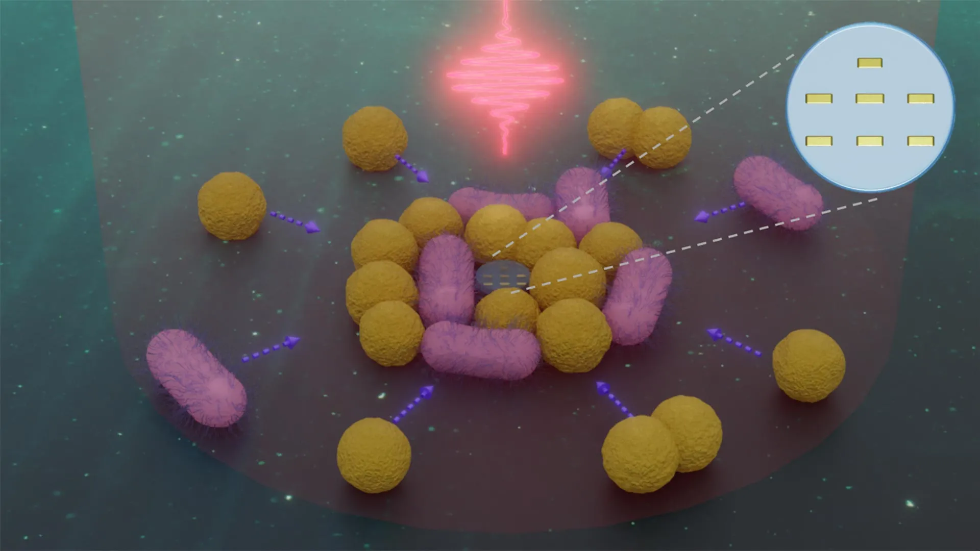
    The devices contain as many as four plasmonic nanoantennas. These antennas absorb light with a particular color and helicity, then emit that light in a specific direction. Redirecting each photon creates a tiny recoil force, similar in principle to the recoil produced when a bullet is fired. Because the microdrones have so little mass, those extremely small forces can generate substantial acceleration and speed.

    For the latest work, the researchers reduced the size of their light-powered robots even further, producing devices smaller than one micrometer. Simplifying the steering system was an important part of achieving that size while retaining propulsion based on photon recoil.

    The new control method takes advantage of nanoscale antenna wires built into the robot. These wires naturally tend to align with the polarization direction of incoming light. By changing the light’s polarization, the researchers can control which direction the nanorobot faces. At the same time, photon recoil continues to propel it forward, creating a steering system that works somewhat like the directional control used in larger vehicles.

    Nanorobots Act as “Microscopic Cleaners”

    “In essence, we have built a light-driven nanorobot that can track down and collect bacteria,” says Jin Qin, lead experimental scientist of the study. “By simplifying the design, we reached a size at which these robots can operate directly in the microbial world – almost like microscopic cleaning devices.”

    The robots are also highly maneuverable. They can make extremely fast 90° turns, helping them scan broad areas of a sample in an organized and efficient way. They can also selectively capture, carry, and release substantial numbers of bacteria.

    Under controlled laboratory conditions, this means the nanorobots can effectively “clean” microscopic environments. They gather bacteria from one area and deposit them at specifically chosen locations.

    “This is a striking example of how light can be used not only to observe the microscopic world, but also to actively shape it,” adds Bert Hecht. “The idea of tiny robotic cleaners may sound futuristic, but we are already demonstrating the physical principles that make it possible.”

    The robots remain fully maneuverable even while carrying larger groups of bacteria, although their speed decreases somewhat under the added load. That ability to keep functioning while transporting larger bacterial clusters points to possible future uses in microbiology, biomedical research, and precise manipulation of materials at the microscopic scale.

  • Scientists turn DNA into a memory device that uses 100x less power

    Scientists turn DNA into a memory device that uses 100x less power

    DNA serves as the genetic blueprint for every living organism, but it is also an extraordinarily dense way to store information. A single gram can hold about 215 million gigabytes of data. Bringing that remarkable storage capacity into electronics could lead to more efficient data centers, faster processing and systems capable of handling increasingly complex information.

    The challenge has been finding a way to make biological DNA function effectively alongside electronic materials. Penn State researchers have now developed an approach designed to overcome that incompatibility.

    The work, published in Advanced Functional Materials and the subject of a patent application, relies on two key components. One is synthetic DNA, made from commercially available, chemically engineered molecules arranged into short genetic sequences tailored for specific electronic requirements. The other is crystalline perovskite, a semiconductor already used in technologies including solar cells, lasers and data storage devices.

    “Biology and electronics are different domains,” said Kavya S. Keremane, co-corresponding author and postdoctoral researcher in materials science and engineering at Penn State. “Bridging these two fields required developing an entirely new materials platform that allows them to function seamlessly together. By combining the information storage capabilities of DNA with the exceptional electronic properties of perovskite semiconductors, we created a bio-hybrid system that fundamentally changes how low-power memory devices can be designed.”

    Building a Low Power Memory Device

    Using these materials, the team created a memory resistor, known as a “memristor,” that operates with very little energy. Unlike ordinary resistors, which maintain a set resistance to electrical current in devices ranging from cell phones to space shuttles and lose their stored information when power disappears, memristors can preserve a record of previous electrical activity. They can remember the direction in which current previously flowed even after the power source is removed.

    That ability allows information to be stored and processed in the same place, resembling the way neurons function in the brain. Such an arrangement could support more simultaneous and sophisticated forms of data processing. According to the researchers, however, practical commercial systems would still require enough storage capacity and electrical power to become costly and inefficient without DNA’s ability to pack enormous amounts of information into a very small space while consuming little energy.

    “As the demand for artificial intelligence (AI) grows, we need a new strategy for low-power, high-storage devices,” said Bed Poudel, co-corresponding author and research professor of materials science and engineering at Penn State.

    Poudel said AI and other emerging technologies are expected to depend increasingly on neuromorphic computing, which is designed to operate more like the human brain. Such systems can evaluate multiple inputs at once while making decisions informed by previous experiences and future priorities.

    “Usually, it takes more power to store more information. Our device, however, consumes 100 times less power and the storage capacity is higher than traditional storage devices, like flash drives.”

    Engineering DNA to Conduct Electricity

    To construct the device, the researchers added silver nanoparticles to a layer of customized DNA sequences — specially designed to be of certain compositions and lengths — that was integrated with thin films of perovskite.

    This technique, called “doping,” involves introducing a small amount of another material to produce specific properties. In this case, adding the silver nanoparticles allowed the DNA to conduct electricity while also helping its molecular units line up in a more orderly arrangement.

    Synthetic DNA offered another important advantage over natural DNA. Unlike natural DNA — long, entangled strands that behave like wet spaghetti when handled — short and rigid pieces of synthetic DNA can be arranged with much greater precision at extremely small scales.

    According to co-author Neela H. Yennawar, research professor and director of the Penn State Huck Institutes of the Life Sciences’ Biomolecular Interactions Core Facility, molecularly engineered DNA can provide structural organization, adjustable electrical conductivity and functional control that natural DNA cannot achieve when incorporated into thin films.

    “We can computationally determine exactly which sequences we need and how long they should be, and then we can rationally design them with synthetic DNA,” Yennawar said. “These structures can be systematically doped with silver and other ions and engineered to interface seamlessly with perovskites — transforming DNA from a biological macromolecule into a programmable, multifunctional nanomaterials platform.”

    DNA and Perovskite Work Better Together

    When combined, the silver-doped DNA and perovskite formed bio-hybrid pathways that directed the flow of electrical current through the device.

    The researchers found that electrons moved reliably when they applied less than 0.1 volt — for comparison, standard U.S. outlets have 120 volts — and the device responded predictably when the direction of the current was changed.

    The carefully designed DNA structures, combined with the perovskite, also helped make the device unusually stable. According to the team, it continued operating consistently at temperatures approaching 250 degrees Fahrenheit and remained functional at room temperature for more than six weeks, substantially exceeding the performance standards of existing perovskite-based memory storage devices.

    The researchers also reported that the new system could perform the same memory function as comparable technologies while consuming only one-tenth as much power. That level of efficiency could make the approach especially attractive for future electronics designed to handle large amounts of information with lower energy demands.

    “Using just the DNA or just perovskite alone did not produce near as robust a result as the combination,” Keremane said. “It’s this combination that enables a very high memory storage density that requires very little power.”

    A New Direction for Bio-Inspired Electronics

    The team now plans to improve the technology further and explore additional uses for bio-inspired electronic systems.

    “Nature has the solution — we just have to find it and apply it,” Poudel said. “This work of integrating DNA into electronics to do amazing things gives a glimpse into what is possible.”

    In addition to Keremane, Yennawar and Poudel, other Penn State co-authors include co-corresponding author Luyao Zheng, postdoctoral research in materials science and engineering; Haodong Wu, doctoral student in materials science and engineering; Jiamao Zheng, who was a master’s student in materials science and engineering at the time of research and has since graduated from Penn State; Shashank Priya, who was a professor of materials science and engineering at the time of research; and Chiranth C. Ravi, who was a master’s student in the Huck Institutes of the Life Sciences at the time of research and has since graduated from Penn State. Abhinav Gorthy and co-corresponding author Rashmi Jha, chemical engineering and materials science, University of Minnesota, also contributed.

    The U.S. National Science Foundation, the National Institutes of Health, Penn State and the University of Minnesota supported this research.

  • When gut microbes run low on fiber, they may start eating you

    When gut microbes run low on fiber, they may start eating you

    Plant based diets support health in several ways, including benefits for the gut, immune system, metabolism, and cardiovascular system. Part of that effect comes from encouraging a diverse population of bacteria in the intestines. Scientists have long known that dietary fiber contributes to these benefits because gut microbes help break it down. Plants also contain colorful “phytochemicals” that help protect them from environmental threats and may influence human health. Even so, researchers are still working to understand exactly how gut bacteria process the many components of plant foods and how those interactions produce beneficial effects.

    Two studies led by Ludwig Princeton’s Jenna AbuSalim and Director Joshua Rabinowitz offer new insight into that process. One appears in the current issue of the Proceedings of the National Academy of Sciences, while the other was published in Nature Metabolism in June. The first study found that plant fiber and certain plant proteins can change microbial metabolism in ways that increase beneficial metabolites while reducing harmful ones. The second showed that several biologically important metabolites usually credited to gut microbes can also be produced in substantial amounts by mammalian metabolism.

    “There’s growing interest across medical disciplines in manipulating the human microbiome or using its metabolic products themselves for therapy,” said Rabinowitz. “Diet holds great promise for controlling the microbiome and its outputs. But to devise effective therapeutic interventions, we need to understand what aspects of the diet control which microbial outputs.”

    How Plant Foods Shift Gut Metabolites

    In the PNAS study, Rabinowitz, AbuSalim and their colleagues examined how plant based foods influence phenol metabolites. Gut bacteria create these compounds when they digest the amino acids tyrosine and phenylalanine, but the resulting metabolites can have very different effects on health.

    Phenylpropionate and hippuric acid are produced when bacteria process phenylalanine, and they are associated with gut health and healthy body weight. By contrast, p-cresol sulfate and phenol sulfate come from tyrosine and have been linked to worse outcomes in cancer patients as well as systemic toxicity in people with kidney disease.

    “Our studies showed that both the fiber and indigestible proteins from plants — which we call ‘proteins imitating fiber,’ or Prif — shift the balance of phenol metabolites from the harmful kind made from tyrosine to the healthful variety derived from phenylalanine,” said AbuSalim.

    Fiber has long been recognized as an important part of a healthy diet, but indigestible plant proteins have received far less attention. AbuSalim, Rabinowitz and their colleagues found that these proteins are processed by gut microbes and can alter both the makeup of the microbiome and the host’s metabolism. Working together with indigestible plant fiber, they can also change the metabolic activity of gut bacteria in ways that favor the production of beneficial phenols.

    When Gut Bacteria Turn to the Gut Lining

    To trace where these compounds came from, the researchers labeled proteins with stable (non-radioactive) isotopes and followed their digestion in the mouse gut. They found that the “bad” phenols were produced when bacteria consumed proteins from the host, including proteins found in the mucus lining of the gut. The good phenols, in contrast, came almost entirely from indigestible proteins in the diet (Prif).

    Fiber reduced the bacterial breakdown of the gut’s mucus lining, which in turn lowered production of the harmful phenols. Prif increased the amount of dietary protein that reached gut microbes, giving them more material to produce the beneficial phenols.

    “We think Prifs represent an emerging class of dietary nutrients that shape the composition of the gut microbiome and could have a far-reaching influence on metabolic health,” said AbuSalim.

    “Food packaging may eventually list Prif right below fiber,” said Rabinowitz.

    Rethinking Where Gut Metabolites Come From

    The Nature Metabolism study focused on the origins of phenol metabolites as well as indole metabolites, which are produced from the amino acid tryptophan. Like phenols, indoles are being studied for their possible therapeutic value.

    Indole metabolites have been connected to a wide range of diseases, including inflammatory bowel disease, neurodegenerative disorders, and cancer. In cancer research, they have been found to affect processes that include cancer metastasis and anti-tumor immune responses.

    Scientists had generally assumed that phenols and indoles were produced only by gut bacteria. AbuSalim, Rabinowitz and their colleagues decided to test that assumption. Researchers have been especially interested in dietary and probiotic approaches that might increase beneficial indole metabolites. But those strategies may need to be reconsidered if mammalian metabolism, rather than microbes, is responsible for much of what circulates in the body.

    Using isotope tracing in mice, rats and human cells, the researchers found that mammalian metabolism can produce many indole and phenol metabolites on its own. These included important compounds such as indole-3-lactate and indole-3-acetate.

    In mice, circulating levels of these metabolites remained high even after antibiotic treatment disrupted the microbiome. A similar pattern appeared in samples from patients taking antibiotics, including cancer patients. At the same time, metabolites made exclusively by microbes, including indole-3-propionate and p-cresol sulfate, declined after antibiotic treatment.

    New Clues for Diet and Microbiome Therapies

    Together, the two studies provide a clearer picture of where phenol and indole metabolites come from and how they are produced. The findings could influence the development of therapies designed to raise or lower specific metabolites.

    They also add important detail to scientists’ understanding of how diet interacts with the microbiome. Knowing which foods influence particular microbial products could eventually help researchers design more precise dietary, probiotic, or metabolic interventions.

    “Beyond that,” said Rabinowitz, “a clearer picture of how different foods interact with the microbiome to modulate the production of bacterial metabolites will help sharpen the guidance nutritionists and doctors can give to people for disease prevention and therapy.”

    These studies were funded by the Ludwig Institute for Cancer Research, the National Institutes of Health, the National Institute of Diabetes and Digestive and Kidney Diseases, the Princeton Alliance for Collaborative Research and Innovation, Princeton University.

    Aside from his post as Director of the Princeton Branch of the Ludwig Institute for Cancer Research, Joshua Rabinowitz is Professor in the Department of Chemistry & Lewis-Sigler Institute for Integrative Genomics and a member of the Rutgers Cancer Institute.

  • Scientists tracked kids for 8 years — the screen time result was unexpected

    Scientists tracked kids for 8 years — the screen time result was unexpected

    A Finnish study has found an unexpected association between screen use and cognitive performance. Children who accumulated more screen time as they grew up tended to show better cognitive processing during adolescence. One of the researchers cautions against viewing screen time as entirely harmful and says the goal should be to balance physical activity with screen use that encourages active thinking.

    Low levels of physical activity among adolescents remain a significant public health concern. Sedentary behavior has been associated with poorer academic achievement, while physical activity is known to support brain function, particularly in adults. Much less is known, however, about how physical activity and sedentary behavior during childhood and adolescence relate to cognitive processing later in adolescence.

    These years are especially important because the brain is still developing. Factors that shape cognitive abilities during childhood and adolescence may have lasting effects that extend into adulthood, potentially influencing education and working life.

    Researchers examined how physical activity, sedentary behavior, and screen time from childhood through adolescence were related to cognitive processing in the teenage years. They also looked for differences between girls and boys and investigated whether the intensity of physical activity played a role.

    More Screen Time Linked to Better Cognitive Processing

    The results showed that greater screen time beginning in childhood was associated with better cognitive processing in adolescence.

    “The findings suggest that screen time can support children’s and adolescents’ cognitive processing. Presumably, the essential point here is what kind of things they do in their screen time. Teachers and parents should encourage children to use devices and screens in such ways that promote active thinking, problem-solving, creativity and learning,” states Doctoral Researcher Petri Jalanko from the University of Jyväskylä.

    The findings suggest that the type of screen activity may be important. Digital activities that involve learning, creativity, problem-solving, or other forms of active mental engagement could potentially help explain the association observed in the study.

    “We should not regard screen time solely as harmful but seek balance between physical activity and screen time that promotes active thinking,” Jalanko summarizes.

    Physical Activity Showed Different Links in Girls and Boys

    The relationship between physical activity and cognition was more complicated.

    Among girls, greater amounts of light intensity physical activity since childhood were associated with better working memory during adolescence. Among boys, greater participation in guided physical activity from childhood through adolescence was linked to better working memory.

    The researchers also found an unexpected pattern involving unsupervised exercise. Lower levels of self-reported unsupervised physical activity were associated with better cognitive processing in adolescence.

    By contrast, physical activity and sedentary time measured using a device that tracked heart rate and movement were not associated with cognitive processing. One possible explanation is that these sensors can measure movement and physiological activity but cannot determine exactly what a person is doing during periods of activity or inactivity.

    “Our study indicates that the connections of physical activity and sedentary behavior to cognitive processing are complex and depend on the sex, the type and assessment method of physical activity and sedentary time. Moreover, boys and girls may benefit from different types of physical activity in view of brain health,” Jalanko adds.

    More Research Is Needed to Determine Cause and Effect

    The researchers emphasize that the findings show associations rather than proving that screen time or particular types of physical activity directly cause changes in cognitive performance.

    “However, we need more intervention studies to find out causal relations and sex differences in this respect. Moreover, it is important to examine more specifically the effects of the intensity of physical activity on changes taking place in cognitive processing.”

    The results come from an eight-year follow-up of the PANIC study on children’s physical activity and nutrition. The current analysis included 124 girls and 136 boys with an average age of 15.8 years.

    Researchers measured physical activity and sedentary behavior using both survey questionnaires and a device that combined heart rate and movement measurements. Learning, attention, and working memory were evaluated using the CogState test battery.

    The research was published in the journal Pediatric Exercise Science.

  • World’s first superconducting quantum heat engine could help unlock massive quantum computers

    World’s first superconducting quantum heat engine could help unlock massive quantum computers

    A newly developed superconducting quantum heat engine could deepen our understanding of thermodynamics while helping advance technologies needed for quantum computers with very large numbers of qubits.

    Scientists are getting a clearer picture of how thermodynamics behaves in the quantum world, and that progress could benefit both quantum technology and our understanding of familiar thermodynamic principles. Researchers at Aalto University have now taken an important step by demonstrating the first cyclic quantum heat engine built inside a superconducting circuit.

    The experiment connects two areas of physics that normally describe very different scales. Quantum mechanics explains the behavior of matter at extremely small scales, even below the size of atoms, while thermodynamics describes how heat and energy behave in much larger systems, from collections of molecules to the universe itself. Bringing the two together raises a fundamental question: what happens to familiar thermodynamic processes when quantum effects such as tunneling, entanglement, and superposition enter the picture?

    A Heat Engine Built for the Quantum World

    Conventional heat engines turn heat into useful work. James Watt’s steam engine is one famous example, but the same basic concept remains central to modern transportation and electricity production, powering cars, ships, planes and many power plants.

    The researchers have now created the world’s first superconducting quantum heat engine. The extremely small device combines a transmon qubit, a resonator and a quantum refrigerator.

    Operating under ultracold quantum conditions, the engine was able to use the tiny amount of available heat to repeatedly produce positive work. Achieving this kind of cyclic operation has been an important objective for researchers working on quantum heat engines. The result provides a proof of concept for superconducting heat engines that could eventually contribute to improved quantum computing technology.

    The study, led by Academy Professor Mikko Möttönen, was published in Nature Communications.

    Recreating an Otto Cycle Near Absolute Zero

    To make the engine operate, the researchers reproduced an Otto cycle inside a superconducting circuit. The Otto cycle is a thermodynamic process also used in car engines and other conventional machines.

    “In our experiment, we built a nanofabricated heat engine using superconducting circuits and operated it in a cryostat near absolute zero. At its heart is a transmon qubit, one of the basic building blocks of modern quantum technologies,” says Tuomas Uusnäkki, the study’s first author.

    The researchers connected the transmon qubit to a quantum circuit refrigerator, allowing them to control heat flow on the quantum scale and demonstrate that this heat could be transformed into measurable work. A conventional heat engine normally relies on separate hot and cold environments. In this system, however, the same quantum refrigerator can supply both heating and cooling.

    “Our quantum-circuit refrigerator can be tuned to both heat and cool the qubit on demand. Using carefully timed control pulses, we drove the engine in an Otto cycle and monitored the qubit state as the engine ran,” explains Uusnäkki.

    Measurements showed that heat passing through the qubit during the cycle was producing positive work.

    “This is the first experimental demonstration of a cyclic quantum heat engine in superconducting circuits. Using a single controllable quantum refrigerator as both the hot and cold environment of the engine makes it simpler and more versatile,” says Uusnäkki.

    Toward Autonomous Quantum Computer Hardware

    The researchers are now trying to improve the design and eventually develop a fully autonomous heat engine. One possible use would be reading out qubits without having to carry a microwave pulse from millikelvin temperatures all the way to room temperature.

    That capability could become especially valuable as quantum computers grow. Autonomous devices integrated directly into superconducting circuits could reduce both the cost and complexity of machines containing very large numbers of qubits.

    “Finland’s Quantum Technology Strategy envisions a quantum computer with one thousand logical qubits by 2035, which probably means hundreds of thousands of physical qubits. Doing that with current technology requires millions of microwave cables costing thousand euros each. The cables also introduce noise into the system. Using autonomous devices instead would mostly eliminate the need for those cables,” Möttönen says.

    Reducing the need for those microwave connections could therefore address two challenges at once: the enormous hardware requirements of large quantum computers and the unwanted noise that cables can introduce into quantum systems.

    The pioneering experiment was carried out using OtaNano, Finland’s national research infrastructure for nano, micro and quantum technology. Funding came from the Research Council of Finland and the Finnish Cultural Foundation.

  • Drinking orange juice every day may change thousands of genes

    Drinking orange juice every day may change thousands of genes

    Most of us think of orange juice as a simple breakfast habit, something you pour without much thought. Yet scientists are discovering that this everyday drink may be doing far more in the body than quenching thirst.

    A recent study has shown that regular orange juice consumption can influence the activity of thousands of genes inside our immune cells. Many of these genes help control blood pressure, calm inflammation and manage the way the body processes sugar, all of which play an important role in long-term heart health.

    The study followed adults who drank 500ml of pure pasteurized orange juice every day for two months. After 60 days, many genes associated with inflammation and higher blood pressure had become less active.

    These included NAMPT, IL6, IL1B and NLRP3, which usually switch on when the body is under stress. Another gene known as SGK1, which affects the kidneys’ ability to hold onto sodium (salt), also became less active.

    Such changes match previous findings that daily orange juice drinking can reduce blood pressure in young adults.

    This is noteworthy because it offers a possible explanation for why orange juice has been linked to better heart health in several trials. The new work shows that the drink does not simply raise blood sugar. Instead, it appears to trigger small shifts in the body’s regulatory systems that reduce inflammation and help blood vessels relax.

    Natural compounds in oranges, particularly hesperidin, a citrus flavonoid known for its antioxidant and anti-inflammatory effects, seem to influence processes related to high blood pressure, cholesterol balance and the way the body handles sugar.

    The response also varies by body size. People carrying more weight tended to show greater changes in genes involved in fat metabolism, while leaner volunteers showed stronger effects on inflammation.

    A systematic review of controlled trials involving 639 participants from 15 studies found that regular orange juice consumption lowered insulin resistance and blood cholesterol levels. Insulin resistance is a key feature of pre-diabetes, and high cholesterol is an established risk factor for heart disease.

    Another analysis focusing on overweight and obese adults found small reductions in systolic blood pressure and increases in high density lipoprotein (HDL), often called the good cholesterol, after several weeks of daily orange juice consumption. Although these changes are modest, even slight improvements in blood pressure and cholesterol can make a meaningful difference when maintained over many years.

    More clues come from studies that examine metabolites, the tiny molecules produced as the body processes food. A recent review found that orange juice influences pathways related to energy use, communication between cells and inflammation. It may also affect the gut microbiome, which is increasingly understood to play a role in heart health.

    One study showed that drinking blood orange juice for a month increased the number of gut bacteria that produce short-chain fatty acids. These compounds help maintain healthy blood pressure and reduce inflammation. Volunteers also showed improved blood sugar control and lower levels of inflammatory markers.

    People with metabolic syndrome, a cluster of risk factors that includes high blood pressure, raised blood sugar and excess body fat, may see particular benefits.

    In one study, daily orange juice consumption improved the function of the lining of blood vessels, known as endothelial function, in 68 obese participants. Endothelial function describes how well blood vessels relax and widen, and better function is associated with a lower risk of heart attacks.

    Not all studies report the same outcomes. A broader analysis of blood fat concentrations found that although levels of low density lipoprotein (LDL), often called the bad cholesterol, often fall, other lipid measurements such as triglycerides and HDL may not change much. Even so, people who regularly drink orange juice may still benefit.

    A study of 129 workers in an orange juice factory in Brazil reported lower blood concentrations of apolipoprotein B, or apo-B, a marker that reflects the number of cholesterol-carrying particles linked to heart attack risk.

    Altogether, the evidence challenges the idea that drinking citrus fruit juice is simply consuming sugar in a glass. Whole fruit remains the better choice because of its fiber, but a modest daily glass of pure orange juice appears to have effects that build up over time.

    These include easing inflammation, supporting healthier blood flow and improving several blood markers linked to long-term heart health. It is a reminder that everyday foods can have more influence on the body than we might expect.The Conversation

  • Scientists find evidence for two origins of life on Earth

    Scientists find evidence for two origins of life on Earth

    Where and how did life first emerge on Earth? Those questions are at the heart of research at the Institute of Molecular Evolution at Heinrich Heine University Düsseldorf (HHU). Now, an international team led by biologists in Düsseldorf has reported new findings in Science Advances that shed light on the chemical reaction network used by the earliest cells to produce the basic ingredients of life, as well as the energy sources that powered those reactions. By tracing the origins of enzymes during the earliest split between bacteria and archaea, the researchers found evidence that free-living cells may have originated independently twice.

    About 4 billion years ago, Earth looked very different from the planet we know today. If it were possible to watch the earliest cells take shape, the scene may have involved two distinct forms of primitive cellular life.

    “We would see two very different kinds of cells emerging, pioneer bacteria and pioneer archaea, making their first attempts at life outside the confines of a hydrothermal vent,” says Natalia Mrnjavac, biologist at the University of Düsseldorf and lead author on the new publication in Science Advances.

    Mrnjavac and an international team examined genomes, protein structures and chemical reactions to investigate some of the earliest stages of microbial evolution, including the period before fully free-living cells existed.

    “These comparisons are giving us unprecedented insights into the phase of evolution when metabolism catalyzed by enzymes was arising from spontaneous reactions catalyzed by metals in the Earth’s crust,” says Düsseldorf biologist William Martin, senior author of the study.

    Reconstructing the Chemistry of Early Life

    Rather than focusing on only selected parts of early metabolism, the researchers examined the complete group of chemical reactions that cells use to manufacture key biological components (amino acids, RNA bases and vitamins) from materials that were available on the early Earth, including hydrogen gas, ammonia and CO2.

    Together, these 420 chemical reactions form the metabolic network known as metabolism. The reactions themselves are extremely ancient and are conserved across life to a degree comparable with the genetic code.

    What surprised the researchers was that the enzymes responsible for carrying out those reactions do not show the same degree of conservation between bacteria and archaea.

    Martin: “The surprise is that the enzymes that catalyze those reactions are not conserved across the evolutionary divide that separates bacteria and archaea. We found that the last universal ancestor of all cells, LUCA, possessed enzymes for only about half of the reactions of metabolism. The other half was catalyzed by metals in the environment where LUCA arose.”

    That finding suggests that the earliest metabolism depended much more heavily on the surrounding environment than modern cellular metabolism does.

    “Metals that naturally occur in hydrothermal vents can replace a surprisingly large number of enzymes in metabolism,” says Harun Tüysüz, inorganic chemist from the Max-Planck-Institut für Kohlenforschung and the IMDEA Materials Institute in Madrid, and co-author on the study.

    “The closer we look, the more clearly we can see that early biochemical evolution was a hybrid of enzymatic and metal catalysts,” says Joseph Moran from the University of Ottawa, Canada, an international leader in the use of metals to catalyze metabolic reactions, replacing enzymes and cofactors.

    From Metal Catalysts to Enzymes

    One of the study’s major advances was the reconstruction of four stages in the early evolution of biological catalysis.

    The process appears to have begun with reactions driven entirely by metals. That was followed by a stage in LUCA in which metals and enzymes worked together. Afterward, bacteria and archaea began moving along separate evolutionary paths. Within each lineage, newly evolved enzymes gradually replaced the inorganic catalysts supplied by the environment where metabolism had first developed.

    Importantly, the researchers identified examples in which bacteria and archaea appear to have independently developed different enzymes capable of performing the same essential metabolic task.

    “We can see cases where the ancestors of bacteria and archaea independently evolved structurally distinct enzymes to catalyze the same essential metabolic reaction,” says Mrnjavac, “such parallel inventions could have paved the way to the independent emergence of free-living bacteria and archaea.”

    That parallel evolution may have been crucial because it could have allowed the two lineages to become less dependent on the chemistry of hydrothermal vents and eventually survive as independent cells.

    How Early Metabolism May Have Been Powered

    Another major question concerns energy. Modern cells rely heavily on ATP to power metabolism, but ATP is itself a complex molecule produced with the help of enzymes. It would not simply have been freely available in ancient hydrothermal environments.

    The researchers therefore investigated what could have supplied energy before ATP-based metabolism existed.

    “We have identified a new source of energy at metabolic origin,” says Manon Schlikker from the Düsseldorf team.

    One possible answer involves palladium, a metal that occurs naturally in hydrothermal vents and has been known to chemists as an effective catalyst for roughly a century.

    The team found that phosphite, a form of phosphorus naturally found in hydrothermal vents, can react with organic compounds in the presence of palladium and produce reactions associated with metabolic phosphorylation.

    “When we react phosphite, a form of phosphorus that naturally occurs in hydrothermal vents, with organic compounds, we get metabolic phosphorylation reactions overnight in water. Phosphite and palladium replace ATP and enzymes; it’s amazing, and it makes early evolution a lot easier to grasp,” says Schlikker.

    The result provides a possible explanation for how some of the earliest metabolic reactions could have obtained the energy needed to proceed before modern biological energy systems evolved.

    Mapping 420 Reactions at the Origin of Metabolism

    The research is the first study specifically focused on the full reaction network known as metabolism.

    That network contains 420 highly connected reactions, and many of the same compounds participate in multiple parts of the system. Because of those connections, reconstructing how the network may have developed over time presents a difficult mathematical problem.

    Prof. Mike Steel, from the University of Canterbury in New Zealand, and Prof. Daniel Huson from the University of Tübingen brought expertise in analyzing complex networks to the project.

    They developed a method that allowed the researchers to arrange metabolic reactions from the simplest to the most complex. The resulting sequence may reflect, at least in part, the order in which those reactions appeared during the earliest stages of biological evolution.

    “The first question,” says Steel, “is whether or not a unique order exists for these reactions. Once we could prove that there is one, the algorithm to order them became tractable.”

    One Genetic Code, but Two Origins of Life

    Understanding how life began addresses one of humanity’s most fundamental questions: where we came from, where the first living systems appeared, and how Earth’s earliest cells were able to survive.

    According to the researchers, the broader implication of their findings is that bacteria and archaea did not make the transition to independent cellular life together. Instead, each lineage appears to have reached the free-living state separately.

    Martin: “The new data leave only one conclusion. The bacterial and archaeal lineages made the transition to the free-living state independently. Only free-living cells are alive. Let’s call it by name: we are looking at one origin of the genetic code, but two origins of life.”

    In addition to researchers from HHU, the international team included scientists from the Universities of Canterbury (New Zealand), Rostock, Constance, Ottawa (Canada), Strasbourg and Tübingen, the Max-Planck-Institute for Terrestrial Microbiology in Marburg and the Max-Planck-Institut für Kohlenforschung in Mülheim/Ruhr plus the IMDEA Materials Institute in Madrid (Spain).

    Background: Bacteria and Archaea

    Biologists broadly separate cellular life into two categories. Eukaryotes are advanced cells that contain a nucleus, while the more ancient cellular lineages without a nucleus are known as prokaryotes.

    Prokaryotes consist of the two primordial branches of life: Bacteria and Archaea. Many of these organisms are capable of surviving under extreme conditions, including very high temperatures and strongly acidic or alkaline environments.

    Many also live around hydrothermal vents on the ocean floor, environments that some theories identify as possible locations where life first arose.