Tag: Translational Medicine

  • Immune Cell Atlas Charts How People’s Aging Paths Diverge

    Studies of the immune system show that two people born on the same day can have immune systems that age differently, leaving some older adults with resilient defense systems and others with dysfunctional ones that put them at increased risk for age-related diseases. Now a new study led by scientists at Washington University School of Medicine in St Louis, Nationwide Children’s Hospital in Ohio, and King’s College London provides a map of human immune aging that could help explain these different trajectories. Details are published in a new Immunity paper titled “Single-cell analyses of global cohorts outline determinants of immune aging and link the ratio of GZMK⁺ to GZMB⁺ Tem cells to health trajectories.”

    According to the paper, a ratio between two immune cell types can reveal whether someone stays healthy or may face risk of future disease. To get to this insight, the scientists first analyzed approximately 12.4 million individual immune cells from the blood of over 2,600 mainly healthy adults ages 20 to 90-plus across eight cohorts in North America, the United Kingdom, Asia, and Australia. 

    Their analysis showed that while the blood of younger people was uniformly enriched with naive immune cells that had not been exposed to specific pathogens and remained adaptable to new infections or vaccines, older immune systems were more diverse. Specifically, some older participants had a greater abundance of immune cells that reflect inflammation.

    To map this shift, the researchers placed each participant on a spectrum based on the ratio between two types of white blood cells: specifically, granzyme B-producing cells and granzyme K-producing cells. Some white blood cells, known as effector memory CD8 T cells, use granzymes to destroy target cells or coordinate immune responses. While granzyme B-producing cells serve as direct destroyers of diseased cells, granzyme K-producing cells have been less studied and may play a role in boosting immune responses. 

    Next, the researchers evaluated whether participants’ positions on the spectrum, which corresponds to a specific type of CD8 T cell, could dictate their risk of getting sick. In other words, they looked at whether having more of one type of CD8 T cell has an impact on the person’s health as they age. To answer that question, the scientists turned to the UK Biobank. 

    While that dataset includes protein information from the participants, it lacks counts of CD8 T cells. To get around this hurdle, the scientists used a smaller dataset of seemingly healthy adults that included data on CD8 T cell counts and blood proteins to create a computer model to map out the protein tracks left behind when either granzyme B-producing or granzyme K-producing cells dominated. They then applied this model to the baseline blood protein samples of 50,000 participants from the UK Biobank, calculating their position on the immune cell spectrum.

    Using this approach, the scientists were able to draw out some useful insights for their work. Specifically, they found that healthy adults whose immune system leaned heavily toward granzyme B cells at baseline tipped the scale toward a higher risk of death a decade later compared to those with more granzyme K cells. The granzyme B-dominant group also was more likely to develop chronic conditions, including Type 2 diabetes, hypertension, liver disease, and renal failure, among others, over the following decade.

    “A higher level of granzyme B cells in a healthy state means the immune system might be already reacting to something,” said Marina Terekhova, MD, a professor in pathology & immunology at WashU Medicine and one of the co-authors on the paper. “It isn’t a formal disease diagnosis, but it may indicate the body is off track.”

    A healthy immune system gradually shifts toward granzyme K cells over time, while an immune system that leans toward granzyme B cells signals unhealthy aging. Because individuals can reach this granzyme B state much earlier in life, their immune systems age on different trajectories, explaining why two people born in the exact same year can face different risks for chronic disease and death decades later.

    These findings point to “clues in the blood that can tell us whether someone is on a healthy or unhealthy aging trajectory,” said Maxim Artyomov, PhD, the alumni endowed professor of pathology and immunology at WashU Medicine and one of the corresponding authors on the paper. Artyomov and his lab are now adapting their research into a simple blood test that can be processed using standard equipment. “Identifying specific biological red flags in the immune system could pave the way for early-warning tests that spot disease risks years before a clinical diagnosis.”
  • ‘Molecular Movie’ of Penicillin Synthesis Could Inform Future Antibiotic Development

    ‘Molecular Movie’ of Penicillin Synthesis Could Inform Future Antibiotic Development

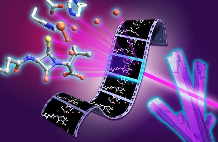
    Molecular movie of penicillin biosynthesis. X-ray free electron laser experiments on isopenicillin N synthase microcrystals capture structural snapshots of key intermediates during the penicillin forming reaction. The film strip illustrates progression through the catalytic cycle, with the monocyclic β-lactam intermediate and reactive high valent ferryl iron species highlighted at the center of the molecular movie. [University of Oxford/Greg Stewart/SLAC National Accelerator Laboratory]

    Researchers from the University of Oxford and international collaborators have revealed previously unseen rapid chemical stages in the formation of β-lactam antibiotics such as penicillin, offering insights that could help to support future antibiotic development.

    Rather than relying on static X-ray crystallographic structures of the enzyme, the Oxford University team, collaborating with partners from Diamond Light Source, Lawrence Berkeley National Laboratory, PAL-XFEL, and SLAC National Accelerator Laboratory, followed the reaction in real time. To do this they used X-ray free-electron lasers (XFEL) to observe the enzyme isopenicillin N synthase, (IPNS) as it converts its linear peptide substrate into the β-lactam ring system of penicillin.

    The study brought together expertise in structural biology, spectroscopy, chemistry, computation and enzyme engineering. The approach, they say, provides a powerful way to study fleeting chemical intermediates that have previously been inaccessible, both in antibiotic biosynthesis and in other biologically important enzyme reactions.

    The team captured several rapid stages in the reaction. These included a thioaldehyde intermediate formed just before the β-lactam ring is created, and a monocyclic β-lactam intermediate, representing the first ring-shaped structure on the way to forming the complete penicillin scaffold. These steps provide the clearest picture yet of how the IPNS enzyme assembles the penicillin scaffold. Subtle movements throughout the enzyme help guide these chemical steps, showing that both the enzyme’s shape and its chemistry work together to control penicillin formation.

    The results show, in unprecedented detail, how the enzyme IPNS achieves an exceptionally complex transformation in a single step. Christopher Schofield, PhD, Professor of Chemistry, University of Oxford, said, “Penicillin has shaped modern medicine, but there is still much to learn about how nature builds this important antibiotic structure. By capturing these fleeting steps, we can better understand how enzymes control complex chemistry with remarkable precision … As rates of antimicrobial resistance continue to rise, understanding this process will ultimately help us make existing antibiotics more efficient and design new antibiotic structures.”

    Allen M Orville, PhD, group leader of the XFEL Hub, further commented, “The XFEL Hub at Diamond brought specialist expertise in time-resolved X-ray methods and XFEL-based structural biology to this study. Working closely with our colleagues in Oxford and at partner facilities, we helped bring together the experimental approaches needed to capture these fleeting stages of enzyme catalysis. The results show the power of combining complementary expertise and X-ray techniques to move beyond static structures and reveal how enzymes work in real time. It is a great example of how time-resolved structural biology can uncover new principles of enzyme function and, ultimately, inform the design of new catalysts and therapeutics.”

    Schofield is co-senior author of the researchers’ published paper in Nature Catalysis, titled “Unanticipated intermediates during isopenicillin N synthase catalysis identified by time-resolved X-ray free-electron laser studies.”

    Ever since penicillin was developed into a working drug at the University of Oxford in the early 1940s, β-lactam antibiotics have been among the most important medicines for treating bacterial infections. Their activity depends on a special ring—the β-lactam ring—a highly strained chemical ring system that interferes with bacterial cell wall synthesis, ultimately causing the cell wall to fail and the bacteria to die. “Isopenicillin N synthase (IPNS) catalyses the biosynthesis of isopenicillin N (IPN), forming the fused four-membered β-lactam and five-membered thiazolidine ring system that is characteristic of the penicillins,” the authors explained.

    Rising rates of antimicrobial resistance are undermining the effectiveness of existing antibiotics. Today, one in six bacterial infections is resistant to antibiotics, the authors noted, citing WHO figures. With too few new antibiotics in development, understanding how nature builds these molecules is an important step towards replenishing the antibiotic pipeline.

    Scientists have been studying how nature constructs this β-lactam ring for decades, but the key fleeting reaction intermediates have been too difficult to observe directly.

    To capture the reaction in real time, the researchers used a system in which thousands of tiny droplets containing anaerobic enzyme microcrystals were deposited onto a moving 2 mm wide tape. As the tape entered an oxygen filled chamber, oxygen rapidly diffused into the crystals and initiated the reaction simultaneously across the sample. By precisely controlling the speed of the tape, the researchers could determine how long each crystal reacted before reaching the X-ray interaction point, where an ultrafast XFEL pulse recorded an atomic resolution snapshot.

    Combining thousands of these snapshots allowed the team to build a frame-by-frame “molecular movie” of penicillin biosynthesis. This method allows individual reaction intermediates that exist only for tiny fractions of a second to be observed at atomic resolution, and under physiological temperature and pressure, before they disappear.

    The newly reported findings help to resolve a long-standing mechanistic question that has remained unanswered for more than four decades. The work also showed that water molecules inside the enzyme play a key role in guiding the reaction. The combined results of their studies, they noted, “… reveal unexpected insight into the IPNS reaction mechanism including the vital roles of active-site water molecules.”

    The study ultimately builds on the work of Nobel prize-winning Oxford chemist Dorothy Hodgkin, PhD, who first solved the structure of penicillin in 1945 using X-ray crystallography.

    Beyond penicillin, IPNS belongs to a large family of iron-dependent oxygenase enzymes involved in human biology, including those that enable us to sense and respond to changes in oxygen availability. The mechanistic insights from this work could therefore have implications beyond antibiotics, offering new principles for enzyme engineering and catalyst design.

    “These findings provide structural insight into IPNS catalysis, inform on the wider Fe(II) oxygenase superfamily and highlight the power of X-ray free-electron laser methods for resolving transient enzymatic intermediates,” the team stated in their paper. First author Patrick Rabe, PhD, Wellcome Career Development Award Investigator, University of Oxford, said, “These insights matter because they can inform how we think about antibiotic biosynthesis and future strategies for developing antibacterial medicines. The ability to capture structural snapshots over milliseconds to seconds of reaction time allows us to connect enzyme motion, iron chemistry and water-mediated proton transfer. This gives us a much richer view of how IPNS controls a difficult chemical transformation. By understanding this process in atomic detail, we can begin to think about engineering these enzymes to produce new or improved antibiotic scaffolds.”

  • DNA Typewriter, PEtracer Map Mouse Development Through Organogenesis

    DNA Typewriter, PEtracer Map Mouse Development Through Organogenesis

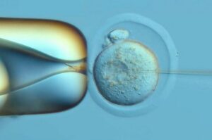
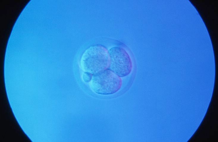
    A mouse embryo at the four cell stage, and polar body. [Clouds Hill Imaging Ltd. / Corbis Documentary / Getty Images]

    Researchers have used two DNA-recording systems to reconstruct cellular family trees across mouse embryonic development. The companion studies—“A DNA typewriter records the cell lineage history of a mouse, from zygote to late organogenesis,” published in Science, and “Comprehensive lineage tracing maps the landscape of cell fate decisions in mouse embryogenesis,” published in Cell—offer high-resolution views of mammalian development, which has been difficult to track because embryos develop inside the uterus and cannot be continuously observed.

    The team from the Science study was led by co-senior authors Jay Shendure, PhD, professor of genome sciences at the University of Washington School of Medicine and a Howard Hughes Medical Institute investigator, and Chengxiang Qiu, PhD, a molecular and systems biologist at Dartmouth College. Their approach centered on DNA Typewriter, a lineage-tracing technology developed by Shendure and Junhong Choi, PhD, that uses a cell’s DNA as a recording medium.

    The researchers redesigned the system’s recording “tape” to make its history easier to recover from individual cells, then inserted it into the genome of a fertilized mouse egg. As cells divided, “a burst of editing unequivocally marks the daughters of the first cleavage, which serve as inline replicates,” the authors wrote in the Science paper. Because daughter cells inherited the marks already written before acquiring new ones, shared patterns revealed common ancestry and the order of developmental branching.

    DNA Typewriter is injected into the genetic material of a fertilized mouse egg. This technology taps into the genetic material in the fertilized egg and resulting embryonic cells as a recording device of the cells' histories. It tracks their relationships as the embryo develops.
    DNA Typewriter is injected into the genetic material of a fertilized mouse egg. This technology taps into the genetic material in the fertilized egg and resulting embryonic cells as a recording device of the cells’ histories. It tracks their relationships as the embryo develops. [Kyle O’Conner/Seattle Hub for Synthetic Biology]

    “Most biological measurements are based on either live imaging, which is limited by the fact that most animal tissues are not transparent, or genomics, which are destructive and only measure a single timepoint,” Shendure said. “Recording techniques like the ones in these studies enable measurements over time including in settings that we can’t directly visualize.”

    The experiment was attempted in 100 fertilized eggs and yielded 10 embryos for examination. One embryo carried the most informative record, allowing the team to trace nearly every profiled cell back to one of the first two cells formed after the fertilized egg divided. Although one founding cell produced more descendants, the two branches generated diverse cell types in nearly equal proportions.

    The ordered recording also allowed the researchers to estimate when cell types diverged from shared developmental paths. Blood and retinal cells committed relatively early, whereas cells forming the skin’s outer layer committed later. The results indicate that cell identity is not fixed in a single coordinated step; instead, different lineages become restricted on distinct schedules.

    Earlier lineage-tracing approaches often relied on DNA-cutting enzymes that could damage cells, erase previous records, exhaust recording capacity, or leave unordered marks whose chronology had to be inferred. “DNA Typewriter avoids all of this,” said co-first author Haedong Kim, PhD, a postdoctoral scientist in genome sciences at UW Medicine. “It writes without fully severing the DNA, keeps recording relatively steadily, and writes everything in strict order, so we can record cell lineages at much higher resolution for a longer time.”

    In the Cell paper, Jonathan Weissman, PhD, and colleagues used a prime-editing system called PEtracer to map lineage dynamics across more than 1.4 million cells from 16 mouse embryos. Together with the DNA Typewriter study in Science, the work demonstrates that mammalian cellular family trees can be reconstructed at unprecedented scale. “It’s really proof of principle that we can do what was done with the roundworm in 1983, but for mammals like you and me,” Weissman said.

    The work provides a framework for investigating how normal organs form and where development can go awry. More complete lineage records could help researchers study congenital malformations, neurodevelopmental and genetic disorders, and cancer, as well as guide stem cell engineering. As Shendure explained, “Recording techniques like the ones in these studies enable measurements over time including in settings that we can’t directly visualize.” Scaling the methods across additional embryos and experimental conditions could support more quantitative—and eventually predictive—models of mammalian development.

  • From Discovery to Development

    From Discovery to Development

    The Era of Modality Convergence: Why Drug Development Is Being Rewritten

    From Discovery to Development: Navigating the New Era of Therapeutic Innovation eBook cover

    The life sciences industry is entering a new era, defined not by a single breakthrough technology but by the convergence of many. Artificial intelligence is accelerating drug discovery, cell and gene therapies are expanding treatment possibilities, and antibody engineering continues to evolve beyond traditional monoclonal antibodies. Meanwhile, mRNA platforms, viral vectors, antibody-drug conjugates (ADCs), multispecific antibodies, and other advanced therapeutic modalities are transforming how developers approach previously intractable diseases. Taken individually, each of these innovations represents a significant advancement. Together, they are reshaping the drug development landscape.

    For decades, therapeutic development followed a familiar path: identify a target, optimize a lead candidate, advance through preclinical studies, and move into clinical evaluation. While the science was challenging, the overall framework remained relatively consistent. Today, that framework is evolving rapidly. Developers are no longer limited to choosing between small molecules and conventional biologics. Instead, they have access to a growing toolkit of therapeutic technologies, each with unique advantages, manufacturing requirements, analytical challenges, and regulatory considerations. At the same time, advances in computational biology and AI-driven discovery are generating more candidates than ever before.

    As a result, the challenge is no longer simply finding promising molecules. Increasingly, success depends on identifying candidates that can be manufactured, scaled, and translated into viable therapies. Decisions once reserved for later stages, including developability, manufacturability, analytical characterization, and clinical strategy, must now be addressed much earlier in the process. Drug development is becoming a continuous, interconnected journey rather than a series of separate steps.

    The boundaries between traditional disciplines are also beginning to blur. Cell therapies may incorporate viral vectors. Gene therapies may leverage lipid nanoparticles. Antibody programs may evolve into multispecific or conjugated formats. The future of drug development is not defined by any single modality, but by the ability to integrate technologies effectively.

    Organizations that combine discovery expertise, modality-specific knowledge, CMC capabilities, and manufacturing execution will be best positioned to navigate this increasingly complex environment. The articles that follow examine key trends driving this transformation, from AI-enabled discovery and experimental validation to advanced cell, gene, and biologic platforms, and explore how the next generation of therapies will move from concept to clinic.