Tag: Bioprocessing

  • ‘Molecular Movie’ of Penicillin Synthesis Could Inform Future Antibiotic Development

    ‘Molecular Movie’ of Penicillin Synthesis Could Inform Future Antibiotic Development

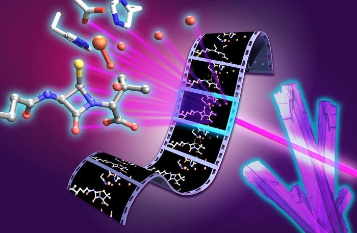
    Molecular movie of penicillin biosynthesis. X-ray free electron laser experiments on isopenicillin N synthase microcrystals capture structural snapshots of key intermediates during the penicillin forming reaction. The film strip illustrates progression through the catalytic cycle, with the monocyclic β-lactam intermediate and reactive high valent ferryl iron species highlighted at the center of the molecular movie. [University of Oxford/Greg Stewart/SLAC National Accelerator Laboratory]

    Researchers from the University of Oxford and international collaborators have revealed previously unseen rapid chemical stages in the formation of β-lactam antibiotics such as penicillin, offering insights that could help to support future antibiotic development.

    Rather than relying on static X-ray crystallographic structures of the enzyme, the Oxford University team, collaborating with partners from Diamond Light Source, Lawrence Berkeley National Laboratory, PAL-XFEL, and SLAC National Accelerator Laboratory, followed the reaction in real time. To do this they used X-ray free-electron lasers (XFEL) to observe the enzyme isopenicillin N synthase, (IPNS) as it converts its linear peptide substrate into the β-lactam ring system of penicillin.

    The study brought together expertise in structural biology, spectroscopy, chemistry, computation and enzyme engineering. The approach, they say, provides a powerful way to study fleeting chemical intermediates that have previously been inaccessible, both in antibiotic biosynthesis and in other biologically important enzyme reactions.

    The team captured several rapid stages in the reaction. These included a thioaldehyde intermediate formed just before the β-lactam ring is created, and a monocyclic β-lactam intermediate, representing the first ring-shaped structure on the way to forming the complete penicillin scaffold. These steps provide the clearest picture yet of how the IPNS enzyme assembles the penicillin scaffold. Subtle movements throughout the enzyme help guide these chemical steps, showing that both the enzyme’s shape and its chemistry work together to control penicillin formation.

    The results show, in unprecedented detail, how the enzyme IPNS achieves an exceptionally complex transformation in a single step. Christopher Schofield, PhD, Professor of Chemistry, University of Oxford, said, “Penicillin has shaped modern medicine, but there is still much to learn about how nature builds this important antibiotic structure. By capturing these fleeting steps, we can better understand how enzymes control complex chemistry with remarkable precision … As rates of antimicrobial resistance continue to rise, understanding this process will ultimately help us make existing antibiotics more efficient and design new antibiotic structures.”

    Allen M Orville, PhD, group leader of the XFEL Hub, further commented, “The XFEL Hub at Diamond brought specialist expertise in time-resolved X-ray methods and XFEL-based structural biology to this study. Working closely with our colleagues in Oxford and at partner facilities, we helped bring together the experimental approaches needed to capture these fleeting stages of enzyme catalysis. The results show the power of combining complementary expertise and X-ray techniques to move beyond static structures and reveal how enzymes work in real time. It is a great example of how time-resolved structural biology can uncover new principles of enzyme function and, ultimately, inform the design of new catalysts and therapeutics.”

    Schofield is co-senior author of the researchers’ published paper in Nature Catalysis, titled “Unanticipated intermediates during isopenicillin N synthase catalysis identified by time-resolved X-ray free-electron laser studies.”

    Ever since penicillin was developed into a working drug at the University of Oxford in the early 1940s, β-lactam antibiotics have been among the most important medicines for treating bacterial infections. Their activity depends on a special ring—the β-lactam ring—a highly strained chemical ring system that interferes with bacterial cell wall synthesis, ultimately causing the cell wall to fail and the bacteria to die. “Isopenicillin N synthase (IPNS) catalyses the biosynthesis of isopenicillin N (IPN), forming the fused four-membered β-lactam and five-membered thiazolidine ring system that is characteristic of the penicillins,” the authors explained.

    Rising rates of antimicrobial resistance are undermining the effectiveness of existing antibiotics. Today, one in six bacterial infections is resistant to antibiotics, the authors noted, citing WHO figures. With too few new antibiotics in development, understanding how nature builds these molecules is an important step towards replenishing the antibiotic pipeline.

    Scientists have been studying how nature constructs this β-lactam ring for decades, but the key fleeting reaction intermediates have been too difficult to observe directly.

    To capture the reaction in real time, the researchers used a system in which thousands of tiny droplets containing anaerobic enzyme microcrystals were deposited onto a moving 2 mm wide tape. As the tape entered an oxygen filled chamber, oxygen rapidly diffused into the crystals and initiated the reaction simultaneously across the sample. By precisely controlling the speed of the tape, the researchers could determine how long each crystal reacted before reaching the X-ray interaction point, where an ultrafast XFEL pulse recorded an atomic resolution snapshot.

    Combining thousands of these snapshots allowed the team to build a frame-by-frame “molecular movie” of penicillin biosynthesis. This method allows individual reaction intermediates that exist only for tiny fractions of a second to be observed at atomic resolution, and under physiological temperature and pressure, before they disappear.

    The newly reported findings help to resolve a long-standing mechanistic question that has remained unanswered for more than four decades. The work also showed that water molecules inside the enzyme play a key role in guiding the reaction. The combined results of their studies, they noted, “… reveal unexpected insight into the IPNS reaction mechanism including the vital roles of active-site water molecules.”

    The study ultimately builds on the work of Nobel prize-winning Oxford chemist Dorothy Hodgkin, PhD, who first solved the structure of penicillin in 1945 using X-ray crystallography.

    Beyond penicillin, IPNS belongs to a large family of iron-dependent oxygenase enzymes involved in human biology, including those that enable us to sense and respond to changes in oxygen availability. The mechanistic insights from this work could therefore have implications beyond antibiotics, offering new principles for enzyme engineering and catalyst design.

    “These findings provide structural insight into IPNS catalysis, inform on the wider Fe(II) oxygenase superfamily and highlight the power of X-ray free-electron laser methods for resolving transient enzymatic intermediates,” the team stated in their paper. First author Patrick Rabe, PhD, Wellcome Career Development Award Investigator, University of Oxford, said, “These insights matter because they can inform how we think about antibiotic biosynthesis and future strategies for developing antibacterial medicines. The ability to capture structural snapshots over milliseconds to seconds of reaction time allows us to connect enzyme motion, iron chemistry and water-mediated proton transfer. This gives us a much richer view of how IPNS controls a difficult chemical transformation. By understanding this process in atomic detail, we can begin to think about engineering these enzymes to produce new or improved antibiotic scaffolds.”

  • From Discovery to Development

    From Discovery to Development

    The Era of Modality Convergence: Why Drug Development Is Being Rewritten

    From Discovery to Development: Navigating the New Era of Therapeutic Innovation eBook cover

    The life sciences industry is entering a new era, defined not by a single breakthrough technology but by the convergence of many. Artificial intelligence is accelerating drug discovery, cell and gene therapies are expanding treatment possibilities, and antibody engineering continues to evolve beyond traditional monoclonal antibodies. Meanwhile, mRNA platforms, viral vectors, antibody-drug conjugates (ADCs), multispecific antibodies, and other advanced therapeutic modalities are transforming how developers approach previously intractable diseases. Taken individually, each of these innovations represents a significant advancement. Together, they are reshaping the drug development landscape.

    For decades, therapeutic development followed a familiar path: identify a target, optimize a lead candidate, advance through preclinical studies, and move into clinical evaluation. While the science was challenging, the overall framework remained relatively consistent. Today, that framework is evolving rapidly. Developers are no longer limited to choosing between small molecules and conventional biologics. Instead, they have access to a growing toolkit of therapeutic technologies, each with unique advantages, manufacturing requirements, analytical challenges, and regulatory considerations. At the same time, advances in computational biology and AI-driven discovery are generating more candidates than ever before.

    As a result, the challenge is no longer simply finding promising molecules. Increasingly, success depends on identifying candidates that can be manufactured, scaled, and translated into viable therapies. Decisions once reserved for later stages, including developability, manufacturability, analytical characterization, and clinical strategy, must now be addressed much earlier in the process. Drug development is becoming a continuous, interconnected journey rather than a series of separate steps.

    The boundaries between traditional disciplines are also beginning to blur. Cell therapies may incorporate viral vectors. Gene therapies may leverage lipid nanoparticles. Antibody programs may evolve into multispecific or conjugated formats. The future of drug development is not defined by any single modality, but by the ability to integrate technologies effectively.

    Organizations that combine discovery expertise, modality-specific knowledge, CMC capabilities, and manufacturing execution will be best positioned to navigate this increasingly complex environment. The articles that follow examine key trends driving this transformation, from AI-enabled discovery and experimental validation to advanced cell, gene, and biologic platforms, and explore how the next generation of therapies will move from concept to clinic.

  • How Preclinical Manufacturing Can De-Risk the Path to GMP-Ready Viral Vector Programs

    How Preclinical Manufacturing Can De-Risk the Path to GMP-Ready Viral Vector Programs

    To move viral vector programs from early research toward the clinic, sponsors have to generate the right material and data during development. Non-GMP production of adeno-associated virus (AAV), lentiviral, and adenoviral vectors can support applications ranging from R&D and animal studies to assay development, toxicology planning, and early manufacturability assessments. But to be successful, sponsors have to align production scale, analytical testing, documentation, and material use with the specific questions their studies aim to answer.

    In this GEN webinar, Chris Nieder, an expert in viral vector programs, will examine how strategically designed preclinical manufacturing campaigns provide a practical foundation for GMP production. He will discuss topics such as how to select from among shake flasks, development-scale bioreactors, and pilot-scale approaches; and how to use early process and analytical data to identify yield and scalability challenges. Attendees will also learn how early CDMO engagement can clarify manufacturing scope, timelines, costs, and GMP-readiness without compromising expectations for non-GMP material. Key takeaways from the webinar include:

    • How early-stage sponsors can use non-GMP AAV, lentiviral, and adenoviral production to support R&D, animal studies, toxicology planning, assay development, and manufacturability assessment
    • How process and analytical data generated before GMP can reduce duplicated prep work, lower technical risk, and streamline later scope definition
    • When shake flasks, development-scale bioreactors, or pilot-scale production can provide the most useful insight for the next development step
    • How to align material use, analytical testing, documentation, and expectations without over-positioning preclinical material as clinical-grade supply

    A live Q&A session will follow the presentation offering you a chance to pose questions to our expert panelist.

    Produced with support from:

    skpharmteco logo

  • ARPA-H’s BoSS Program Funds Three Teams Tackling Cold Chain Challenges for Cell Therapies

    ARPA-H’s BoSS Program Funds Three Teams Tackling Cold Chain Challenges for Cell Therapies

    The Advanced Research Projects Agency for Health, (ARPA-H), an agency within the United States’ Department of Health and Human Services (HHS), recently announced awards and teams involved in its BioStabilization Systems (BoSS) program, an initiative that aims to develop new methods for producing, storing, and transporting cell-based biologic medicines at room temperature. ARPA-H has committed to provide up to $87 million across the four-year program. This round of funding will support Phase I of the program.

    The teams have been tasked with pursuing new technologies that confer long-term, shelf-life stability to cell-based therapies and biologics making it possible to store and ship them without refrigeration. To accomplish this goal, BoSS teams will work across two technical areas. They will work on interventions to stabilize cells for storage and then reactivate them on demand as well as on creating novel processing systems that ensure these interventions are efficient and scalable for manufacturing. 

    This week, members of the Reversible Ambient Temperature (RAB) team said that they have been awarded up to $7.3 million initially from ARPA-H. The RAB team includes representatives from Draper, a non-profit research, development, and manufacturing company, which serves as the administrative lead,and Likarda, a biotechnology company that develops hydrogel technologies for delivering and preserving cell therapies and biologics, which serves as the technical lead for the team. Other members of the team include scientists from CaseBioscience, CPSI Biotech, Fresenius Kabi, MiNK Therapeutics, and University of Colorado. 

    For their part, this team will use AI-assisted design of experiments to optimize formulation, quiescence induction parameters, hydrogel composition, and recovery media as well as freeze-assisted droplet drying to achieve a stable product. Specifically, the RAB team’s biostabilization strategy, led by Likarda, will focus on simultaneous control of cell state, physical environment, and the cellular stress responses activated during freezing, dehydration, storage, and recovery. Meanwhile, the bioprocessing engineering team, led by Draper, will convert the manual steps of biostabilization into an automated, commercially viable workflow and will develop custom instruments to scale up the processes.

    Likarda will contribute its proprietary Core-Shell Spherification hydrogel technology, which is designed to encapsulate and protect living cells while maintaining viability and function. CaseBioscience will provide expertise in clinical media formulation, biopreservation, and cellular metabolism to the development and evaluation of novel approaches for long-term ambient stabilization of therapeutic cells.

    “The RAB team brings together deep expertise in cryoprotection, encapsulation, and cell state, with a focus on protecting cell viability throughout the stabilization and reanimation process, and the team has direct experience bringing products to market,” said Jason Fiering, distinguished technical staff at Draper. “Working in parallel, our interdisciplinary engineering team will develop the customized instruments needed to transfer these processes to clinical manufacturing environments.” 

    In his comments, John Baust, PhD, an expert in biopreservation and collaborator on the project with CPSI Biotech noted that “the concept of preserving living cells in a dry state has been pursued for more than two decades, but significant scientific and engineering barriers have yielded limited success.” The BoSS program is an opportunity “to tackle this from multiple scientific and engineering directions simultaneously” that if successful “could represent a paradigm shift in how biologics are preserved, stored, and distributed.”

    Other teams announced by ARPA-H include one led by DesiCorp. This team includes members from the University of Louisville, University of Texas at Austin, Via Therapeutics, VeriSIM Life, and Ossium Health. They will leverage high-throughput screening to identify biocompatible formulations that slow cellular metabolism and AI-assisted process optimization to develop a thin-film freeze-drying (TFFD) system that stabilizes cells at room temperature. 

    DesiCorp will lead integration of the overall workflow by using a high-throughput screening platform to rapidly identify biocompatible formulations that enable cells to survive TFFD and resume function after rehydration. Meanwhile, UT Austin researchers will help optimize the TFFD critical process parameters and composition/material attributes, while also contributing expertise in aseptic processing and scaling up the technology. 

    A third team is led by the University of California, Davis and includes scientists from Mayo Clinic, Case Western Reserve University, University of Georgia, and Sersense. The team will induce a low metabolic state in cells using light-responsive gels and microfluidic processing techniques to produce cells that are stable at room temperature. 

    Recently, that team announced that it had been awarded up to $6 million, initially,  from ARPA-H to fund its project dubbed CYBORGEL. Their efforts will leverage research done in the laboratory of Cheemeng Tan, PhD, a professor of biomedical engineering at UC Davis. Previously, he and his collaborators formed water-rich polymer networks inside living cells to create cell-material hybrids. This process, called intracellular gelation, forms a gel inside the cell and is capable of halting cell division while preserving cellular function. Tan and his team dubbed this cellular product a “cyborg” since the gelation process combines living cells with synthetic materials. 

    Through CYBORGEL, the collaborators will investigate whether this intracellular gelation process can be extended to reduce the cost and simplify the storage, transportation and delivery of cellular therapies. The researchers will also test whether CYBORGEL cells remain therapeutically useful after being returned to their original state. For instance, one study will test the effectiveness of reanimated CAR T cells for existing clinical trials on cancer immunotherapies. Another will test it on placental mesenchymal stem cells used to treat spina bifida, a medical intervention being developed by UC Davis researchers. 

    As part of the initiative, the American Type Culture Collection (ATCC) has been tapped to serve as the IV&V partner. ATCC will provide working cell banks of government-selected cell types that will be used for demonstrations. They will also provide unbiased assessments of both the viability and function of the preserved products and the design features of the preservation systems.  

    The first phase of the BoSS program kicked off in September and is expected to run for 15 months. Over the four-year multi-phase program, the teams’ approaches will be evaluated against increasingly stringent benchmarks for cell viability, production speed, and shelf-life stability. If successful, work under the BoSS program could enable lower costs for cell-based drugs, reduce the risk of therapeutic and pharmaceutical product losses, and enable domestic reserves for public health preparedness.