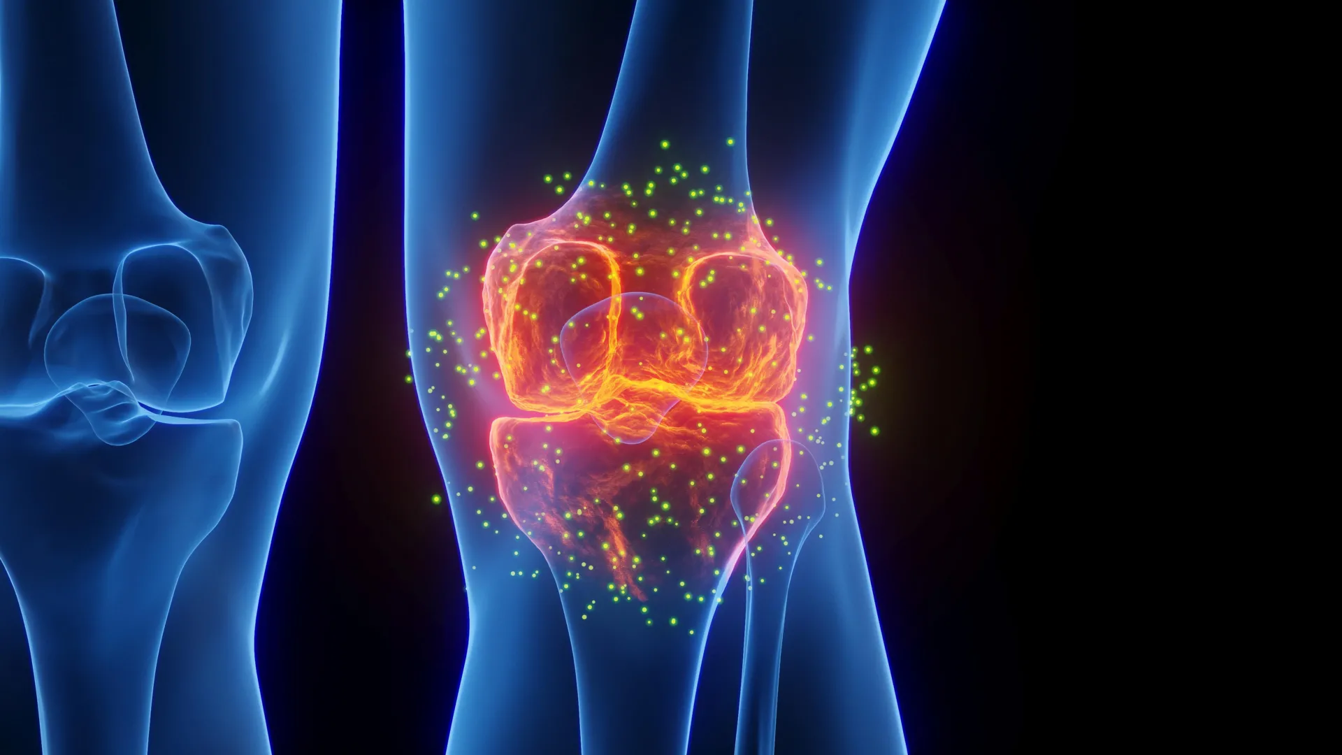
For millions of people with osteoarthritis, pain and stiffness can make everyday activities increasingly difficult. Common treatments, including over-the-counter medications and steroid injections, may reduce symptoms for a time, but they do not prevent the underlying joint damage from progressing.
New research from Yale suggests a different approach. In a study published in Bioactive Materials, scientists found that the medication lacosamide can serve two purposes in osteoarthritis, reducing joint pain while also reversing cartilage damage. The effects were particularly strong when the drug was delivered directly into the joint using a specialized hydrogel.
Why osteoarthritis damages cartilage
Osteoarthritis is often called a condition caused by wear and tear, but that description leaves out much of the biology involved.
Within a healthy joint, cells known as chondrocytes help maintain cartilage by continuously balancing the creation of new tissue with the removal of old material. Osteoarthritis disrupts that balance. Cartilage begins to break down faster than it can be replaced, eventually allowing bones to rub against one another. As the disease advances, some patients may ultimately require joint reconstruction procedures such as a total knee replacement.
“There is a major unmet need in osteoarthritis,” says the study’s principal investigator and Charles W. Ohse Professor of Orthopaedics & Rehabilitation, Chuan-Ju Liu, PhD. “We need therapies that don’t just mask pain but actually change how the disease progresses.”
At present, no medicine approved by the U.S. Food and Drug Administration can both stop osteoarthritis pain and prevent the structural breakdown of cartilage. The work led by Liu suggests that it may be possible to address both problems with a single therapeutic strategy.
The researchers repurposed an existing medication and paired it with an advanced gel designed to keep the drug inside the joint. The approach could potentially preserve joint tissue while delivering sustained pain relief without relying on addictive opioids.
A protein linked to pain and cartilage loss
The research focuses on Nav1.7, a protein that functions as a sodium channel. These channels act as microscopic gates in cell membranes and play an important role in electrical signaling.
Nav1.7 was long thought to operate primarily in specialized nerve cells that transmit pain signals to the brain. More recent work from Liu and his team, however, revealed that the protein is also highly active in chondrocytes, the cells responsible for maintaining cartilage.
Nav1.7 is relatively quiet in healthy joints. In osteoarthritis, however, its activity increases substantially. The researchers found that this heightened activity can intensify pain signaling while also pushing chondrocytes toward breaking down the cartilage they would normally help preserve.
That makes Nav1.7 an unusual therapeutic target because it appears to influence both the sensation of pain and the physical deterioration of joint tissue.
“When Nav1.7 becomes dysregulated, it contributes to both joint degeneration and pain,” Liu says. “Our findings suggest that Nav1.7 is a dual-acting target. By blocking this single protein, we can potentially quiet the pain nerves and tell the cartilage cells to not only stop breaking down but start repairing as well.”
Epilepsy drug shows potential for cartilage repair
Instead of creating an entirely new medication, Liu’s team tested drugs that already inhibit sodium channels. Among them, lacosamide produced strong biological effects at considerably lower concentrations and offered a better safety profile than older drugs in the same class.
Lacosamide is already used as a treatment for epilepsy, but the researchers found that its effects on cartilage depended heavily on the dose.
More was not necessarily better. At an ideal low concentration, lacosamide encouraged cells to produce proteins involved in building cartilage while also suppressing processes that break tissue down. When the concentration was either too high or too low, those benefits began to fade.
“This tells us the system is finely tuned,” Liu notes. “There is an optimal range where the drug helps restore balance without overcorrecting. What stood out was not just its effectiveness, but how little of a dose was needed.”
Further investigation revealed that lacosamide also changes the way cells communicate. The drug stimulated the release of two beneficial signaling proteins, HSP70 and midkine.
HSP70 helps cells respond to stress and supports tissue repair, while midkine helps regulate inflammation and protect joint tissue from degeneration. Together, the proteins appear to create conditions that are more favorable for maintaining cartilage.
“These proteins create a supportive environment for cartilage maintenance,” Liu explains. “They allow the effects of the drug to extend beyond individual cells and influence the entire tissue.”
Smart hydrogel keeps the drug inside the joint
Oral treatment with lacosamide was effective in preclinical testing, but drugs taken by mouth circulate throughout the body, increasing the potential for unwanted effects elsewhere.
The researchers therefore investigated intra-articular injection, which allows the medication to be placed directly inside the affected joint.
There was still a significant problem to overcome. “The knee joint, which is also the most common location for osteoarthritis, naturally acts like a leaky bucket,” Liu says. “The body’s drainage system can clear out liquids injected into the knee within hours.”
To keep the medication in place longer, the team created a specialized hydrogel made from Collagen II. The material responds to temperature. It remains liquid while inside a cool syringe, then becomes a firm, jelly-like material after reaching body temperature.
Once inside the joint, the gel acts as a storage site for lacosamide, keeping the medication concentrated in the affected area while gradually releasing it over several weeks.
“The hydrogel acts as a local reservoir,” Liu says. “It holds the drug in place in the location it is needed most and releases it slowly over time. It transforms a daily pill into a long-lasting, local treatment that stays active for a month or longer.”
In the same preclinical studies, one injection of the lacosamide-containing gel every four weeks prevented cartilage loss more effectively than taking an oral dose every day.
Existing drug could speed clinical testing
One important advantage of lacosamide is that the medication is already approved for use in people. That could make it possible to move toward clinical studies in osteoarthritis patients more quickly than would be possible with a completely new drug.
Lacosamide has also been tested in humans with certain nerve-related pain conditions caused by Nav1.7 mutations. Those results give the researchers greater confidence that the effects seen in laboratory studies could eventually translate into meaningful pain relief for patients.
The work also reflects a broader direction in medicine that combines drugs with advanced biomaterials to control exactly where and how treatments are delivered. If the approach proves successful in people, it could eventually reduce the number of procedures patients need, limit unwanted side effects, and provide longer-lasting protection against structural joint damage.
“We are not just developing a treatment,” Liu concludes. “We are developing a system that allows the medicine to work more effectively where it matters most. Our goal is to move beyond symptom control and towards true disease modification. This effort brings us closer to that reality.”