Category: Biotechnology News

  • X-Chromosome Gene Linked to Increased Autism Diagnosis in Males

    X-Chromosome Gene Linked to Increased Autism Diagnosis in Males

    Researchers at Baylor College of Medicine, the Duncan Neurological Research Institute (Duncan NRI) at Texas Children’s Hospital, and collaborating institutions have identified new genetic candidates that help explain why autism is diagnosed about four times more often in boys than it is in girls. The study, involving tests in induced pluripotent stem cell (iPSC)-derived neurons (iNeurons), revealed that small changes in the gene MECP2 on the X chromosome might help explain part of this difference.

    Huda Zoghbi, MD, Distinguished Service Professor at Baylor, director of Duncan NRI, and a Howard Hughes Medical Institute investigator, is senior and corresponding author of the team’s published paper in The American Journal of Human Genetics, titled “A massively parallel reporter assay of MECP2 cis-regulatory elements reveals genetic candidates for male-biased autism.” In their paper, the team concluded, “These findings suggest noncoding MECP2 variants can cause idiopathic, male-biased autism and provide a framework for uncovering regulatory variants in other X chromosome neurodevelopmental genes that may contribute to autism’s missing heritability.”

    “Autism is a common, heritable neurodevelopmental trait that affects how a person communicates, interacts with others, and experiences the world, and is characterized by altered social interactions and restricted, repetitive behaviors or interests,” Zoghbi explained. “It is diagnosed about four times more in male individuals than in female individuals, suggesting sex-specific factors may increase male vulnerability,” the researchers added in their paper.

    In the current study, the researchers drew from their long experience with gene MECP2, which is on the X chromosome and has been associated with neurodevelopmental conditions, including autism. “We tested the idea that the predominance of autism in males could be explained in part by mild mutations in an X chromosome gene’s regulatory regions,” commented Rebecca Meyer-Schuman, PhD, a postdoctoral associate in the Zoghbi lab. “Females might be protected from such a mild mutation because of their second X chromosome, but males, who have only one copy, would be more vulnerable. The regulatory regions are like dials that control whether a gene will be expressed and by how much. And what better place to start looking at these regulatory regions than the gene MECP2, where brain health depends on how much of the gene is expressed, and where mutations can cause neurodevelopmental disorders that include autism.”

    Previous murine studies by Zoghbi lab and others have taught researchers that MECP2 produces a Goldilocks protein, meaning that it must maintain a “just right” concentration or activity level for a healthy brain. Having too little—about 50% or less of the normal levels—leads to Rett syndrome, and having twice the normal levels causes a different neurodevelopmental condition called MECP2 duplication syndrome. “Both loss-of-function and gain-of-function variants lead to severe neurodevelopmental conditions—Rett syndrome (MIM: 312750) and MECP2 duplication syndrome (MDS; MIM: 300260[intellectual developmental disorder, X-linked, syndromic, Lubs type (MRXSL)]), respectively,” the authors commented. “Because MECP2 is on the X chromosome, hemizygous male individuals have a more severe phenotype than heterozygous female carriers with the same variant.”

    Meyer-Schuman also noted, “Previous studies in mice also showed us that milder changes in MECP2 levels, for instance reducing the expression by 30% or increasing it by 50%, do not cause the more severe features of Rett syndrome or MECP2 duplication syndrome like seizures or motor problems, but lead to autism-like behaviors.”

    For their current study the team used a massively parallel reporter assay (MPRA) to screen the regulatory regions of the MECP2 gene in induced pluripotent stem-cell (iPC)-derived neurons. “This allowed us to identify and manipulate the regulatory regions and determine what kinds of changes would alter their function,” Meyer-Schuman said. The researchers also screened MECP2 regulatory regions in autistic individuals and tested the genetic variants they identified using the MPRA assays.

    “We identified two regulatory regions where male autistic individuals inherited a variant from their unaffected mother that altered regulatory activity,” Meyer-Schuman said. “One of these variants reduced MECP2 expression by approximately 30% in human neurons, a magnitude that produces social deficits, hyperactivity and anxiety-like characteristics in mice. As predicted by the mouse model, this individual was diagnosed with autism and ADHD but had none of the defining characteristics of Rett syndrome.”

    In their paper, the authors commented that their work supports a broader hypothesis that altered dosage or expression of sex chromosome genes are a major source of the male bias observed in autism. Zoghbi added, “These findings, a result of integrating human and animal studies, suggest that MECP2 regulatory variants can contribute to male-biased autism and reveal just the tip of the iceberg, providing a framework for uncovering regulatory variants in other X chromosome neurodevelopmental genes that may contribute to autism’s missing heritability.”

  • Enodia Therapeutics Inks Pfizer Deal to Develop Sec61-Targeting Small Molecules

    Enodia Therapeutics Inks Pfizer Deal to Develop Sec61-Targeting Small Molecules

    Biotechnology firm Enodia Therapeutics has announced a research collaboration and licensing agreement with pharma giant Pfizer to discover and develop small molecule inhibitors of Sec61, using Enodia’s proprietary platform. 

    Under the terms of the agreement, Enodia will perform discovery activities to screen, optimize, and develop Sec61 inhibitor candidates against multiple collaboration targets. Following the collaboration term, Pfizer will be responsible for all clinical, manufacturing, regulatory, and commercial activities. Furthermore, Enodia will receive an upfront payment and is eligible for future research, development, and sales milestone payments, as well as tiered royalties on net sales, bringing the total potential value to more than $500 million.

    Enodia develops novel small-molecule therapies for targeted protein degradation using its platform, which is designed to identify small molecules that enable selective Sec61 translocon modulation, a novel approach to inhibit disease-driving proteins when they are synthesized and before pathological effects occur. To date, the company has built a broad chemical space that spans multiple Sec61 inhibitor families and is supported by biological datasets including proteomics and cryo-EM data as well as tailored cell-line libraries. 

    “Enodia has built a broad platform that enables selective Sec61 translocon modulation, a novel approach to inhibit disease-driving proteins while preserving essential physiological functions,” said Yves Ribeill, PhD, Enodia’s CEO. “We look forward to collaborating with Pfizer, combining their deep expertise and clinical resources with our approach for precisely targeting pathological proteins to bring forth potential new medicines.”

    Earlier this year, Enodia announced that it had purchased assets from Kezar Life Sciences’ Sec61-based discovery and development program. At the time, Enodia said that the acquisition helped advance its understanding of Sec61 selectivity mechanisms. The company also said that insights from Kezar’s programs would further strengthen Enodia’s core focus on Sec61-driven selectivity for targeted protein degradation. Under the terms of the purchase agreement, Kezar received an initial upfront payment totaling $1 million, and was expected to receive future payments upon achievement of certain development, regulatory and commercialization milestones. 

  • The Top 10 Questions Shaping the Future of Optogenetic Biomanufacturing

    The Top 10 Questions Shaping the Future of Optogenetic Biomanufacturing

    Maximilian Hoerner
    Maximilian Hoerner, PhD

    The emergence of increasingly complex biologics such as multi‑specific antibodies, cytokines, fusion proteins, and other structurally demanding modalities has exposed the limits of conventional bioprocessing. Many of these molecules are difficult to express at commercially viable yields, and their quality attributes can be highly sensitive to the cellular environment. As a result, productivity, consistency, and cost remain major constraints across the industry.

    Traditional cell line development (CLD) relies on constitutive promoters that drive static expression throughout development and production. Because production is always on, cells must divide and manufacture protein simultaneously, even when the product is burdensome or toxic. While this model worked relatively well for easy-to-express monoclonal antibodies, it does not work well for next-generation biologics, where the inherent trade-off between higher specific productivity and rapid cell growth, both competing for the same cellular resources, is increased. Light‑controlled gene expression using optogenetics is emerging as a credible alternative.

    This article explores the ten most important questions facing optogenetic biomanufacturing as it moves from proof‑of‑concept demonstrations toward mainstream adoption in modern bioprocessing.

    1. What is optogenetics, and why is it relevant to biologics manufacturing?

    Optogenetics uses light‑responsive proteins to control specific cellular functions. In biomanufacturing, the key application is regulating gene expression with light instead of relying on constitutive promoters or chemical inducers. When light‑controlled gene expression systems are engineered into production cell lines, illumination at defined wavelengths can switch expression on, off, or to intermediate levels.

    This is important because it introduces a fundamentally different control modality. Conventional systems provide static expression, whereas optogenetics enables dynamic, reversible, and tunable control. As biologics become more complex and more sensitive to expression conditions, having a way to precisely modulate transcription directly, rather than perturbing the entire cell by modulating process parameters such as temperature, will become increasingly valuable.

    2. What core limitation in biologics manufacturing does light‑based control address?

    The main limitation is the inherent competition between cell growth and protein production. In constitutive systems, cells must divide and manufacture protein at the same time, drawing on the same finite pool of metabolic resources. This creates a structural trade‑off: clones that produce strongly have a slow doubling time and/or do not grow to viable densities, while clones that grow well often produce only modest titers. This trade‑off becomes much more pronounced for challenging molecules such as multi‑specific antibodies, cytokines, and fusion proteins that are aggregation‑prone or place a significant burden on the host cell.

    Optogenetics addresses this by separating the growth and production phases. Cells can grow in an uninduced, low-burden state and then transition into a productive state upon illumination. As light acts directly on transcriptional regulators in a dose-dependent manner, expression can be tuned precisely rather than modulated indirectly through temperature shifts or coarsely and irreversibly by chemical induction. Chemical inducers can also negatively affect cell performance through off-target effects and are difficult to remove during downstream processing, which has limited their adoption in mammalian biomanufacturing. The direct, tunable control by light also enables more deliberate modulation of pathways that influence critical quality attributes such as post-translational modifications (PTMs), which is increasingly important for complex biologics and biosimilars.

    3. How does optogenetic light exposure work, and is it safe for cells and media?

    Optogenetics uses light as an informational cue rather than an energy source. Because photoreceptors respond to brief, low-intensity pulses and stay “on” for a tunable period of time before reverting to the off state, optogenetic systems can maintain precise control without continuous or high-power illumination.

    The wavelengths used in bioprocessing are in the visible range and non-ionizing, operating well under established phototoxicity thresholds. At these levels, light does not harm cells or DNA, nor does it disrupt cellular physiology. Although certain media components are known to be light-sensitive, the illumination used for optogenetic induction has not shown detrimental effects on cellular performance.

    4. What is the impact of optogenetics on lab operations?

    Optogenetic workflows use standard lab equipment with the addition of illumination devices compatible with existing infrastructure, such as plates, shake flasks, or bioreactors. The main operational difference occurs during pool generation and single-cell cloning, where cells must remain uninduced. This is achieved by working under “safe light” conditions, visible wavelengths that do not activate the specific optogenetic system but allow the operator to handle the cell culture. Only small areas of the lab need to be adapted for this purpose. For all other operations, including biomanufacturing, local interventions to shield cells from room light are typically sufficient.

    5. How many cellular functions can be controlled with light in a single cell line?

    Current systems can independently regulate cellular functions using two or three different wavelengths. For example, one color may control production while another modulates a critical quality‑related pathway. Functions that require coordinated timing can be linked to the same wavelength. As genetic circuit design advances, the number of independently controllable functions is expected to increase.

    6. When might optogenetically manufactured biologics reach clinical and commercial stages?

    Optogenetic cell line development is already being explored for multiple programs. Regulatory agencies have established pathways for evaluating emerging bioprocess technologies, and optogenetic systems are beginning to move through these mechanisms. If early development proceeds as expected, optogenetically controlled processes could support clinical filings in the next two years, with commercial adoption following thereafter.

    7. Can optogenetics be integrated into existing facilities?

    Yes. Optogenetic manufacturing does not require new bioreactor designs; illumination hardware can be added to standard single‑use or stainless‑steel systems. For single‑use bags, light is delivered externally through the bag without breaching the sterility barrier; for stainless‑steel tanks, cultures can be circulated through illuminated loops. No cleanroom redesign is required.

    8. How does optogenetic manufacturing scale to larger bioreactors?

    Scaling optogenetic systems depends on three factors: the sensitivity of the photoreceptors, the lifetime of their activated state in the dark, and the mixing and light-scattering characteristics of the bioreactor. Modern optogenetic systems for biomanufacturing are designed to respond to very low light intensities, and once activated, they gradually return to the off state in the dark at a characteristic rate, typically over tens of minutes.

    Since stirred-tank bioreactors mix continuously and scatter light throughout the vessel, cells receive sufficient illumination over the photoreceptor’s characteristic activation lifetime even at large volumes. Simulation studies and early pilot-scale experiments up to the 200 L scale suggest that optogenetic activation can be achieved at commercially relevant scales, including stainless steel and single-use systems.

    9. What has convinced established players to evaluate this technology?

    Biologics development and manufacturing are intentionally conservative, with new technologies adopted only when they demonstrate clear, reproducible benefits and maintain patient safety. For any emerging bioprocess platform, the threshold for evaluation is data showing that it can deliver meaningful improvements without introducing new risks or operational complexity.

    In comparative studies, optogenetic induction has produced more than two‑fold increases in titer for standard monoclonal antibodies as well as for more difficult‑to‑express therapeutic proteins. These gains have been achieved while maintaining or improving critical quality attributes, including those influenced by folding, assembly, and post‑translational modification.

    As the next generation of therapeutics become increasingly complex, leading biopharma and CDMO players see the need for novel biomanufacturing platforms capable of meeting that complexity without compromising titers, and for that reason several key players are actively evaluating optogenetic technology for potential integration.

    10. Where is optogenetic biomanufacturing headed in the next decade?

    If current trends continue, optogenetic control may become a standard tool for managing expression, quality, and other cellular functions in biologics manufacturing. The ability to modulate gene expression dynamically opens possibilities for more efficient production of complex therapeutics, more predictable quality profiles, and more flexible facility design. As the technology matures, it is likely to be adopted across CDMOs, biotech, and biopharma companies seeking greater control over their manufacturing processes.

    Maximilian Hoerner, PhD, is the head of optogenetics at Prolific Machines

  • UCB Dedicates Future U.S. Biologics Manufacturing Campus in Georgia

    UCB Dedicates Future U.S. Biologics Manufacturing Campus in Georgia

    UCB has dedicated the Rowen, GA, site where it plans to build a $2 billion biologics manufacturing campus that is set to open as the Belgian-based drug developer’s first U.S. manufacturing site.

    When completed in 2032 or 2033, the campus is expected to support commercial production of recently approved and future biologic medicines, applying artificial intelligence, automation, and advanced manufacturing technologies. The campus is intended to further integrate UCB’s U.S. patient value chain from research and development through manufacturing and delivery.

    Some 330 permanent jobs are expected to be based at the approximately 460,000-square-foot campus, which will occupy 79 acres within the Rowen innovation district in Georgia’s Gwinnett County. UCB bought the 79 acres from the Rowen Foundation, which is developing a 1,900-acre campus on Highway 316, launched in 2020 as a “knowledge” community for biopharma and other innovation-based fields.

    In its announcement of the campus site dedication, UCB said it chose to build its first U.S. manufacturing campus in Georgia because of what it called the Peach State’s strong life sciences ecosystem, highly skilled workforce, robust infrastructure, and access to leading academic and research institutions.

    Another factor in the decision: Gwinnett County has committed $174 million in incentives and infrastructure investments to support UCB’s project.

    UCB’s project is the brightest recent success of Georgia life sciences leaders, who worked through industry group Georgia Life Sciences to lay out a 10-year “Roadmap” last year with the aim of “positioning Georgia as the scale-up manufacturing hub of the Southeast.” The UCB project, announced in March, helped place Georgia among the “Next 5 States” within GEN’s Next 10 U.S. Biopharma Clusters A-List.

    “Long-term commitment”

    “Our manufacturing investment in the United States reflects our long-term commitment to patients, strengthening our ability to serve them while contributing to scientific innovation, highly skilled jobs, and economic growth in Georgia,” UCB CEO Jean-Christophe Tellier said in a statement.

    UCB executives and dignitaries cut a ceremonial ribbon to mark the dedication of UCB’s future biologics manufacturing campus in Rowen, GA. Jeffrey Kessler, U.S. Department of Commerce Under Secretary for Industry and Security, stands at left. At the center are Georgia Gov. Brian Kemp (R); UCB CEO Jean-Christophe Tellier; Katherine Raeymaekers, Consul General of Belgium in Atlanta; and Nicole Love Hendrickson (D), Chairwoman of the Gwinnett County Board of Commissioners. [UCB]

    Tellier and other UCB leaders were joined at a dedication ceremony by dignitaries that included Georgia Gov. Brian Kemp (R); Jeffrey Kessler, U.S. Department of Commerce Under Secretary for Industry and Security; Nicole Love Hendrickson (D), Chairwoman of the Gwinnett County Board of Commissioners; and Katherine Raeymaekers, Consul General of Belgium in Atlanta.

    During the ceremony, the dignitaries joined top UCB executives in unveiling the Emmanuel Janssen Circle, a commemorative stonework installation just completed within the roundabout to the entrance of the future campus. Janssen (1879-1955) was a Belgian businessman who founded Union Chimique Belge (UCB) on January 18, 1928. The company initially focused on industrial chemicals before evolving into a developer of biologics for indications in neurology and immunology.

    “UCB’s new biomanufacturing campus builds on the entrepreneurial legacy of Baron Emmanuel Janssen, founder of UCB, while strengthening the transatlantic collaboration that drives economic growth, scientific innovation, and improving people’s lives,” Raeymaekers stated. “Today’s site dedication reflects the strength of the partnership between Belgium, the United States, and the State of Georgia—and our shared commitment to enduring progress, innovation, and opportunity.”

    UCB worked with numerous public and private partners to locate its biologics manufacturing campus in Rowen—including the Metro Atlanta Chamber; Georgia Quick Start, which provides customized workforce training through the Technical College System of Georgia; Gwinnett County Government; Georgia Power Company; the public-private economic development initiative Partnership Gwinnett; and the Georgia Department of Economic Development.

    Headquartered in Brussels, UCB employs more than 10,000 people in nearly 40 countries—including the United States, where the company employs more than 2,000 people in research, development, manufacturing, commercial operations, and corporate functions.

    UCB has maintained a U.S. presence for nearly a century. The company now employs more than 400 people in Georgia, where it established its Stateside headquarters in Atlanta in 1993.

    More than 1,000 construction jobs are expected to be created in connection with the campus during the six- to seven-year projected timeframe for design and construction.

    “At UCB, our purpose is to create value for people living with severe diseases, and that purpose guides the science we pursue, where we invest, and the choices we make for the long term,” Tellier added.

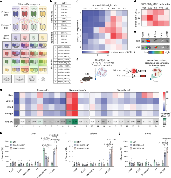
  • In vivo engineering of CAR natural killer cells for immunotherapy using ligand-directed lipid nanoparticles

    In vivo engineering of CAR natural killer cells for immunotherapy using ligand-directed lipid nanoparticles

    Data availability

    All data that support the findings of this study are provided within the paper and its Supplementary Information.

    Code availability

    References

    1. Irvine, D. J., Maus, M. V., Mooney, D. J. & Wong, W. W. The future of engineered immune cell therapies. Science 378, 853–858 (2022).

      Article  CAS  PubMed  PubMed Central  Google Scholar 

    2. Li, Y. R., Zhu, Y., Halladay, T. & Yang, L. In vivo CAR engineering for immunotherapy. Nat. Rev. Immunol. 25, 725–744 (2025).

      Article  CAS  PubMed  Google Scholar 

    3. June, C. H., O’Connor, R. S., Kawalekar, O. U., Ghassemi, S. & Milone, M. C. CAR T cell immunotherapy for human cancer. Science 359, 1361–1365 (2018).

      Article  CAS  PubMed  Google Scholar 

    4. Maude, S. L. et al. Chimeric antigen receptor T cells for sustained remissions in leukemia. N. Engl. J. Med. 371, 1507–1517 (2014).

      Article  PubMed  PubMed Central  Google Scholar 

    5. Wang, V., Gauthier, M., Decot, V., Reppel, L. & Bensoussan, D. Systematic review on CAR-T cell clinical trials up to 2022: academic center input. Cancers (Basel) 15, 1003 (2023).

      Article  CAS  PubMed  PubMed Central  Google Scholar 

    6. Brudno, J. N., Maus, M. V. & Hinrichs, C. S. CAR T cells and T-cell therapies for cancer: a translational science review. JAMA 332, 1924–1935 (2024).

      Article  CAS  PubMed  PubMed Central  Google Scholar 

    7. Biederstadt, A. & Rezvani, K. Engineered natural killer cells for cancer therapy. Cancer Cell 43, 1987–2013 (2025).

      Article  CAS  PubMed  PubMed Central  Google Scholar 

    8. Liu, E. et al. Use of CAR-transduced natural killer cells in CD19-positive lymphoid tumors. N. Engl. J. Med. 382, 545–553 (2020).

      Article  CAS  PubMed  PubMed Central  Google Scholar 

    9. Marin, D. et al. Safety, efficacy and determinants of response of allogeneic CD19-specific CAR-NK cells in CD19+ B cell tumors: a phase 1/2 trial. Nat. Med. 30, 772–784 (2024).

      Article  CAS  PubMed  PubMed Central  Google Scholar 

    10. Ghobadi, A. et al. Induced pluripotent stem-cell-derived CD19-directed chimeric antigen receptor natural killer cells in B-cell lymphoma: a phase 1, first-in-human trial. Lancet 405, 127–136 (2025).

      Article  CAS  PubMed  PubMed Central  Google Scholar 

    11. Sterner, R. C. & Sterner, R. M. CAR-T cell therapy: current limitations and potential strategies. Blood Cancer J. 11, 69 (2021).

      Article  PubMed  PubMed Central  Google Scholar 

    12. Laskowski, T. J., Biederstadt, A. & Rezvani, K. Natural killer cells in antitumour adoptive cell immunotherapy. Nat. Rev. Cancer 22, 557–575 (2022).

      Article  CAS  PubMed  PubMed Central  Google Scholar 

    13. Wolf, N. K., Kissiov, D. U. & Raulet, D. H. Roles of natural killer cells in immunity to cancer, and applications to immunotherapy. Nat. Rev. Immunol. 23, 90–105 (2023).

      Article  CAS  PubMed  Google Scholar 

    14. Schett, G., Mackensen, A. & Mougiakakos, D. CAR T-cell therapy in autoimmune diseases. Lancet 402, 2034–2044 (2023).

      Article  CAS  PubMed  Google Scholar 

    15. Blache, U., Popp, G., Dunkel, A., Koehl, U. & Fricke, S. Potential solutions for manufacture of CAR T cells in cancer immunotherapy. Nat. Commun. 13, 5225 (2022).

      Article  CAS  PubMed  PubMed Central  Google Scholar 

    16. Cliff, E. R. S. et al. High cost of chimeric antigen receptor T-cells: challenges and solutions. Am. Soc. Clin. Oncol. Educ. Book 43, e397912 (2023).

      Article  PubMed  Google Scholar 

    17. Rurik, J. G. et al. CAR T cells produced in vivo to treat cardiac injury. Science 375, 91–96 (2022).

      Article  CAS  PubMed  PubMed Central  Google Scholar 

    18. Billingsley, M. M. et al. In vivo mRNA CAR T cell engineering via targeted ionizable lipid nanoparticles with extrahepatic tropism. Small 20, e2304378 (2024).

      Article  PubMed  Google Scholar 

    19. Hunter, T. L. et al. In vivo CAR T cell generation to treat cancer and autoimmune disease. Science 388, 1311–1317 (2025).

      Article  CAS  PubMed  Google Scholar 

    20. Mullard, A. In vivo CAR T cells move into clinical trials. Nat. Rev. Drug Discov. 23, 727–730 (2024).

      Article  CAS  PubMed  Google Scholar 

    21. Schoenmaker, L. et al. mRNA–lipid nanoparticle COVID-19 vaccines: structure and stability. Int. J. Pharm. 601, 120586 (2021).

      Article  CAS  PubMed  PubMed Central  Google Scholar 

    22. Carrasco, M. J. et al. Ionization and structural properties of mRNA lipid nanoparticles influence expression in intramuscular and intravascular administration. Commun. Biol. 4, 956 (2021).

      Article  CAS  PubMed  PubMed Central  Google Scholar 

    23. Matosevic, S. Viral and nonviral engineering of natural killer cells as emerging adoptive cancer immunotherapies. J. Immunol. Res. 2018, 4054815 (2018).

      Article  PubMed  PubMed Central  Google Scholar 

    24. Schmidt, P., Raftery, M. J. & Pecher, G. Engineering NK cells for CAR therapy—recent advances in gene transfer methodology. Front. Immunol. 11, 611163 (2020).

      Article  CAS  PubMed  Google Scholar 

    25. Robbins, G. M., Wang, M., Pomeroy, E. J. & Moriarity, B. S. Nonviral genome engineering of natural killer cells. Stem Cell Res. Ther. 12, 350 (2021).

      Article  CAS  PubMed  PubMed Central  Google Scholar 

    26. Bi, J. & Tian, Z. NK cell exhaustion. Front. Immunol. 8, 760 (2017).

      Article  PubMed  PubMed Central  Google Scholar 

    27. Borst, L., van der Burg, S. H. & van Hall, T. The NKG2A–HLA-E axis as a novel checkpoint in the tumor microenvironment. Clin. Cancer Res. 26, 5549–5556 (2020).

      Article  CAS  PubMed  Google Scholar 

    28. Vivier, E. et al. Natural killer cell therapies. Nature 626, 727–736 (2024).

      Article  CAS  PubMed  Google Scholar 

    29. Sun, C. et al. High NKG2A expression contributes to NK cell exhaustion and predicts a poor prognosis of patients with liver cancer. Oncoimmunology 6, e1264562 (2017).

      Article  PubMed  Google Scholar 

    30. Bournazos, S., Gupta, A. & Ravetch, J. V. The role of IgG Fc receptors in antibody-dependent enhancement. Nat. Rev. Immunol. 20, 633–643 (2020).

      Article  CAS  PubMed  PubMed Central  Google Scholar 

    31. Hassett, K. J. et al. Optimization of lipid nanoparticles for intramuscular administration of mRNA vaccines. Mol. Ther. Nucleic Acids 15, 1–11 (2019).

      Article  CAS  PubMed  PubMed Central  Google Scholar 

    32. Hagemeyer, C. E. et al. Particle generation, functionalization and sortase A-mediated modification with targeting of single-chain antibodies for diagnostic and therapeutic use. Nat. Protoc. 10, 90–105 (2015).

      Article  CAS  PubMed  Google Scholar 

    33. Beerli, R. R., Hell, T., Merkel, A. S. & Grawunder, U. Sortase enzyme-mediated generation of site-specifically conjugated antibody drug conjugates with high in vitro and in vivo potency. PLoS ONE 10, e0131177 (2015).

      Article  PubMed  PubMed Central  Google Scholar 

    34. Waggoner, L. E., Miyasaki, K. F. & Kwon, E. J. Analysis of PEG-lipid anchor length on lipid nanoparticle pharmacokinetics and activity in a mouse model of traumatic brain injury. Biomater. Sci. 11, 4238–4253 (2023).

      Article  CAS  PubMed  PubMed Central  Google Scholar 

    35. Madisen, L. et al. A robust and high-throughput Cre reporting and characterization system for the whole mouse brain. Nat. Neurosci. 13, 133–140 (2010).

      Article  CAS  PubMed  Google Scholar 

    36. Cheng, Q. et al. Selective organ targeting (SORT) nanoparticles for tissue-specific mRNA delivery and CRISPR–Cas gene editing. Nat. Nanotechnol. 15, 313–320 (2020).

      Article  CAS  PubMed  PubMed Central  Google Scholar 

    37. Dilliard, S. A., Cheng, Q. & Siegwart, D. J. On the mechanism of tissue-specific mRNA delivery by selective organ targeting nanoparticles. Proc. Natl Acad. Sci. USA 118, e2109256118 (2021).

      Article  CAS  PubMed  PubMed Central  Google Scholar 

    38. Chiesa, R. et al. Base-edited CAR7 T cells for relapsed T-cell acute lymphoblastic leukemia. N. Engl. J. Med. 389, 899–910 (2023).

      Article  CAS  PubMed  Google Scholar 

    39. Cwynarski, K. et al. TRBC1-CAR T cell therapy in peripheral T cell lymphoma: a phase 1/2 trial. Nat. Med. 31, 137–143 (2025).

      Article  CAS  PubMed  Google Scholar 

    40. Terziroli Beretta-Piccoli, B., Mieli-Vergani, G. & Vergani, D. Autoimmune hepatitis. Cell. Mol. Immunol. 19, 158–176 (2022).

    41. Dubrot, J. et al. In vivo CRISPR screens reveal the landscape of immune evasion pathways across cancer. Nat. Immunol. 23, 1495–1506 (2022).

      Article  CAS  PubMed  Google Scholar 

    42. Zhang, X. et al. Qa-1b modulates resistance to anti-PD-1 immune checkpoint blockade in tumors with defects in antigen processing. Mol. Cancer Res. 19, 1076–1084 (2021).

      Article  CAS  PubMed  PubMed Central  Google Scholar 

    43. Li, P., McDermott, G. & Strong, R. K. Crystal structures of RAE-1β and its complex with the activating immunoreceptor NKG2D. Immunity 16, 77–86 (2002).

      Article  CAS  PubMed  Google Scholar 

    44. Jencks, W. P. On the attribution and additivity of binding energies. Proc. Natl Acad. Sci. USA 78, 4046–4050 (1981).

      Article  CAS  PubMed  PubMed Central  Google Scholar 

    45. Aryee, K. E. et al. Enhanced development of functional human NK cells in NOD-scid-IL2rgnull mice expressing human IL15. FASEB J. 36, e22476 (2022).

      Article  CAS  PubMed  PubMed Central  Google Scholar 

    46. Board, N. L. et al. Bispecific antibodies promote natural killer cell-mediated elimination of HIV-1 reservoir cells. Nat. Immunol. 25, 462–470 (2024).

      Article  CAS  PubMed  PubMed Central  Google Scholar 

    47. Hervier, B., Russick, J., Cremer, I. & Vieillard, V. NK cells in the human lungs. Front. Immunol. 10, 1263 (2019).

      Article  CAS  PubMed  PubMed Central  Google Scholar 

    48. Li, J., Xiao, C., Li, C. & He, J. Tissue-resident immune cells: from defining characteristics to roles in diseases. Signal Transduct. Target. Ther. 10, 12 (2025).

      Article  CAS  PubMed  PubMed Central  Google Scholar 

    49. Basar, R., Daher, M. & Rezvani, K. Next-generation cell therapies: the emerging role of CAR-NK cells. Blood Adv. 4, 5868–5876 (2020).

      Article  CAS  PubMed  PubMed Central  Google Scholar 

    50. Ran, G. H. et al. Natural killer cell homing and trafficking in tissues and tumors: from biology to application. Signal Transduct. Target. Ther. 7, 205 (2022).

      Article  CAS  PubMed  PubMed Central  Google Scholar 

    51. Sun, L., Su, Y., Jiao, A., Wang, X. & Zhang, B. T cells in health and disease. Signal Transduct. Target. Ther. 8, 235 (2023).

      Article  CAS  PubMed  PubMed Central  Google Scholar 

    52. Kroeze, E., Loeffen, J. L. C., Poort, V. M. & Meijerink, J. P. P. T-cell lymphoblastic lymphoma and leukemia: different diseases from a common premalignant progenitor? Blood Adv. 4, 3466–3473 (2020).

      Article  CAS  PubMed  PubMed Central  Google Scholar 

    53. Aljobaily, N. et al. Autoimmune CD4+ T cells fine-tune TCF1 expression to maintain function and survive persistent antigen exposure during diabetes. Immunity 57, 2583–2596 (2024).

      Article  CAS  PubMed  PubMed Central  Google Scholar 

    54. Arnold, F. et al. Tissue-resident memory T cells break tolerance to renal autoantigens and orchestrate immune-mediated nephritis. Cell. Mol. Immunol. 21, 1066–1081 (2024).

      Article  CAS  PubMed  PubMed Central  Google Scholar 

    55. Liu, E. et al. Cord blood NK cells engineered to express IL-15 and a CD19-targeted CAR show long-term persistence and potent antitumor activity. Leukemia 32, 520–531 (2018).

      Article  CAS  PubMed  Google Scholar 

    56. Myers, J. A. & Miller, J. S. Exploring the NK cell platform for cancer immunotherapy. Nat. Rev. Clin. Oncol. 18, 85–100 (2021).

      Article  PubMed  Google Scholar 

    57. Holdener, M. et al. Breaking tolerance to the natural human liver autoantigen cytochrome P450 2D6 by virus infection. J. Exp. Med. 205, 1409–1422 (2008).

      Article  CAS  PubMed  PubMed Central  Google Scholar 

    58. Guimaraes, C. P. et al. Site-specific C-terminal and internal loop labeling of proteins using sortase-mediated reactions. Nat. Protoc. 8, 1787–1799 (2013).

      Article  PubMed  PubMed Central  Google Scholar 

    59. Ta, H. T. et al. Enzymatic single-chain antibody tagging: a universal approach to targeted molecular imaging and cell homing in cardiovascular disease. Circ. Res. 109, 365–373 (2011).

      Article  CAS  PubMed  Google Scholar 

    60. Guo, X., Wu, Z. & Guo, Z. New method for site-specific modification of liposomes with proteins using sortase A-mediated transpeptidation. Bioconjug. Chem. 23, 650–655 (2012).

      Article  CAS  PubMed  PubMed Central  Google Scholar 

    61. Chen, Y. et al. Targeting Xkr8 via nanoparticle-mediated in situ co-delivery of siRNA and chemotherapy drugs for cancer immunochemotherapy. Nat. Nanotechnol. 18, 193–204 (2023).

      Article  PubMed  Google Scholar 

    62. Hintermann, E., Ehser, J. & Christen, U. The CYP2D6 animal model: how to induce autoimmune hepatitis in mice. J. Vis. Exp. (60), 3644 (2012).

    63. Chen, Y. et al. An oncolytic virus–T cell chimera for cancer immunotherapy. Nat. Biotechnol. 42, 1876–1887 (2024).

      Article  CAS  PubMed  Google Scholar 

    64. Eastman, P. et al. OpenMM 8: molecular dynamics simulation with machine learning potentials. J. Phys. Chem. B 128, 109–116 (2024).

      Article  CAS  PubMed  Google Scholar 

    65. Maier, J. A. et al. ff14SB: improving the accuracy of protein side chain and backbone parameters from ff99SB. J. Chem. Theory Comput. 11, 3696–3713 (2015).

      Article  CAS  PubMed  PubMed Central  Google Scholar 

    66. Shaw, D. E. et al. Atomic-level characterization of the structural dynamics of proteins. Science 330, 341–346 (2010).

      Article  CAS  PubMed  Google Scholar 

    67. Michaud-Agrawal, N., Denning, E. J., Woolf, T. B. & Beckstein, O. MDAnalysis: a toolkit for the analysis of molecular dynamics simulations. J. Comput. Chem. 32, 2319–2327 (2011).

      Article  CAS  PubMed  PubMed Central  Google Scholar 

    68. Thadhani, E. NK-MD-Analysis: MD interface analysis notebooks, v1.0.0. Zenodo https://doi.org/10.5281/zenodo.21983910 (2026).

    69. Shan, L., Flavell, R. A. & Herndler-Brandstetter, D. Development of humanized mouse models for studying human NK cells in health and disease. Methods Mol. Biol. 2463, 53–66 (2022).

      Article  PubMed  PubMed Central  Google Scholar 

    70. Cooper, M. L. et al. An ‘off-the-shelf’ fratricide-resistant CAR-T for the treatment of T cell hematologic malignancies. Leukemia 32, 1970–1983 (2018).

      Article  CAS  PubMed  PubMed Central  Google Scholar 

    71. Lei, W. et al. Safety and feasibility of 4-1BB co-stimulated CD19-specific CAR-NK cell therapy in refractory/relapsed large B cell lymphoma: a phase 1 trial. Nat. Cancer 6, 786–800 (2025).

      Article  CAS  PubMed  PubMed Central  Google Scholar 

    Download references

    Acknowledgements

    We thank the Koch Institute’s Robert A. Swanson (1969) Biotechnology Center for technical support, specifically the Flow Cytometry Core, the Hope Babette Tang (1983) Histology Core and the Animal Imaging and Preclinical Testing Core.

    Funding

    This work was funded by Sanofi (formerly Translate Bio) (D.G.A.), the Marble Center for Cancer Nanomedicine (to D.G.A. and R.L.) and Koch Institute Support (core) grant 5P30-CA014051 from the National Cancer Institute.

    Author information

    Author notes

    1. These authors contributed equally: Yuang Chen, Julia Monkovic, Haseeb Mughal.

    Authors and Affiliations

    1. David H. Koch Institute for Integrative Cancer Research, Massachusetts Institute of Technology, Cambridge, MA, USA

      Yuang Chen, Julia Monkovic, Haseeb Mughal, Binlong Chen, Elina Thadhani, Patrick McMullen, Ava Self, Yuxuan Chen, Yizong Hu, Samuel Detmer, Min Chen, Anna Lapteva, Ria Kamat, Ashley Vernon, Robert Langer & Daniel G. Anderson

    2. Department of Chemical Engineering, Massachusetts Institute of Technology, Cambridge, MA, USA

      Yuang Chen, Julia Monkovic, Binlong Chen, Patrick McMullen, Ava Self, Yuxuan Chen, Yizong Hu, Min Chen, Robert Langer & Daniel G. Anderson

    3. Department of Chemistry, Massachusetts Institute of Technology, Cambridge, MA, USA

      Haseeb Mughal, Samuel Detmer & Robert Langer

    4. Harvard and MIT Division of Health Sciences and Technology, Massachusetts Institute of Technology, Cambridge, MA, USA

      Elina Thadhani, Robert Langer & Daniel G. Anderson

    5. Department of Anesthesiology, Critical Care and Pain Medicine, Boston Children’s Hospital, Boston, MA, USA

      Patrick McMullen, Robert Langer & Daniel G. Anderson

    6. Department of Biological Engineering, Massachusetts Institute of Technology, Cambridge, MA, USA

      Anna Lapteva, Ria Kamat & Robert Langer

    7. Institute for Medical Engineering and Science, Massachusetts Institute of Technology, Cambridge, MA, USA

      Robert Langer & Daniel G. Anderson

    Authors

    1. Yuang Chen
    2. Julia Monkovic
    3. Haseeb Mughal
    4. Binlong Chen
    5. Elina Thadhani
    6. Patrick McMullen
    7. Ava Self
    8. Yuxuan Chen
    9. Yizong Hu
    10. Samuel Detmer
    11. Min Chen
    12. Anna Lapteva
    13. Ria Kamat
    14. Ashley Vernon
    15. Robert Langer
    16. Daniel G. Anderson

    Contributions

    Yuang Chen, J.M., H.M., B.C., E.T., P.M., A.S., Yuxuan Chen, Y.H., S.D., M.C., A.L., R.K. and A.V. performed the experiments and analyzed data. Yuang Chen, J.M., H.M. and D.G.A. discussed the results and wrote the paper with input from all authors. R.L. and D.G.A. acquired funding and supervised the project.

    Corresponding author

    Correspondence to Daniel G. Anderson.

    Ethics declarations

    Competing interests

    D.G.A. discloses potential competing interests because of his advising (past and present) for Alnylam, Sanofi, Translate Bio, CRISPR Tx, oRNA Therapeutics, Soufflé Therapeutics and Combined Therapeutics. R.L. discloses potential competing interests because of his advising (past and present) for Alnylam, Moderna, Soufflé Therapeutics and Combined Therapeutics. A list of entities with which R.L. is involved with, compensated or uncompensated, is available on Dropbox (www.dropbox.com/s/yc3xqb5s8s94v7x/Rev%20Langer%20COI.pdf?dl=0). A patent application covering aspects of this work has been filed by D.G.A., Yuang C., J.M., H.M. and R.L. (application no. 64/059,548) and is currently pending. The other authors declare no competing interests.

    Peer review

    Peer review information

    Nature Biotechnology thanks Rafet Basar, Yizhou Dong, Katy Rezvani and the other, anonymous, reviewer(s) for their contribution to the peer review of this work.

    Additional information

    Publisher’s note Springer Nature remains neutral with regard to jurisdictional claims in published maps and institutional affiliations.

    Extended data

    Extended Data Fig. 1 Qa-1b-dependent enhancement of NK cell function by NKG2A targeting.

    a, Quantification of Qa-1b expression on resting, activated B cell, T cell and B16F10 tumor cell line. b,c, In vitro co-culture of NK cells with B16F10 melanoma cells (Qa-1b high) after pretreatment with unmodified LNP, BiNKG2A-LNP, or BiNKG2D-LNP encapsulating luciferase mRNA. Frequencies of NK cells’ degranulation (CD107a+) and corresponding tumor cell killing are shown. d,e, Co-culture of NK cells with Qa-1b knockout B16F10 cells (B16F10Qa-1b−/−) under the same treatment conditions. f, In vivo growth of parental B16F10 and engineered B16F10-hCD19 tumors after subcutaneous inoculation. g, Quantification and representative flow cytometry plots of CD69 expression on CAR NK cells following treatment of mice bearing parental B16F10 or B16F10-hCD19 tumors. h,i, Individual tumor growth curves of mice under the indicated treatment conditions. Each line represents one mouse. n = 3 for in vitro experiment. Data are mean ± s.d. Statistical significance was assessed by one-way and two-way ANOVA followed by Dunnett’s multiple comparisons test. n = 5 biologically independent mice per group. Data are mean ± s.d. Statistical significance was assessed by one-way ANOVA followed by Dunnett’s multiple comparisons test.

    Extended Data Fig. 2 Safety and immunogenicity profile of repeated intravenous NtLNP administration.

    a, Serum cytokine profiling collected at acute (6 h) time point after repeated intravenous treatment with PBS, unmodified LNP, BiNKG2A-LNP, or BiNKG2D-LNP. b, Longitudinal body weight monitoring during the treatment course. c, Blood chemistry analysis of liver and kidney function collected at short (24 h) and long-term (14 d) time points after repeated treatment. d, Hepatocyte-tumor cell binding was quantified as PB+ APC+ events within a doublet-enriched flow cytometry gate. e, Representative H&E staining of major organs, including heart, liver, spleen, lung, and kidney, collected at short and long-term time points after repeated treatment. f, Anti-scFv antibody titers in serum collected after repeated administration of BiNKG2A-LNP or BiNKG2D-LNP. g,h, Functional evaluation of serum neutralizing activity after repeated NtLNP administration, measured by MFI of scFv binding to primary NK cells. i,j, Functional evaluation of serum dissociated NKG2D scFv on NKG2D-dependent NK cell cytotoxicity after repeated NtLNP administration, measured by CD107a. n = 5 biologically independent mice per group. Data are mean ± s.d. Statistical significance was assessed by one-way ANOVA followed by Dunnett’s multiple comparisons test or by two-tailed Student’s t-test for comparison.

    Extended Data Fig. 3 Structural features and cross-species activity of NtLNPs.

    a, Pairwise human-murine sequence identity, alignment score, and normalized score for the NKG2D homodimer and NKG2A:CD94 heterodimer. b, Conservation maps across receptor domains (dark blue = high, gray = low) show continuous conservation for NKG2D versus fragmented conservation in NKG2A and CD94. c, RAE-1β:NKG2D and BiNKG2D:NKG2D MD structures aligned on murine NKG2D (mNKG2D); side (top), top (bottom) views. d, Residue-level H-bond occupancy across RAE-1β, BiNKG2D, and the NKG2DE2 (single-epitope scFv):NKG2D complexes (avg. frequency, 1 μs trajectory). e, Epitope mapping of RAE-1β (left) vs BiNKG2D (right) on NKG2D. f, Representative H-bond networks at the NKG2D interface; BiNKG2D engages more residues, likely stabilizing binding. g,h, Average H-bond occupancy for NKG2D residues implicated in native binding (g) or unique to engineered ligands (h). n = 3 independent MD simulation replicates per complex, each initiated from the same starting structure with a distinct random seed for initial atomic velocities; mean ± s.d., one-way ANOVA with Dunnett’s test. i, Structural alignment of BiNKG2D with human (purple) and murine (teal) NKG2D. j, Close-up of conserved BiNKG2D interface residues (H174, D173, Y154, G152) through hydrogen bonds, salt bridges, and cation–π interactions. k, MD-structure of the human MICA-NKG2D complex. l, Close-up of the native MICA:hNKG2D interface: salt bridges (A.K197-D66/D164) and hydrogen bonding (A.Y199-D164). m, Residue-level H-bond occupancy heat map for hNKG2D with MICA or BiNKG2D. n,o, Average H-bond occupancy for hNKG2D residues implicated in MICA binding (n) or newly engaged by BiNKG2D (o). n = 3 independent MD simulation replicates per complex, each initiated from the same starting structure with a distinct random seed for initial atomic velocities; mean ± s.d., one-way ANOVA with Dunnett’s test.

    Extended Data Fig. 4 Extended assessment of tumor recurrence, CAR NK kinetics and T cell depletion in humanized xenograft models.

    a, Continued bioluminescence imaging of Raji-luciferase-GFP tumor burden from day 36 to day 48 in NSG-IL-15 mice treated with ex vivo hCD19-hCAR NK cells or the indicated LNP formulations. b, Percentages of hCD19-hCAR+ human NK cells in blood at 1, 6, 24, 48, 72 and 96 h after intravenous administration of BiNKG2D-LNP. c, Representative spleen and liver images at day 50. PBS-treated mice inoculated with Raji-luciferase-GFP cells on day 30 served as tumor-bearing positive control. d,e, Flow cytometric analysis of GFP+ Raji tumor cells in spleen, liver, bone marrow and blood at day 50. f, Continued bioluminescence imaging of CCRF-CEM-luciferase-GFP tumor burden from day 36 to day 48 in NSG-IL-15 mice treated with the indicated LNP formulations. g, CAR positivity of infused ex vivo CAR NK cells before each treatment. h,i, Validation of human T cell depletion in the Raji model. Representative flow cytometry plots and quantification of human T cells and NK cells among live hCD45+ peripheral immune cells in mice treated with BiNKG2D-LNP or BiNKG2D-LNP with T cell depletion. n = 5 biologically independent mice per group. Data are mean ± s.d.

    Supplementary information

    About this article

    Check for updates. Verify currency and authenticity via CrossMark

    Cite this article

    Chen, Y., Monkovic, J., Mughal, H. et al. In vivo engineering of CAR natural killer cells for immunotherapy using ligand-directed lipid nanoparticles. Nat Biotechnol (2026). https://doi.org/10.1038/s41587-026-03310-1

    Download citation

    • Received:

    • Accepted:

    • Published:

    • Version of record:

    • DOI: https://doi.org/10.1038/s41587-026-03310-1

  • Proteomic aging clocks go to trial

    Proteomic aging clocks go to trial

    • News & Views
    • Published:

    Biomarkers

    Nature Biotechnology (2026) Cite this article

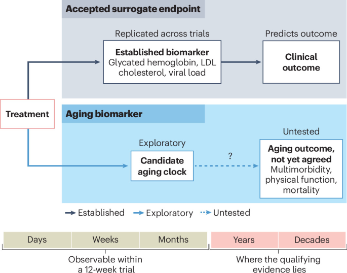
    Six proteomic aging clocks are deployed in a clinical trial for the first time, revealing more about the clocks than about the drug.

    This is a preview of subscription content, access via your institution

    Access options

    Access through your institution

    Access Nature and 54 other Nature Portfolio journals

    Get Nature+, our best-value online-access subscription

    $32.99 / 30 days

    cancel any time

    Subscribe to this journal

    Receive 12 print issues and online access

    $259.00 per year

    only $21.58 per issue

    Rent or buy this article

    Prices vary by article type

    from$1.95

    to$39.95

    Prices may be subject to local taxes which are calculated during checkout

    Fig. 1: The conceptual leap in aging biomarker capabilities.

    Subjects

    References

    1. Zhavoronkov, A. et al. Nat. Biotechnol. https://doi.org/10.1038/s41587-026-03286-y (2026).

      Article  PubMed  Google Scholar 

    2. Fleming, T. R. & DeMets, D. L. Ann. Intern. Med. 125, 605–613 https://doi.org/10.7326/0003-4819-125-7-199610010-00011 (1996).

      Article  CAS  PubMed  Google Scholar 

    3. Argentieri, M. A. et al. Nat. Med. 30, 2450–2460 https://doi.org/10.1038/s41591-024-03164-7 (2024).

      Article  CAS  PubMed  PubMed Central  Google Scholar 

    4. Xu, Z. et al. Nat. Med. 31, 2602–2610 https://doi.org/10.1038/s41591-025-03743-2 (2025).

      Article  CAS  PubMed  PubMed Central  Google Scholar 

    5. Ren, F. et al. Nat. Biotechnol. 43, 63–75 https://doi.org/10.1038/s41587-024-02143-0 (2025).

      Article  CAS  PubMed  Google Scholar 

    6. Dabrowski, J. K. et al. Nat. Aging 4, 1493–1507 https://doi.org/10.1038/s43587-024-00700-5 (2024).

      Article  CAS  PubMed  PubMed Central  Google Scholar 

    7. López-Otín, C. et al. Cell 186, 243–278 https://doi.org/10.1016/j.cell.2022.11.001 (2023).

      Article  CAS  PubMed  Google Scholar 

    8. Moqri, M. et al. Cell 186, 3758–3775 https://doi.org/10.1016/j.cell.2023.08.003 (2023).

      Article  CAS  PubMed  PubMed Central  Google Scholar 

    9. Justice, J. N. et al. Geroscience 40, 419–436 https://doi.org/10.1007/s11357-018-0042-y (2018).

      Article  CAS  PubMed  PubMed Central  Google Scholar 

    Download references

    Author information

    Authors and Affiliations

    1. Robert and Arlene Kogod Center for Aging, Mayo Clinic, Rochester, MN, USA

      Neil A. Robertson & Tamir Chandra

    2. Department of Quantitative Health Science, Mayo Clinic, Rochester, MN, USA

      Neil A. Robertson & Tamir Chandra

    3. Department of Biochemistry and Molecular Biology, Mayo Clinic, Rochester, MN, USA

      Tamir Chandra

    Authors

    1. Neil A. Robertson
    2. Tamir Chandra

    Corresponding authors

    Correspondence to Neil A. Robertson or Tamir Chandra.

    About this article

    Check for updates. Verify currency and authenticity via CrossMark

    Cite this article

    Robertson, N.A., Chandra, T. Proteomic aging clocks go to trial. Nat Biotechnol (2026). https://doi.org/10.1038/s41587-026-03361-4

    Download citation

    • Published:

    • Version of record:

    • DOI: https://doi.org/10.1038/s41587-026-03361-4

  • StockWatch: Caribou Plunges as It Pursues Strategic Alternatives

    StockWatch: Caribou Plunges as It Pursues Strategic Alternatives

    Caribou Biosciences (Nasdaq: CRBU), whose co-founders include Jennifer Doudna, PhD, capped a one-year 83% freefall of its stock after announcing plans to evaluate strategic alternatives, as investors responded with a selloff that sent shares of the CRISPR genome-editing cell therapy developer plunging 62% this past week.

    Caribou announced plans to carry out an unspecified “substantial” reduction of its workforce—which numbered 97 people as of February 27, according to its most recent Form 10-K Annual report, filed March 5—as well as a halt to trials of its two clinical-phase pipeline candidates, both allogeneic chimeric antigen receptor (CAR) T-cell therapy programs, and other cost reduction measures also yet to be specified.

    Rachel Haurwitz, PhD, Caribou Biosciences co-founder, president, and CEO

    “Caribou’s story has taken a heartbreaking turn,” co-founder, president, and CEO Rachel Haurwitz, PhD, posted on LinkedIn, announcing the company’s strategic review. “This decision is not a loss of belief in our science, our clinical data, or what these programs could mean for patients. It reflects the difficult reality that the current financing environment for allogeneic CAR T-cell therapies has made it impossible to secure the capital required to responsibly advance them.”

    Haurwitz co-established Caribou in 2011 along with Doudna and two other co-founders: Martin Jínek, PhD, a former postdoctoral fellow in Doudna’s lab, now at the University of Zurich, who led the seminal 2012 study leading to the Nobel Prize; and James Berger, PhD, professor in the department of biophysics and biophysical chemistry at Johns Hopkins University School of Medicine.

    Caribou became operational in 2012, which would make it the oldest CRISPR therapeutics company as far as Haurwitz could recall in a 2021 interview. That year, Caribou went public through an upsized initial public offering (IPO) that raised $321 million in aggregate net proceeds after deducting underwriting discounts and commissions and other offering expenses. Caribou sold 21.85 million shares at an IPO price of $16 a share, up from earlier plans to sell 13.5 million shares.

    Two years later, Haurwitz shared with GEN her journey from graduate student in Doudna’s lab to launching a biotech start-up: “This is the only job I’ve ever had!”

    Trials halted

    As a result of the strategic review, Caribou has halted trials of vispacabtagene regedleucel (vispa-cel, formerly CB-010) a treatment for relapsed or refractory (r/r) B cell non-Hodgkin lymphoma. Caribou has said vispa-cel is the first allogeneic CAR T-cell therapy “to our knowledge” that has reached the clinic with a PD-1 knockout, a genome-editing strategy designed to improve antitumor activity by limiting premature CAR T-cell exhaustion.

    To manufacture vispa-cel, Caribou applies Cas9 hybrid RNA-DNA guide or “chardonnay” (chRDNA) to make three edits: A knockout of the TRAC gene to remove the T cell receptor; a site-specific insertion of an anti-CD19 CAR into the TRAC gene; and a knockout of PD-1.

    Also halted were clinical studies of CB-011, an allogeneic anti-BCMA CAR-T cell therapy for the treatment of patients with r/r multiple myeloma (RRMM). To manufacture CB-011, Caribou uses Cas12a chRDNAs designed to direct precise genome editing, in order to make four edits:

    • A knockout of the TRAC gene to remove the T-cell receptor
    • A site-specific insertion of a humanized anti-BCMA CAR into the TRAC gene
    • A site-specific insertion of a gene encoding a B2M–HLA-E-peptide fusion into the B2M gene
    • The resulting knockout of the endogenous B2M

    “This method simultaneously eliminates endogenous HLA class I presentation on the surface of the CAR T cells and expresses HLA-E, a minor HLA class I antigen, to blunt both T and NK cell-mediated rejection of the CAR T-cell therapy by the patient’s immune system,” Caribou explained on its website.

    Strategic pivot

    The development of CB-011 and vispa-cel followed a strategic pivot by Caribou from a platform company for biopharma partners applying genome editing in research or product development, to a developer of genome-edited therapies based on its own next-generation CRISPR technology.

    While that pivot has generated positive data readouts, it has not shielded Caribou from the financial challenges faced by numerous gene-edited therapy developers. In April 2025, Caribou chopped its workforce by 32% and pruned its pipeline by ending all preclinical research, the Phase I GALLOP trial (NCT06752876) of vispa-cel in lupus (before the first patient could be dosed), and development of CB-012, including the Phase I AMpLify trial (NCT06128044) assessing the candidate in relapsed or refractory acute myeloid leukemia (AML).

    “It was really about right sizing the organization relative to our focused priorities,” Haurwitz explained last year in an interview. Asked how hopeful she was about an improving economic environment over the next 12 months, she replied: “I remain a believer and I remain an optimist. I’d probably be in the wrong job in the wrong line of work if I weren’t.”

    “I certainly think for the work that we are doing, I continue to fully believe that doing right by patients is the North Star, because business success falls out from that in terms of nearer term clinical catalysts and opportunities that come from doing right by patients. And longer-term commercial opportunity also comes from doing right by patients,” Haurwitz added.

    Caribou has retained Wedbush Securities as its exclusive financial advisor to assist in evaluating strategic options.

    Caribou announced its pursuit of strategic alternatives after the closure of financial markets on October 6. The following day, Caribou shares began three days of stock declines, nosediving 46% from $1.14 to 62 cents. Those shares tumbled another 22% Thursday to 48 cents, but closed Friday with another 4% decline, finishing the week at 46 cents.

    Cash crunch

    Daina M. Graybosch, PhD, senior managing director, immuno-oncology and a senior research analyst with Leerink Partners, reported in a research note that Caribou was facing an increasingly pressing cash crunch at a time when the company needed significant capital to fund its planned Phase III ANTLER-3 trial, designed to assess vispa-cel in second-line auto-CAR T ineligible diffuse large B-cell lymphoma (DLBCL).

    Caribou never enrolled patients in the pivotal study, though it reached agreement with the FDA in May on a design for the trial, which was expected to be a randomized, controlled clinical study enrolling approximately 250 CD19-naïve second line (2L) large B cell lymphoma patients ineligible for transplant and not candidates or not eligible for autologous CAR T-cell therapy.

    “We are sympathetic to management’s stated rationale for exploring strategic alternatives,” Graybosch commented. “Evidently, investors were unwilling to fund the trial on what would have been highly dilutive terms, especially in the context of lingering clinical risk and uncertainty around the strength of vispa-cel’s Ph[ase I/II] outcomes and partial HLA-matching strategy, as well as substantial in- and out-of-class competition.”

    That competition, she wrote, included epcoritamab, a bispecific T-cell engager (BiTE) therapy already marketed as Epkinly® by Genmab (Nasdaq Copenhagen and Nasdaq: GMAB) and AbbVie (NYSE: ABBV).

    On October 5—the day before Caribou’s strategic alternatives announcement—Genmab and AbbVie reported positive topline data from the Phase III EPCORE DLBCL-2 trial (NCT05578976) showing that the combination of epcoritamab, rituximab, the corticosteroid prednisone, and three chemotherapy drugs showed statistically significant and clinically meaningful improvement in progression-free survival (PFS) in frontline diagnosed DLBCL patients.

    The combination therapy, nicknamed R-CHOP, consists of rituximab—the CD20-directed cytolytic antibody marketed by Genentech, a member of the Roche Group (SIX Swiss: RO and ROP) and Biogen (Nasdaq: BIIB) as Rituxan®—plus cyclophosphamide, doxorubicin hydrochloride, vincristine, and prednisone. In DLBCL patients with an International Prognostic Index (IPI) score of 2 to 5, R-CHOP reduced the risk of disease progression or death by 51% (HR 0.49 [95% CI 0.36, 0.67]; p-value < 0.0001).

    “Interesting” timing

    “The timing of [Caribou’s] initiative is interesting,” Graybosch observed. “We believe there is enhanced strategic value and clinical rationale for allogeneic cell therapies in a world where TCEs [T-cell engagers] become entrenched in the 1L [first-line] setting.”

    As for CB-011, it began development much earlier than vispa-cel, “with an appreciably more challenging competitive setup in RRMM, where we saw elevated clinical risk for its post-BCMA therapy development path due to potential efficacy-compromising loss-of-antigen,” the Leerink Partners analyst added.

    Single target allogeneic BCMA CAR-T treatments have posed a potential risk of RRMM relapse through antigen loss as myeloma cells mutate or downregulate BCMA to evade the CAR T cells. To minimize that risk, Caribou designed its Phase I CaMMouflage trial (NCT05722418) to limit the therapy dosing to BCMA-naïve patients—an approach that proved successful when the company last year announced positive data from a 12-patient, BCMA-naïve cohort showing a 92% overall response rate (11/12 patients), a 75% or greater complete response rate (9/12), and 91% minimal residual disease (MRD) negativity (10/11 evaluable patients).

    To avoid the risk of antigen loss, several developers of allogeneic BCMA CAR-Ts have designed dual targeting CAR Ts. For example, Allogene Therapeutics (Nasdaq: ALLO) is developing ALLO-329, which targets CD19 on B cells and CD70 on activated T cells. ALLO-329 generated positive preclinical data in April, with a data update from the Phase I RESOLUTION trial (NCT07085104) expected later in Q4.

    Another challenge to CB-011 and other allogenic therapies for RRMM: The multiple myeloma space has been reshaped by autologous BCMA CAR-T therapies that have won approval by demonstrating safety and effectiveness—including Carvykti® (ciltacabtagene autoleucel), co-marketed by Legend Biotech (Nasdaq: LEGN) and Johnson & Johnson (NYSE: JNJ); and Abecma® (idecabtagene vicleucel), marketed by Bristol Myers Squibb (NYSE: BMY).

    Souring on the stock

    Graybosch downgraded Caribou’s rating from “Outperform” to “Market Perform” and chopped the firm’s 12-month price target 75%, from $4 to $1. Graybosch was among several Wall Street analysts who responded to Caribou’s news by souring on the company’s stock, downgrading their ratings:

    • Brookline Capital Markets (Leah Ruch Cann)—From “Buy” to “Hold,” no price target.
    • H.C. Wainwright (Robert Burns)—From “Buy” to “Neutral,” no price target.
    • RBC Capital Markets (Luca Issi, PhD)—From “Outperform” to “Sector Perform,” price target slashed 90% from $10 to $1.

    Caribou reported $113.8 million in cash, cash equivalents, and marketable securities as of the end of the second quarter on June 30—a 4% dip from the $118.6 million reported at the end of Q1, but a 20% drop from the $142.8 million Caribou had as of December 31, 2025, and a 38% slide from the $183.9 million reported June 30, 2025.

    “We expect that our existing cash, cash equivalents, and marketable securities will be sufficient to fund our operations for at least the next 12 months from the date this Form 10-Q is filed with the SEC [U.S. Securities and Exchange Commission],” Caribou stated in its most recent Form 10-Q quarterly report detailing second-quarter results, filed August 13.

    Leaders and laggards

    • Arrivent BioPharma (Nasdaq: AVBP) shares plummeted 47% October 6 from $28.46 to $15.09 after the cancer drug developer announced that its lead pipeline candidate firmonertinib failed the Phase III FURVENT trial (NCT05607550) designed to assess the oral, mutation-selective epidermal growth factor receptor (EGFR) inhibitor in previously untreated, locally advanced or metastatic non-squamous non-small cell lung cancer (NSCLC) harboring EGFR exon 20 insertion mutations. Firmonertinib did not meet its primary endpoint of meaningful progression free survival (PFS) compared to chemotherapy as evaluated by blinded independent central review (BICR). Arrivent said that clinical benefit was seen in secondary endpoints such as PFS by investigator’s assessment and confirmed objective response rate by BICR. A trend toward improved overall survival was also seen, the company added.
    • Pacira BioSciences (Nasdaq: PCRX) shares soared 44% Thursday from $25.20 to $36.39 after Viatris (Nasdaq: VTRS) agreed to acquire the non-opioid pain drug developer for $1.65 billion. The deal adds to Pacira’s portfolio with two marketed drugs—the postsurgical pain relief therapy Exparel® (bupivacaine liposome injectable suspension), and the osteoarthritis knee pain treatment Zilretta® (triamcinolone acetonide extended-release injectable suspension). Viatris said it plans to apply its intellectual property expertise and ability to extend product lifecycles and sustain meaningful sales post-competition “to maximize the long-term value of the Pacira portfolio” and expects to expand its reach across select markets. Pacira generated approximately $746 million in revenue and approximately $177 million in adjusted earnings before interest, taxes, depreciation, and amortization (EBITDA) during the 12 months ending June 30. Viatris plans to commence a tender offer for all outstanding Pacira common stock shares for $36.50 per share cash. The deal is set to close by year’s end.
  • Five questions with Bowen Li

    Five questions with Bowen Li

    A true multidisciplinary scientist, Bowen Li finds that some of the most interesting questions are located precisely at the boundaries between fields.

    Access options

    Access through your institution

    Access Nature and 54 other Nature Portfolio journals

    Get Nature+, our best-value online-access subscription

    $32.99 / 30 days

    cancel any time

    Subscribe to this journal

    Receive 12 print issues and online access

    $259.00 per year

    only $21.58 per issue

    Buy this article

    • Purchase on SpringerLink
    • Instant access to the full article PDF.

    USD 39.95

    Prices may be subject to local taxes which are calculated during checkout

    Author information

    About this article

    Check for updates. Verify currency and authenticity via CrossMark

    Cite this article

    Francisco, M. Five questions with Bowen Li. Nat Biotechnol 44, 1741–1742 (2026). https://doi.org/10.1038/s41587-026-03340-9

    Download citation

    • Published:

    • Version of record:

    • Issue date:

    • DOI: https://doi.org/10.1038/s41587-026-03340-9

  • Biotech news from around the world

    Biotech news from around the world

    • News in Brief
    • Published:

    Nature Biotechnology volume 44, page 1594 (2026) Cite this article

    Scientists from Ataturk University use gene editing to increase the growth of rainbow trout. Injecting two guide RNAs targeting the first exon of the myostatin gene mstnb1 into fertilized rainbow trout eggs, the team was able to increase the trouts’ body weight by 56%. The results suggest that CRISPR–Cas9 could help shorten production time and improve production efficiency in aquaculture.

    Seoul-based pharma DuChemBio will build Korea’s first plant dedicated to radiopharmaceuticals after agreeing to a $25.5 million contract with North Jeolla Province and the city of Jeongeup. The facility will combine radioisotope production, drug manufacturing and export. Construction is scheduled to begin in mid-2027, targeting a 2029 opening.

    This is a preview of subscription content, access via your institution

    Access options

    Access through your institution

    Access Nature and 54 other Nature Portfolio journals

    Get Nature+, our best-value online-access subscription

    $32.99 / 30 days

    cancel any time

    Subscribe to this journal

    Receive 12 print issues and online access

    $259.00 per year

    only $21.58 per issue

    Buy this article

    • Purchase on SpringerLink
    • Instant access to the full article PDF.

    USD 39.95

    Prices may be subject to local taxes which are calculated during checkout

    About this article

    Check for updates. Verify currency and authenticity via CrossMark

    Cite this article

    Biotech news from around the world. Nat Biotechnol 44, 1594 (2026). https://doi.org/10.1038/s41587-026-03347-2

    Download citation

    • Published:

    • Version of record:

    • Issue date:

    • DOI: https://doi.org/10.1038/s41587-026-03347-2

  • Live-cell imaging goes WHOLISTIC

    • Research Highlight
    • Published:

    Imaging

    Nature Biotechnology volume 44, page 1617 (2026) Cite this article

    Coordinated cellular communication is at the basis of organismal physiology, but approaches for monitoring cellular dynamics across multiple tissues and organs simultaneously remain limited. Writing in Nature, Ruetten and colleagues describe WHOLISTIC (Whole-Organism Live-Imaging System for Recording Tissue and Intracellular Activity), an imaging platform that captures cellular activities across the entire vertebrate body at single-cell resolution.

    WHOLISTIC combines the expression of genetically encoded calcium sensors with high-speed volumetric fluorescence imaging, computational motion correction and tissue-scale activity analysis in transparent vertebrate models. The authors developed transgenic zebrafish that express a calcium indicator in nearly all cell types and demonstrated the applicability of the approach in adult Danionella cerebrum. Whole-body expansion microscopy and functional imaging enabled the molecular and anatomical characterization of cell populations.

    This is a preview of subscription content, access via your institution

    Access options

    Access through your institution

    Access Nature and 54 other Nature Portfolio journals

    Get Nature+, our best-value online-access subscription

    $32.99 / 30 days

    cancel any time

    Subscribe to this journal

    Receive 12 print issues and online access

    $259.00 per year

    only $21.58 per issue

    Buy this article

    • Purchase on SpringerLink
    • Instant access to the full article PDF.

    USD 39.95

    Prices may be subject to local taxes which are calculated during checkout

    Author information

    Authors and Affiliations

    1. Nature Biotechnology https://www.nature.com/nbt/

      Dario Ummarino

    Corresponding author

    Correspondence to Dario Ummarino.

    About this article

    Check for updates. Verify currency and authenticity via CrossMark

    Cite this article

    Ummarino, D. Live-cell imaging goes WHOLISTIC. Nat Biotechnol 44, 1617 (2026). https://doi.org/10.1038/s41587-026-03354-3

    Download citation

    • Published:

    • Version of record:

    • Issue date:

    • DOI: https://doi.org/10.1038/s41587-026-03354-3