
Category: Bioprocessing

  • The Top 10 Questions Shaping the Future of Optogenetic Biomanufacturing

    The Top 10 Questions Shaping the Future of Optogenetic Biomanufacturing

    Maximilian Hoerner
    Maximilian Hoerner, PhD

    The emergence of increasingly complex biologics such as multi‑specific antibodies, cytokines, fusion proteins, and other structurally demanding modalities has exposed the limits of conventional bioprocessing. Many of these molecules are difficult to express at commercially viable yields, and their quality attributes can be highly sensitive to the cellular environment. As a result, productivity, consistency, and cost remain major constraints across the industry.

    Traditional cell line development (CLD) relies on constitutive promoters that drive static expression throughout development and production. Because production is always on, cells must divide and manufacture protein simultaneously, even when the product is burdensome or toxic. While this model worked relatively well for easy-to-express monoclonal antibodies, it does not work well for next-generation biologics, where the inherent trade-off between higher specific productivity and rapid cell growth, both competing for the same cellular resources, is increased. Light‑controlled gene expression using optogenetics is emerging as a credible alternative.

    This article explores the ten most important questions facing optogenetic biomanufacturing as it moves from proof‑of‑concept demonstrations toward mainstream adoption in modern bioprocessing.

    1. What is optogenetics, and why is it relevant to biologics manufacturing?

    Optogenetics uses light‑responsive proteins to control specific cellular functions. In biomanufacturing, the key application is regulating gene expression with light instead of relying on constitutive promoters or chemical inducers. When light‑controlled gene expression systems are engineered into production cell lines, illumination at defined wavelengths can switch expression on, off, or to intermediate levels.

    This is important because it introduces a fundamentally different control modality. Conventional systems provide static expression, whereas optogenetics enables dynamic, reversible, and tunable control. As biologics become more complex and more sensitive to expression conditions, having a way to precisely modulate transcription directly, rather than perturbing the entire cell by modulating process parameters such as temperature, will become increasingly valuable.

    2. What core limitation in biologics manufacturing does light‑based control address?

    The main limitation is the inherent competition between cell growth and protein production. In constitutive systems, cells must divide and manufacture protein at the same time, drawing on the same finite pool of metabolic resources. This creates a structural trade‑off: clones that produce strongly have a slow doubling time and/or do not grow to viable densities, while clones that grow well often produce only modest titers. This trade‑off becomes much more pronounced for challenging molecules such as multi‑specific antibodies, cytokines, and fusion proteins that are aggregation‑prone or place a significant burden on the host cell.

    Optogenetics addresses this by separating the growth and production phases. Cells can grow in an uninduced, low-burden state and then transition into a productive state upon illumination. As light acts directly on transcriptional regulators in a dose-dependent manner, expression can be tuned precisely rather than modulated indirectly through temperature shifts or coarsely and irreversibly by chemical induction. Chemical inducers can also negatively affect cell performance through off-target effects and are difficult to remove during downstream processing, which has limited their adoption in mammalian biomanufacturing. The direct, tunable control by light also enables more deliberate modulation of pathways that influence critical quality attributes such as post-translational modifications (PTMs), which is increasingly important for complex biologics and biosimilars.

    3. How does optogenetic light exposure work, and is it safe for cells and media?

    Optogenetics uses light as an informational cue rather than an energy source. Because photoreceptors respond to brief, low-intensity pulses and stay “on” for a tunable period of time before reverting to the off state, optogenetic systems can maintain precise control without continuous or high-power illumination.

    The wavelengths used in bioprocessing are in the visible range and non-ionizing, operating well under established phototoxicity thresholds. At these levels, light does not harm cells or DNA, nor does it disrupt cellular physiology. Although certain media components are known to be light-sensitive, the illumination used for optogenetic induction has not shown detrimental effects on cellular performance.

    4. What is the impact of optogenetics on lab operations?

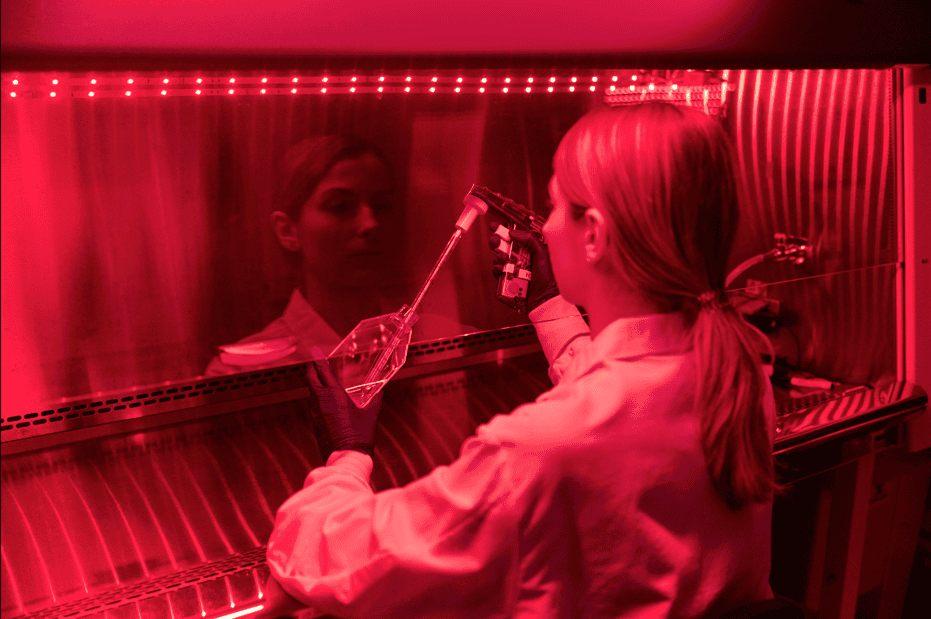
    Optogenetic workflows use standard lab equipment with the addition of illumination devices compatible with existing infrastructure, such as plates, shake flasks, or bioreactors. The main operational difference occurs during pool generation and single-cell cloning, where cells must remain uninduced. This is achieved by working under “safe light” conditions, visible wavelengths that do not activate the specific optogenetic system but allow the operator to handle the cell culture. Only small areas of the lab need to be adapted for this purpose. For all other operations, including biomanufacturing, local interventions to shield cells from room light are typically sufficient.

    5. How many cellular functions can be controlled with light in a single cell line?

    Current systems can independently regulate cellular functions using two or three different wavelengths. For example, one color may control production while another modulates a critical quality‑related pathway. Functions that require coordinated timing can be linked to the same wavelength. As genetic circuit design advances, the number of independently controllable functions is expected to increase.

    6. When might optogenetically manufactured biologics reach clinical and commercial stages?

    Optogenetic cell line development is already being explored for multiple programs. Regulatory agencies have established pathways for evaluating emerging bioprocess technologies, and optogenetic systems are beginning to move through these mechanisms. If early development proceeds as expected, optogenetically controlled processes could support clinical filings in the next two years, with commercial adoption following thereafter.

    7. Can optogenetics be integrated into existing facilities?

    Yes. Optogenetic manufacturing does not require new bioreactor designs; illumination hardware can be added to standard single‑use or stainless‑steel systems. For single‑use bags, light is delivered externally through the bag without breaching the sterility barrier; for stainless‑steel tanks, cultures can be circulated through illuminated loops. No cleanroom redesign is required.

    8. How does optogenetic manufacturing scale to larger bioreactors?

    Scaling optogenetic systems depends on three factors: the sensitivity of the photoreceptors, the lifetime of their activated state in the dark, and the mixing and light-scattering characteristics of the bioreactor. Modern optogenetic systems for biomanufacturing are designed to respond to very low light intensities, and once activated, they gradually return to the off state in the dark at a characteristic rate, typically over tens of minutes.

    Since stirred-tank bioreactors mix continuously and scatter light throughout the vessel, cells receive sufficient illumination over the photoreceptor’s characteristic activation lifetime even at large volumes. Simulation studies and early pilot-scale experiments up to the 200 L scale suggest that optogenetic activation can be achieved at commercially relevant scales, including stainless steel and single-use systems.

    9. What has convinced established players to evaluate this technology?

    Biologics development and manufacturing are intentionally conservative, with new technologies adopted only when they demonstrate clear, reproducible benefits and maintain patient safety. For any emerging bioprocess platform, the threshold for evaluation is data showing that it can deliver meaningful improvements without introducing new risks or operational complexity.

    In comparative studies, optogenetic induction has produced more than two‑fold increases in titer for standard monoclonal antibodies as well as for more difficult‑to‑express therapeutic proteins. These gains have been achieved while maintaining or improving critical quality attributes, including those influenced by folding, assembly, and post‑translational modification.

    As the next generation of therapeutics become increasingly complex, leading biopharma and CDMO players see the need for novel biomanufacturing platforms capable of meeting that complexity without compromising titers, and for that reason several key players are actively evaluating optogenetic technology for potential integration.

    10. Where is optogenetic biomanufacturing headed in the next decade?

    If current trends continue, optogenetic control may become a standard tool for managing expression, quality, and other cellular functions in biologics manufacturing. The ability to modulate gene expression dynamically opens possibilities for more efficient production of complex therapeutics, more predictable quality profiles, and more flexible facility design. As the technology matures, it is likely to be adopted across CDMOs, biotech, and biopharma companies seeking greater control over their manufacturing processes.

    Maximilian Hoerner, PhD, is the head of optogenetics at Prolific Machines

  • UCB Dedicates Future U.S. Biologics Manufacturing Campus in Georgia

    UCB Dedicates Future U.S. Biologics Manufacturing Campus in Georgia

    UCB has dedicated the Rowen, GA, site where it plans to build a $2 billion biologics manufacturing campus that is set to open as the Belgian-based drug developer’s first U.S. manufacturing site.

    When completed in 2032 or 2033, the campus is expected to support commercial production of recently approved and future biologic medicines, applying artificial intelligence, automation, and advanced manufacturing technologies. The campus is intended to further integrate UCB’s U.S. patient value chain from research and development through manufacturing and delivery.

    Some 330 permanent jobs are expected to be based at the approximately 460,000-square-foot campus, which will occupy 79 acres within the Rowen innovation district in Georgia’s Gwinnett County. UCB bought the 79 acres from the Rowen Foundation, which is developing a 1,900-acre campus on Highway 316, launched in 2020 as a “knowledge” community for biopharma and other innovation-based fields.

    In its announcement of the campus site dedication, UCB said it chose to build its first U.S. manufacturing campus in Georgia because of what it called the Peach State’s strong life sciences ecosystem, highly skilled workforce, robust infrastructure, and access to leading academic and research institutions.

    Another factor in the decision: Gwinnett County has committed $174 million in incentives and infrastructure investments to support UCB’s project.

    UCB’s project is the brightest recent success of Georgia life sciences leaders, who worked through industry group Georgia Life Sciences to lay out a 10-year “Roadmap” last year with the aim of “positioning Georgia as the scale-up manufacturing hub of the Southeast.” The UCB project, announced in March, helped place Georgia among the “Next 5 States” within GEN’s Next 10 U.S. Biopharma Clusters A-List.

    “Long-term commitment”

    “Our manufacturing investment in the United States reflects our long-term commitment to patients, strengthening our ability to serve them while contributing to scientific innovation, highly skilled jobs, and economic growth in Georgia,” UCB CEO Jean-Christophe Tellier said in a statement.

    UCB executives and dignitaries cut a ceremonial ribbon to mark the dedication of UCB’s future biologics manufacturing campus in Rowen, GA. Jeffrey Kessler, U.S. Department of Commerce Under Secretary for Industry and Security, stands at left. At the center are Georgia Gov. Brian Kemp (R); UCB CEO Jean-Christophe Tellier; Katherine Raeymaekers, Consul General of Belgium in Atlanta; and Nicole Love Hendrickson (D), Chairwoman of the Gwinnett County Board of Commissioners. [UCB]

    Tellier and other UCB leaders were joined at a dedication ceremony by dignitaries that included Georgia Gov. Brian Kemp (R); Jeffrey Kessler, U.S. Department of Commerce Under Secretary for Industry and Security; Nicole Love Hendrickson (D), Chairwoman of the Gwinnett County Board of Commissioners; and Katherine Raeymaekers, Consul General of Belgium in Atlanta.

    During the ceremony, the dignitaries joined top UCB executives in unveiling the Emmanuel Janssen Circle, a commemorative stonework installation just completed within the roundabout to the entrance of the future campus. Janssen (1879-1955) was a Belgian businessman who founded Union Chimique Belge (UCB) on January 18, 1928. The company initially focused on industrial chemicals before evolving into a developer of biologics for indications in neurology and immunology.

    “UCB’s new biomanufacturing campus builds on the entrepreneurial legacy of Baron Emmanuel Janssen, founder of UCB, while strengthening the transatlantic collaboration that drives economic growth, scientific innovation, and improving people’s lives,” Raeymaekers stated. “Today’s site dedication reflects the strength of the partnership between Belgium, the United States, and the State of Georgia—and our shared commitment to enduring progress, innovation, and opportunity.”

    UCB worked with numerous public and private partners to locate its biologics manufacturing campus in Rowen—including the Metro Atlanta Chamber; Georgia Quick Start, which provides customized workforce training through the Technical College System of Georgia; Gwinnett County Government; Georgia Power Company; the public-private economic development initiative Partnership Gwinnett; and the Georgia Department of Economic Development.

    Headquartered in Brussels, UCB employs more than 10,000 people in nearly 40 countries—including the United States, where the company employs more than 2,000 people in research, development, manufacturing, commercial operations, and corporate functions.

    UCB has maintained a U.S. presence for nearly a century. The company now employs more than 400 people in Georgia, where it established its Stateside headquarters in Atlanta in 1993.

    More than 1,000 construction jobs are expected to be created in connection with the campus during the six- to seven-year projected timeframe for design and construction.

    “At UCB, our purpose is to create value for people living with severe diseases, and that purpose guides the science we pursue, where we invest, and the choices we make for the long term,” Tellier added.

  • ‘Molecular Movie’ of Penicillin Synthesis Could Inform Future Antibiotic Development

    ‘Molecular Movie’ of Penicillin Synthesis Could Inform Future Antibiotic Development

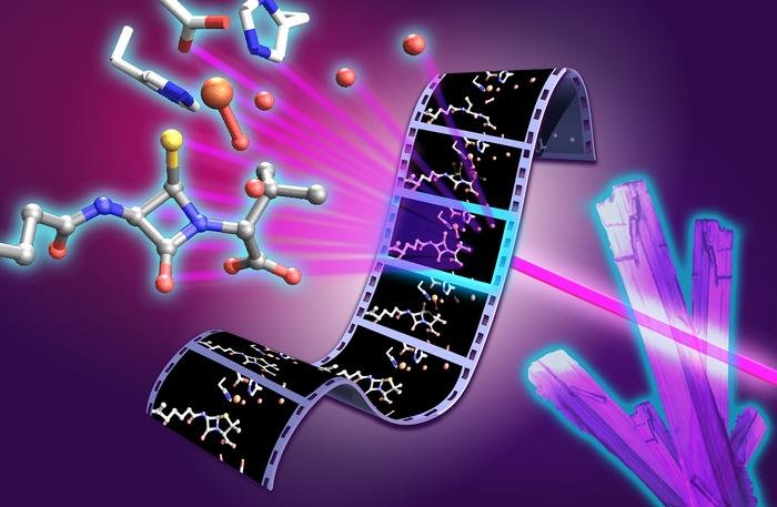
    Molecular movie of penicillin biosynthesis. X-ray free electron laser experiments on isopenicillin N synthase microcrystals capture structural snapshots of key intermediates during the penicillin forming reaction. The film strip illustrates progression through the catalytic cycle, with the monocyclic β-lactam intermediate and reactive high valent ferryl iron species highlighted at the center of the molecular movie. [University of Oxford/Greg Stewart/SLAC National Accelerator Laboratory]

    Researchers from the University of Oxford and international collaborators have revealed previously unseen rapid chemical stages in the formation of β-lactam antibiotics such as penicillin, offering insights that could help to support future antibiotic development.

    Rather than relying on static X-ray crystallographic structures of the enzyme, the Oxford University team, collaborating with partners from Diamond Light Source, Lawrence Berkeley National Laboratory, PAL-XFEL, and SLAC National Accelerator Laboratory, followed the reaction in real time. To do this they used X-ray free-electron lasers (XFEL) to observe the enzyme isopenicillin N synthase, (IPNS) as it converts its linear peptide substrate into the β-lactam ring system of penicillin.

    The study brought together expertise in structural biology, spectroscopy, chemistry, computation and enzyme engineering. The approach, they say, provides a powerful way to study fleeting chemical intermediates that have previously been inaccessible, both in antibiotic biosynthesis and in other biologically important enzyme reactions.

    The team captured several rapid stages in the reaction. These included a thioaldehyde intermediate formed just before the β-lactam ring is created, and a monocyclic β-lactam intermediate, representing the first ring-shaped structure on the way to forming the complete penicillin scaffold. These steps provide the clearest picture yet of how the IPNS enzyme assembles the penicillin scaffold. Subtle movements throughout the enzyme help guide these chemical steps, showing that both the enzyme’s shape and its chemistry work together to control penicillin formation.

    The results show, in unprecedented detail, how the enzyme IPNS achieves an exceptionally complex transformation in a single step. Christopher Schofield, PhD, Professor of Chemistry, University of Oxford, said, “Penicillin has shaped modern medicine, but there is still much to learn about how nature builds this important antibiotic structure. By capturing these fleeting steps, we can better understand how enzymes control complex chemistry with remarkable precision … As rates of antimicrobial resistance continue to rise, understanding this process will ultimately help us make existing antibiotics more efficient and design new antibiotic structures.”

    Allen M Orville, PhD, group leader of the XFEL Hub, further commented, “The XFEL Hub at Diamond brought specialist expertise in time-resolved X-ray methods and XFEL-based structural biology to this study. Working closely with our colleagues in Oxford and at partner facilities, we helped bring together the experimental approaches needed to capture these fleeting stages of enzyme catalysis. The results show the power of combining complementary expertise and X-ray techniques to move beyond static structures and reveal how enzymes work in real time. It is a great example of how time-resolved structural biology can uncover new principles of enzyme function and, ultimately, inform the design of new catalysts and therapeutics.”

    Schofield is co-senior author of the researchers’ published paper in Nature Catalysis, titled “Unanticipated intermediates during isopenicillin N synthase catalysis identified by time-resolved X-ray free-electron laser studies.”

    Ever since penicillin was developed into a working drug at the University of Oxford in the early 1940s, β-lactam antibiotics have been among the most important medicines for treating bacterial infections. Their activity depends on a special ring—the β-lactam ring—a highly strained chemical ring system that interferes with bacterial cell wall synthesis, ultimately causing the cell wall to fail and the bacteria to die. “Isopenicillin N synthase (IPNS) catalyses the biosynthesis of isopenicillin N (IPN), forming the fused four-membered β-lactam and five-membered thiazolidine ring system that is characteristic of the penicillins,” the authors explained.

    Rising rates of antimicrobial resistance are undermining the effectiveness of existing antibiotics. Today, one in six bacterial infections is resistant to antibiotics, the authors noted, citing WHO figures. With too few new antibiotics in development, understanding how nature builds these molecules is an important step towards replenishing the antibiotic pipeline.

    Scientists have been studying how nature constructs this β-lactam ring for decades, but the key fleeting reaction intermediates have been too difficult to observe directly.

    To capture the reaction in real time, the researchers used a system in which thousands of tiny droplets containing anaerobic enzyme microcrystals were deposited onto a moving 2 mm wide tape. As the tape entered an oxygen filled chamber, oxygen rapidly diffused into the crystals and initiated the reaction simultaneously across the sample. By precisely controlling the speed of the tape, the researchers could determine how long each crystal reacted before reaching the X-ray interaction point, where an ultrafast XFEL pulse recorded an atomic resolution snapshot.

    Combining thousands of these snapshots allowed the team to build a frame-by-frame “molecular movie” of penicillin biosynthesis. This method allows individual reaction intermediates that exist only for tiny fractions of a second to be observed at atomic resolution, and under physiological temperature and pressure, before they disappear.

    The newly reported findings help to resolve a long-standing mechanistic question that has remained unanswered for more than four decades. The work also showed that water molecules inside the enzyme play a key role in guiding the reaction. The combined results of their studies, they noted, “… reveal unexpected insight into the IPNS reaction mechanism including the vital roles of active-site water molecules.”

    The study ultimately builds on the work of Nobel prize-winning Oxford chemist Dorothy Hodgkin, PhD, who first solved the structure of penicillin in 1945 using X-ray crystallography.

    Beyond penicillin, IPNS belongs to a large family of iron-dependent oxygenase enzymes involved in human biology, including those that enable us to sense and respond to changes in oxygen availability. The mechanistic insights from this work could therefore have implications beyond antibiotics, offering new principles for enzyme engineering and catalyst design.

    “These findings provide structural insight into IPNS catalysis, inform on the wider Fe(II) oxygenase superfamily and highlight the power of X-ray free-electron laser methods for resolving transient enzymatic intermediates,” the team stated in their paper. First author Patrick Rabe, PhD, Wellcome Career Development Award Investigator, University of Oxford, said, “These insights matter because they can inform how we think about antibiotic biosynthesis and future strategies for developing antibacterial medicines. The ability to capture structural snapshots over milliseconds to seconds of reaction time allows us to connect enzyme motion, iron chemistry and water-mediated proton transfer. This gives us a much richer view of how IPNS controls a difficult chemical transformation. By understanding this process in atomic detail, we can begin to think about engineering these enzymes to produce new or improved antibiotic scaffolds.”

  • How COVID, Characterization, and Commerce Are Driving an RNA Revolution

    How COVID, Characterization, and Commerce Are Driving an RNA Revolution

    The technology underpinning RNA vaccines1 and therapeutics2 has been around for decades. However, until recently, only a handful of products were on the market.

    Everything changed in 2020 when mRNA vaccines proved3 to be effective against SARS-CoV-2, the virus that causes COVID-19. In the years since, many more RNA products entered the clinic as developers recognized both their therapeutic and commercial potential.

    The pandemic also kick-started efforts to optimize RNA manufacturing that will likely shape the sector for decades to come, according to David Sokolowski, global workflow manager for nucleic acid therapeutics at Cytiva.

    “The COVID vaccines heralded the rapid industrialization of RNA and LNP [lipid nanoparticle] technologies, and due to the nature of a pandemic, large amounts of vaccine safety data were generated. However, it remains a nascent industry with limited applications commercially available, and this space is where developers are rushing to build.

    “Manufacturing good quality RNA and LNP became easier with the proliferation of high-quality reports from successful studies, trials, and campaigns occurring all over the world.

    “Now, we’re supporting the development of therapeutics with even grander aspirations and increasingly exotic biology—whether they are encoding and producing mAb via RNA, or enabling next-generation in vivo cell therapy, LNP-mediated therapeutics have no shortage of potential advances ahead,” he continued.

    Manufacturability

    The key to developing any RNA-based product—be it a vaccine or a therapeutic—is to ensure it can be produced in bulk in a cost-effective and efficient manner, says Luis Avila, PhD, a scientist at services firm Novoprotein Scientific.

    “RNA design fundamentally determines both manufacturability and biological performance, but optimizing for one often comes at the expense of the other.” The best approach is to design for manufacturability from the start,” he says, adding “the right template architecture can contribute with optimized promoter design, transcription initiation sequences, encoded polyA tails, transcript boundaries, and sequence composition to improve IVT [in vitro transcription] performance while minimizing problematic secondary structures.”

    “Combined with other factors, such as engineered RNA polymerases, optimized reaction chemistries and advanced capping technologies, these design strategies will improve RNA yield, integrity, and translational performance all while reducing the generation of dsRNA,” he tells GEN.

    The type of RNA being made should be the key consideration in any assessment of manufacturability, according to Avila, who cites the differences between messenger RNA and self-amplifying RNA to illustrate the point.

    “For mRNA, coding-sequence composition, transcript length, GC content, and secondary structure strongly influence IVT efficiency. Whereas optimized UTRs [untranslated regions], cap structures, and poly(A) tails primarily govern RNA stability and translational efficiency.

    “For saRNA, the incorporation of the large alphavirus replicase cassette substantially increases transcript length and structural complexity, thus making IVT more challenging while enabling intracellular RNA amplification that dramatically lowers the therapeutic dose required,” he says.

    Producing circular RNA—a covalently closed-loop structure that is stable and resistant to exonuclease degradation—can be even harder, according to Avila.

    “circRNA presents an even greater manufacturing challenge because linear IVT is followed by an additional circularization step whose efficiency remains a major bottleneck. Following circularization, translation relies on efficient cap-independent initiation through optimized IRES [internal ribosome entry site] elements and RNA architecture.”

    Characterization

    So, designing an RNA therapeutic or vaccine with manufacturing in mind is critical. However, the ability to realize that optimized blueprint on the factory floor is entirely dependent on being able to characterize the product in real-time, as it is being made.

    And, unfortunately for the industry, RNA characterization is a complex challenge, says Matthew Ranaghan, PhD, applications scientist at analysis technology firm Refeyn.

    TwoMP with model
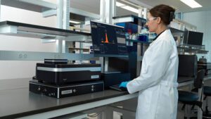
    Analyzing multiple RNA molecules in a single pass reduces the experimentation burden on RNA developers. Being able to visualize characteristics within minutes—as shown here—would benefit any developer, says Refeyn. [Refeyn]

    “Thorough Characterization of RNA is critical to ensuring the safety, stability, and efficacy of next-generation RNA therapeutics, yet it remains a significant analytical bottleneck.”

    “Current methods are limited to various types of chromatography, which struggle with poor resolution for large RNAs, or mass spectrometry, which requires digesting samples into smaller oligos with RNases. While both have advantages, they demand extensive and time-consuming method development.”

    Traditional RNA analysis usually consists of separate assays looking at specific characteristics. For example, size exclusion chromatography is used to assess aggregation while enzyme-linked immunosorbent assays are used to check for trace amounts of double-stranded RNA.

    One promising alternative is mass photometry (MP), a technique that can analyze RNA molecules within minutes based on only nanograms of material within minutes in a single experiment. There are multiple potential advantages according to Ranaghan.

    “A single readout replaces the need for multiple instruments, drastically reducing hands-on time, reagent costs, and the need for extensive method development across different platforms. A single readout also enables at-line process monitoring, allowing for immediate go/no-go decisions during bioprocessing and purification.”

    “We envision the faster speed and higher resolution of MP to enable its use as a multi-attribute tool for at-line assessment of the various critical quality attributes (CQAs) of RNA therapeutics,” he says.

    “We have already demonstrated how MP can rapidly assess integrity, fragments, multimer, and purity in a single measurement. We are also actively building out more applications,” Ranaghan adds.

    Commercial concerns

    Manufacturers of RNA products also need to develop commercialization plans as early as possible—in parallel with process development—to ensure they reach the patients who need them and garner the revenues needed to fund future research.

    Rajiv Gangurde
    Rajiv Gangurde, PhD
    Vice President
    Parexel International

    And, for some firms, developing such plans can be a steep learning curve, says Rajiv Gangurde, PhD, vice president of chemistry, manufacturing, and controls (CMC) for cell and gene therapies at contract research organization Parexel International.

    “Like many newer modalities, RNA-based therapies typically emerge out of academia or start-up biotech, and transitioning from academia to industry can be challenging due to differences in objectives, differences in expertise, fundraising, etc.”

    “In addition, newer modalities require heavier than usual investment in CMC activities to establish proof-of-concept as well as manufacturability. There is heavy reliance on external partners, creating capability and capacity constraints.

    “Most importantly,” Gangurde continued, “a phase-appropriate CMC strategy aimed at global approvals can significantly derisk development and help minimize costs and timelines.”

    Yet preparing a CMC strategy that will win approval in multiple markets is no small undertaking, particularly, as Gangurde points out, because regulatory guidelines for RNA-based therapies are still evolving.

    Another challenge is the lack of consistency. For instance, the U.S. Food and Drug Administration (FDA) and EU EMA use different definitions and guidelines for CMC.

    Gangurde told us, “Harmonization of guidelines and expectations across the two agencies will be hugely beneficial to developers of RNA therapies. For example, while both agencies recognize potency/efficacy assessment as a critical quality attribute (CQA), there are some differences in method validation requirements, timing, and regulatory expectations across clinical phases.”

    “While the FDA accepts real-world experience and surrogate endpoints, particularly for rare or life-threatening diseases, EMA requires more comprehensive clinical data and demonstration of long-term efficacy for approval. The EMA also mandates an appropriate degree of method validation at each stage of development, whereas the FDA does not expect validation of the potency assays during Phase I,” he added.

    The product component designations used by regulatory agencies can also differ. For example, the lipid nanoparticle for the COVID-19 vaccine, Spikevax, consists of four lipids: SM-102, PEG2000-DMG, DSPC, and cholesterol.

    The FDA designates each of these components as a “drug substance,” whereas the EMA designates them as “novel excipients”—each designation has differing data submission requirements.

    Continuous process

    In the future, RNA manufacturing methods are likely to keep improving, according to Novoprotein’s Avila, who says continuous production processes are likely to have a major impact.

    “One of the most exciting developments is the emergence of continuous or flow-based IVT manufacturing. This has the potential to improve productivity, reduce costs, and enable more consistent large-scale RNA production through continuous enzyme utilization and process integration.”

    “As equally transformative are advances in engineered low-dsRNA T7 RNA polymerases, improved co-transcriptional capping chemistries, and integrated continuous purification. All of which will enable improvements in RNA quality, simplify downstream processing, and accelerate GMP manufacturing,” he added.

    TwoMP- Auto with model
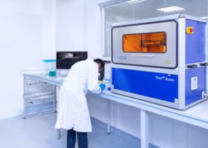
    RNA characterization is a challenge using current technologies. Refeyn claims its mass photometry system—seen here at lab in Waltham, Massachusetts—reduces the number of assays required. [Refeyn]

    This view is shared by Ranaghan from Refeyn, who cites the shift toward continuous flow manufacturing as one of the most exciting advances in the field.

    “Moving away from traditional batch processing to a continuous flow model is highly logical because it minimizes the time that the fragile mRNA product spends exposed to active reaction components that drive fragmentation and aggregation. This shift not only improves overall yield and intactness, especially for massive transcripts like saRNA, but it also creates a mandate for faster, real-time analytics.”

    For Parexel’s Gangurde, efforts to make production more efficient and sustainable will drive innovation, with the WHO’s recently launched mRNA Technology Transfer Programme Phase 2.04 being a case in point.

    “This is a major global initiative aimed at scaling up commercially viable mRNA-based vaccine and therapeutic production in low- and middle-income countries—where the need for these therapies is most critical.”

    Launched in 2021 in collaboration with the Medicines Patent Pool (MPP), the initiative has enabled 15 partners across Latin America, Africa, Eastern Europe, and Asia to receive foundational mRNA technology.

    Phase 2.0 focuses on building local manufacturing capacity so countries can make mRNA vaccines and therapeutics for future outbreaks and diseases like influenza, TB, HIV, malaria, dengue, and leishmaniasis, as well as oncology drugs and mAbs.

    Gangurde says, “By decentralizing production, the program reduces reliance on a few high-income countries for mRNA supply, supporting global access for mRNA-based therapies.”

    References

    1. Beyrer C. The Long History of mRNA Vaccines. Johns Hopkins Bloomberg School of Public Health; 2021. Accessed August 12, 2026.
    2. Chatterjee S, Bhattacharya M, Lee SS, Chakraborty C. An insight of different classes of RNA-based therapeutic, nanodelivery and clinical status: Current landscape. Current Research in Biotechnology 2023;6:100150-100150. doi:10.1016/j.crbiot.2023.100150
    3. Parvin N, Mandal TK, Joo SW. The Impact of COVID-19 on RNA Therapeutics: A Surge in Lipid Nanoparticles and Alternative Delivery Systems, Systems. Pharmaceutics 2024; 16(11): 1366. Doi: 10.3390/pharmaceutics16111366
    4. World Health Organization: WHO. mRNA Technology Transfer Programme’s Phase 2.0 discussed with partners on the sidelines of G20 Summit. Who. June 13, 2025. Accessed August 12, 2026.
  • From Discovery to Development

    From Discovery to Development

    The Era of Modality Convergence: Why Drug Development Is Being Rewritten

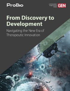
    From Discovery to Development: Navigating the New Era of Therapeutic Innovation eBook cover

    The life sciences industry is entering a new era, defined not by a single breakthrough technology but by the convergence of many. Artificial intelligence is accelerating drug discovery, cell and gene therapies are expanding treatment possibilities, and antibody engineering continues to evolve beyond traditional monoclonal antibodies. Meanwhile, mRNA platforms, viral vectors, antibody-drug conjugates (ADCs), multispecific antibodies, and other advanced therapeutic modalities are transforming how developers approach previously intractable diseases. Taken individually, each of these innovations represents a significant advancement. Together, they are reshaping the drug development landscape.

    For decades, therapeutic development followed a familiar path: identify a target, optimize a lead candidate, advance through preclinical studies, and move into clinical evaluation. While the science was challenging, the overall framework remained relatively consistent. Today, that framework is evolving rapidly. Developers are no longer limited to choosing between small molecules and conventional biologics. Instead, they have access to a growing toolkit of therapeutic technologies, each with unique advantages, manufacturing requirements, analytical challenges, and regulatory considerations. At the same time, advances in computational biology and AI-driven discovery are generating more candidates than ever before.

    As a result, the challenge is no longer simply finding promising molecules. Increasingly, success depends on identifying candidates that can be manufactured, scaled, and translated into viable therapies. Decisions once reserved for later stages, including developability, manufacturability, analytical characterization, and clinical strategy, must now be addressed much earlier in the process. Drug development is becoming a continuous, interconnected journey rather than a series of separate steps.

    The boundaries between traditional disciplines are also beginning to blur. Cell therapies may incorporate viral vectors. Gene therapies may leverage lipid nanoparticles. Antibody programs may evolve into multispecific or conjugated formats. The future of drug development is not defined by any single modality, but by the ability to integrate technologies effectively.

    Organizations that combine discovery expertise, modality-specific knowledge, CMC capabilities, and manufacturing execution will be best positioned to navigate this increasingly complex environment. The articles that follow examine key trends driving this transformation, from AI-enabled discovery and experimental validation to advanced cell, gene, and biologic platforms, and explore how the next generation of therapies will move from concept to clinic.

  • How Preclinical Manufacturing Can De-Risk the Path to GMP-Ready Viral Vector Programs

    How Preclinical Manufacturing Can De-Risk the Path to GMP-Ready Viral Vector Programs

    To move viral vector programs from early research toward the clinic, sponsors have to generate the right material and data during development. Non-GMP production of adeno-associated virus (AAV), lentiviral, and adenoviral vectors can support applications ranging from R&D and animal studies to assay development, toxicology planning, and early manufacturability assessments. But to be successful, sponsors have to align production scale, analytical testing, documentation, and material use with the specific questions their studies aim to answer.

    In this GEN webinar, Chris Nieder, an expert in viral vector programs, will examine how strategically designed preclinical manufacturing campaigns provide a practical foundation for GMP production. He will discuss topics such as how to select from among shake flasks, development-scale bioreactors, and pilot-scale approaches; and how to use early process and analytical data to identify yield and scalability challenges. Attendees will also learn how early CDMO engagement can clarify manufacturing scope, timelines, costs, and GMP-readiness without compromising expectations for non-GMP material. Key takeaways from the webinar include:

    • How early-stage sponsors can use non-GMP AAV, lentiviral, and adenoviral production to support R&D, animal studies, toxicology planning, assay development, and manufacturability assessment
    • How process and analytical data generated before GMP can reduce duplicated prep work, lower technical risk, and streamline later scope definition
    • When shake flasks, development-scale bioreactors, or pilot-scale production can provide the most useful insight for the next development step
    • How to align material use, analytical testing, documentation, and expectations without over-positioning preclinical material as clinical-grade supply

    A live Q&A session will follow the presentation offering you a chance to pose questions to our expert panelist.

    Produced with support from:

    skpharmteco logo

  • ARPA-H’s BoSS Program Funds Three Teams Tackling Cold Chain Challenges for Cell Therapies

    ARPA-H’s BoSS Program Funds Three Teams Tackling Cold Chain Challenges for Cell Therapies

    The Advanced Research Projects Agency for Health, (ARPA-H), an agency within the United States’ Department of Health and Human Services (HHS), recently announced awards and teams involved in its BioStabilization Systems (BoSS) program, an initiative that aims to develop new methods for producing, storing, and transporting cell-based biologic medicines at room temperature. ARPA-H has committed to provide up to $87 million across the four-year program. This round of funding will support Phase I of the program.

    The teams have been tasked with pursuing new technologies that confer long-term, shelf-life stability to cell-based therapies and biologics making it possible to store and ship them without refrigeration. To accomplish this goal, BoSS teams will work across two technical areas. They will work on interventions to stabilize cells for storage and then reactivate them on demand as well as on creating novel processing systems that ensure these interventions are efficient and scalable for manufacturing. 

    This week, members of the Reversible Ambient Temperature (RAB) team said that they have been awarded up to $7.3 million initially from ARPA-H. The RAB team includes representatives from Draper, a non-profit research, development, and manufacturing company, which serves as the administrative lead,and Likarda, a biotechnology company that develops hydrogel technologies for delivering and preserving cell therapies and biologics, which serves as the technical lead for the team. Other members of the team include scientists from CaseBioscience, CPSI Biotech, Fresenius Kabi, MiNK Therapeutics, and University of Colorado. 

    For their part, this team will use AI-assisted design of experiments to optimize formulation, quiescence induction parameters, hydrogel composition, and recovery media as well as freeze-assisted droplet drying to achieve a stable product. Specifically, the RAB team’s biostabilization strategy, led by Likarda, will focus on simultaneous control of cell state, physical environment, and the cellular stress responses activated during freezing, dehydration, storage, and recovery. Meanwhile, the bioprocessing engineering team, led by Draper, will convert the manual steps of biostabilization into an automated, commercially viable workflow and will develop custom instruments to scale up the processes.

    Likarda will contribute its proprietary Core-Shell Spherification hydrogel technology, which is designed to encapsulate and protect living cells while maintaining viability and function. CaseBioscience will provide expertise in clinical media formulation, biopreservation, and cellular metabolism to the development and evaluation of novel approaches for long-term ambient stabilization of therapeutic cells.

    “The RAB team brings together deep expertise in cryoprotection, encapsulation, and cell state, with a focus on protecting cell viability throughout the stabilization and reanimation process, and the team has direct experience bringing products to market,” said Jason Fiering, distinguished technical staff at Draper. “Working in parallel, our interdisciplinary engineering team will develop the customized instruments needed to transfer these processes to clinical manufacturing environments.” 

    In his comments, John Baust, PhD, an expert in biopreservation and collaborator on the project with CPSI Biotech noted that “the concept of preserving living cells in a dry state has been pursued for more than two decades, but significant scientific and engineering barriers have yielded limited success.” The BoSS program is an opportunity “to tackle this from multiple scientific and engineering directions simultaneously” that if successful “could represent a paradigm shift in how biologics are preserved, stored, and distributed.”

    Other teams announced by ARPA-H include one led by DesiCorp. This team includes members from the University of Louisville, University of Texas at Austin, Via Therapeutics, VeriSIM Life, and Ossium Health. They will leverage high-throughput screening to identify biocompatible formulations that slow cellular metabolism and AI-assisted process optimization to develop a thin-film freeze-drying (TFFD) system that stabilizes cells at room temperature. 

    DesiCorp will lead integration of the overall workflow by using a high-throughput screening platform to rapidly identify biocompatible formulations that enable cells to survive TFFD and resume function after rehydration. Meanwhile, UT Austin researchers will help optimize the TFFD critical process parameters and composition/material attributes, while also contributing expertise in aseptic processing and scaling up the technology. 

    A third team is led by the University of California, Davis and includes scientists from Mayo Clinic, Case Western Reserve University, University of Georgia, and Sersense. The team will induce a low metabolic state in cells using light-responsive gels and microfluidic processing techniques to produce cells that are stable at room temperature. 

    Recently, that team announced that it had been awarded up to $6 million, initially,  from ARPA-H to fund its project dubbed CYBORGEL. Their efforts will leverage research done in the laboratory of Cheemeng Tan, PhD, a professor of biomedical engineering at UC Davis. Previously, he and his collaborators formed water-rich polymer networks inside living cells to create cell-material hybrids. This process, called intracellular gelation, forms a gel inside the cell and is capable of halting cell division while preserving cellular function. Tan and his team dubbed this cellular product a “cyborg” since the gelation process combines living cells with synthetic materials. 

    Through CYBORGEL, the collaborators will investigate whether this intracellular gelation process can be extended to reduce the cost and simplify the storage, transportation and delivery of cellular therapies. The researchers will also test whether CYBORGEL cells remain therapeutically useful after being returned to their original state. For instance, one study will test the effectiveness of reanimated CAR T cells for existing clinical trials on cancer immunotherapies. Another will test it on placental mesenchymal stem cells used to treat spina bifida, a medical intervention being developed by UC Davis researchers. 

    As part of the initiative, the American Type Culture Collection (ATCC) has been tapped to serve as the IV&V partner. ATCC will provide working cell banks of government-selected cell types that will be used for demonstrations. They will also provide unbiased assessments of both the viability and function of the preserved products and the design features of the preservation systems.  

    The first phase of the BoSS program kicked off in September and is expected to run for 15 months. Over the four-year multi-phase program, the teams’ approaches will be evaluated against increasingly stringent benchmarks for cell viability, production speed, and shelf-life stability. If successful, work under the BoSS program could enable lower costs for cell-based drugs, reduce the risk of therapeutic and pharmaceutical product losses, and enable domestic reserves for public health preparedness.