Category: Artificial

  • Artificial neurons successfully communicate with living brain cells

    Artificial neurons successfully communicate with living brain cells

    Engineers at Northwestern University have created printed artificial neurons that go beyond imitation and can directly interact with real brain cells. These flexible, low-cost devices produce electrical signals that closely resemble those generated by living neurons, allowing them to activate biological brain tissue.

    In experiments using slices of mouse brain, the artificial neurons successfully triggered responses in real neurons. This result shows a new level of compatibility between electronic devices and living neural systems.

    Toward Brain Interfaces and Energy-Efficient AI

    This advance moves researchers closer to electronics that can directly interface with the nervous system. Potential uses include brain-machine interfaces and neuroprosthetics, such as implants that could help restore hearing, vision, or movement.

    The technology also points toward a new generation of computing systems inspired by the brain. By replicating how neurons communicate, future hardware could perform complex tasks using far less energy. The brain remains the most energy-efficient computing system known, and scientists hope to apply its principles to modern technology.

    The study will be published on April 15 in the journal Nature Nanotechnology.

    “The world we live in today is dominated by artificial intelligence (AI),” said Northwestern’s Mark C. Hersam, who led the study. “The way you make AI smarter is by training it on more and more data. This data-intensive training leads to a massive power-consumption problem. Therefore, we have to come up with more efficient hardware to handle big data and AI. Because the brain is five orders of magnitude more energy efficient than a digital computer, it makes sense to look to the brain for inspiration for next-generation computing.”

    Hersam is an expert in brain-inspired computing and holds multiple roles at Northwestern University, including the Walter P. Murphy Professor of Materials Science and Engineering at the McCormick School of Engineering. He also is a professor of medicine at Northwestern University Feinberg School of Medicine and a professor of chemistry at the Weinberg College of Arts and Sciences. In addition, he serves as chair of the department of materials science and engineering, director of the Materials Research Science and Engineering Center, and a member of the International Institute for Nanotechnology. He co-led the study with Vinod K. Sangwan, a research associate professor at McCormick.

    Why the Brain Outperforms Traditional Silicon

    Modern computers handle increasing workloads by packing billions of identical transistors onto rigid, two-dimensional silicon chips. Each component behaves the same way, and once manufactured, the system remains fixed.

    The brain works very differently. It consists of many types of neurons, each with specialized roles, arranged in soft, three-dimensional networks. These networks are constantly changing, forming and adjusting connections as learning occurs.

    “Silicon achieves complexity by having billions of identical devices,” Hersam said. “Everything is the same, rigid and fixed once it’s fabricated. The brain is the opposite. It’s heterogeneous, dynamic and three-dimensional. To move in that direction, we need new materials and new ways to build electronics.”

    Although artificial neurons have been developed before, most produce overly simple signals. To achieve more complex behavior, engineers typically need large networks of devices, which increases energy use.

    Printable Materials Enable Brain-Like Behavior

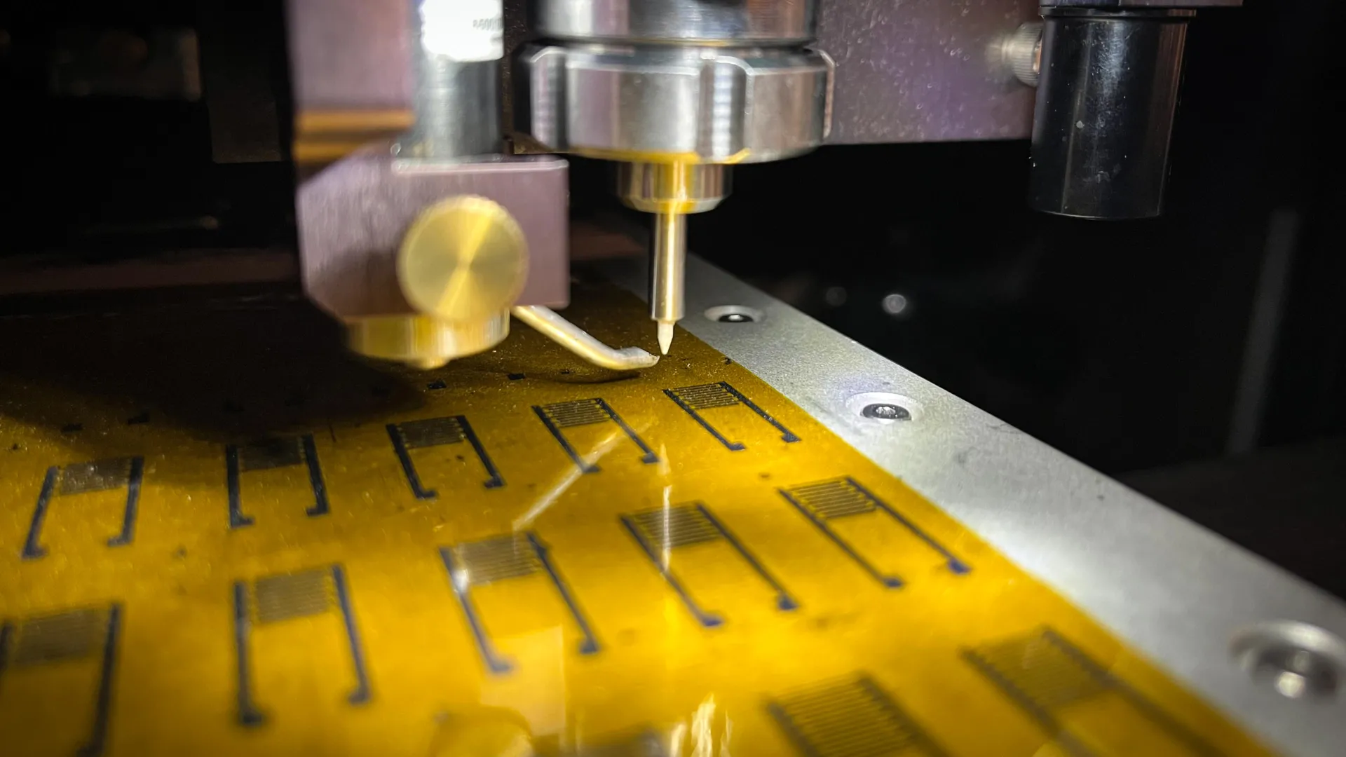
    To better replicate real neural activity, Hersam’s team built artificial neurons using soft, printable materials that more closely match the brain’s structure. Their approach relies on electronic inks made from nanoscale flakes of molybdenum disulfide (MoS2), which acts as a semiconductor, and graphene, which serves as an electrical conductor. These materials were deposited onto flexible polymer surfaces using aerosol jet printing.

    Previously, researchers treated the polymer in these inks as a flaw because it interfered with electrical performance. As a result, they removed it after printing. In this work, the team used that same feature to enhance the device.

    “Instead of fully removing the polymer, we partially decompose it,” he said. “Then, when we pass current through the device, we drive further decomposition of the polymer. This decomposition occurs in a spatially inhomogeneous manner, leading to formation of a conductive filament, such that all the current is constricted into a narrow region in space.”

    That narrow conductive path produces a sudden electrical response similar to a neuron firing. The resulting device can generate a wide variety of signals, including single spikes, continuous firing, and bursting patterns, closely resembling real neural communication.

    Because each artificial neuron can produce more complex signals, fewer components are needed to perform advanced tasks. This could significantly improve computing efficiency.

    Testing Artificial Neurons on Real Brain Tissue

    To evaluate whether the artificial neurons could truly interact with living systems, the researchers partnered with Indira M. Raman, the Bill and Gayle Cook Professor of Neurobiology at Weinberg. Her team applied the artificial signals to slices of mouse cerebellum.

    The results showed that the electrical spikes matched key biological properties, including their timing and duration. These signals reliably activated real neurons and triggered neural circuits in a way similar to natural brain activity.

    “Other labs have tried to make artificial neurons with organic materials, and they spiked too slowly,” Hersam said. “Or they used metal oxides, which are too fast. We are within a temporal range that was not previously demonstrated for artificial neurons. You can see the living neurons respond to our artificial neuron. So, we’ve demonstrated signals that are not only the right timescale but also the right spike shape to interact directly with living neurons.”

    Low-Cost, Sustainable Manufacturing and AI Implications

    Beyond performance, the new approach offers environmental and practical advantages. The manufacturing process is simple and inexpensive, and the additive printing method places material only where it is needed, reducing waste.

    Improving energy efficiency is especially important as artificial intelligence systems grow more demanding. Large data centers already consume vast amounts of power and require significant water for cooling.

    “To meet the energy demands of AI, tech companies are building gigawatt data centers powered by dedicated nuclear power plants,” Hersam said. “It is evident that this massive power consumption will limit further scaling of computing since it’s hard to imagine a next-generation data center requiring 100 nuclear power plants. The other issue is that when you’re dissipating gigawatts of power, there’s a lot of heat. Because data centers are cooled with water, AI is putting severe stress on the water supply. However you look at it, we need to come up with more energy-efficient hardware for AI.”

    The study, “Multi-order complexity spiking neurons enabled by printed MoS2 memristive nanosheet networks,” was supported by the National Science Foundation.


  • Artificial intelligence and genetics can help farmers grow corn with less fertilizer

    Artificial intelligence and genetics can help farmers grow corn with less fertilizer

    New York University scientists are using artificial intelligence to determine which genes collectively govern nitrogen use efficiency in plants such as corn, with the goal of helping farmers improve their crop yields and minimize the cost of nitrogen fertilizers.

    “By identifying genes-of-importance to nitrogen utilization, we can select for or even modify certain genes to enhance nitrogen use efficiency in major US crops like corn,” said Gloria Coruzzi, the Carroll & Milton Petrie Professor in NYU’s Department of Biology and Center for Genomics and Systems Biology and the senior author of the study, which appears in the journal The Plant Cell.

    In the last 50 years, farmers have been able to grow larger crop yields thanks to major improvements in plant breeding and fertilizers, including how efficiently crops uptake and use nitrogen, the key component of fertilizers.

    Still, most crops only use roughly 55 percent of the nitrogen in fertilizer that farmers apply to their fields, while the remainder ends up in the surrounding soil. When nitrogen seeps into groundwater, it can contaminate drinking water and cause harmful algae blooms in lakes, rivers, reservoirs, and warm ocean waters. Furthermore, the unused nitrogen that remains in the soil is converted by bacteria into nitrous oxide, a potent greenhouse gas that is 265 times more effective at trapping heat over a 100-year period than is carbon dioxide.

    The United States is the world’s leading producer of corn. This major cash crop requires large amounts of nitrogen to grow, but much of the fertilizer fed to corn is not taken up or used. Corn’s low nitrogen use efficiency presents a financial challenge for farmers, given the increasing costs of fertilizer — the majority of which is imported — and also risks harming the soil, water, air, and climate.

    To address this challenge in corn and other crops, NYU researchers have developed a novel process to improve nitrogen use efficiency that integrates plant genetics with machine learning, a type of artificial intelligence that detects patterns in data — in this case, to associate genes with a trait (nitrogen use efficiency).

    Using a model-to-crop approach, NYU researchers tracked the evolutionary history of corn genes that are shared with Arabidopsis, a small flowering weed often used as a model organism in plant biology due to the ease of studying it in the lab using the power of molecular genetic approaches. In a previous study published in Nature Communications, Coruzzi’s team identified genes whose responsiveness to nitrogen was conserved between corn and Arabidopsis and validated their role in plants.

    In The Plant Cell study, their most recent on this topic, the NYU researchers built upon their work in corn and Arabidopsis to identify how nitrogen use efficiency is governed by groups of genes — also known as “regulons” — that are activated or repressed by the same transcription factor (a regulatory protein).

    “Traits like nitrogen use efficiency or photosynthesis are never controlled by one single gene. The beauty of the machine learning process is it learns sets of genes that are collectively responsible for a trait, and can also identify the transcription factor or factors that control these sets of genes,” said Coruzzi.

    The researchers first used RNA sequencing to measure how genes in corn and Arabidopsis respond to nitrogen treatment. Using these data, they trained machine learning models to identify nitrogen-responsive genes conserved across corn and Arabidopsis varieties, as well as the transcription factors that regulate the genes-of-importance to nitrogen use efficiency (NUE). For each “NUE Regulon” — the transcription factor and corresponding set of regulated NUE genes — the researchers calculated a collective machine learning score and then ranked the top performers based on how well the combined expression levels could accurately predict how efficiently nitrogen is used in field-grown varieties of corn.

    For the top-ranked NUE Regulons, the researchers used cell-based studies in both corn and Arabidopsis to validate the machine learning predictions for the set of genes in the genome that are regulated by each transcription factor. These experiments confirmed NUE Regulons for two corn transcription factors (ZmMYB34/R3) that regulate 24 genes controlling nitrogen use as well as for a closely related transcription factor in Arabidopsis (AtDIV1), which regulates 23 target genes sharing a genetic history with corn that also control nitrogen use. When fed back into the machine learning models, these model-to-crop conserved NUE Regulons significantly enhanced the ability of AI to predict nitrogen use efficiency across field-grown corn varieties.

    Identifying NUE Regulons of collective genes and related transcription factors that govern nitrogen use will enable crop scientists to breed or engineer corn that needs less fertilizer.

    “By looking at corn hybrids at the seedling stage to see if expression of the identified genes-of-importance to nitrogen use efficiency is high, rather than planting them in the field and measuring their nitrogen use, we can use molecular markers to select the hybrids at the seedling stage that are most efficient in nitrogen use, and then plant those varieties,” said Coruzzi. “This will not only result in a cost savings for farmers, but also reduce the harmful effects of nitrogen pollution of groundwaters and nitrous oxide greenhouse gas emissions.”

    New York University has filed a patent application covering the research and findings described in this paper. Additional study authors include Ji Huang, Tim Jeffers, Nathan Doner, Hung-Jui Shih, Samantha Frangos, and Manpreet Singh Katari of NYU; Chia-Yi Cheng of NYU and National Taiwan University, and Matthew Brooks of the US Department of Agriculture’s Agricultural Research Service. The research was supported by the National Science Foundation Plant Genome Research Program (IOS-1339362) and the National Institutes of Health (R01-GM121753, F32GM116347).

  • Artificial intelligence tools make education materials more patient friendly

    Artificial intelligence tools make education materials more patient friendly

    Artificial intelligence (AI) tools significantly improve the readability of online patient education materials (PEMs), making them more accessible, a new study shows.

    Led by researchers at NYU Langone Health, the study focused on the readability of PEMs available on the websites of the American Heart Association (AHA), American Cancer Society (ACS), and American Stroke Association (ASA). According to the researchers, these materials help patients make decisions about their health care but often exceed the recommended reading level of grade 6, making them difficult for many patients to understand.

    For the study, researchers evaluated the capabilities of three large language models (LLMs) — ChatGPT, Gemini, and Claude — to optimize the readability of PEMs without compromising accuracy. These generative AI tools are designed to simplify complex texts by predicting the next word in a sentence based on extensive Internet data. This next-word prediction gives such models the ability to rewrite any article in simpler language as directed.

    Published online April 10 in the Journal of Medical Internet Research, the study involved 60 randomly selected PEMs from the AHA, ACS, and ASA websites. Researchers prompted the LLMs to simplify the reading level of the materials. Results showed that the original readability scores were significantly above the recommended level of grade 6, with mean grade-level scores of 10.7, 10, and 9.6, respectively.

    After optimization by the LLMs, readability scores improved significantly across all three websites. ChatGPT improved readability to a mean grade level of 7.6, Gemini to 6.6, and Claude to 5.6. Word counts were also significantly reduced, making the materials more concise.

    “Our study shows that widely used large language models have the potential to transform patient education materials into more readable content, which is essential for patient empowerment and better health outcomes,” said study senior author Jonah Feldman, MD, medical director of transformation and informatics at NYU Langone.

    “Our findings demonstrate that even expert-composed education materials, which are already patient-directed, can benefit from AI-driven improvements,” said Feldman, who also serves as an assistant professor at NYU Grossman Long Island School of Medicine.

    This study, the researchers say, provides an example of how healthcare organizations can apply AI to make clinical communication more patient friendly. Prior studies demonstrated the capabilities of AI models to create patient-focused explanations of heart test results, to draft responses to electronic advice queries, and to generate human-friendly summaries of complex medical reports.

    “The breadth of possible AI offerings shows how technology can be leveraged to transform the patient experience across health care systems, and not just in the United States,” said study co-author Paul Testa, MD, JD, MPH, chief health informatics officer at NYU Langone.

    “These studies are not just theoretical — after demonstrating their effectiveness, we are actively putting these AI tools into practice,” said Testa, who is also a clinical professor at NYU Grossman School of Medicine.

    According to Testa, the NYU Langone team is already using the same AI tools in a randomized controlled trial that incorporates AI-generated, patient-friendly summaries for hospital discharge instructions, with the goal to evaluate their effectiveness in improving patient comprehension and satisfaction. The researchers hope to show that providing clear and accessible discharge instructions will help ensure better postdischarge care and smoother transitions.

    “Generating real-world evidence through randomized trials is crucial for validating the effectiveness of AI tools in clinical settings,” said study co-author Jonah Zaretsky, MD, associate chief of medicine at NYU Langone Hospital — Brooklyn. “This approach ensures that the AI-generated documentation is not only accurate but also genuinely beneficial for patients and their families,” added Zaretsky, a clinical assistant professor at NYU Grossman School of Medicine.

    The study was self-funded by NYU Langone. Besides Feldman, Testa, and Zaretsky, NYU Langone researchers involved in the study were lead author John Will, and co-authors Mahin Gupta and Aliesha Dowlath.

  • A new artificial intelligence tool for cancer

    A new artificial intelligence tool for cancer

    Scientists at Harvard Medical School have designed a versatile, ChatGPT-like AI model capable of performing an array of diagnostic tasks across multiple forms of cancers.

    The new AI system, described Sept. 4 in Nature, goes a step beyond many current AI approaches to cancer diagnosis, the researchers said. (DOI 10.1038/s41586-024-07894-z)

    Current AI systems are typically trained to perform specific tasks — such as detecting cancer presence or predicting a tumor’s genetic profile — and they tend to work only in a handful of cancer types. By contrast, the new model can perform a wide array of tasks and was tested on 19 cancer types, giving it a flexibility like that of large language models such as ChatGPT.

    While other foundation AI models for medical diagnosis based on pathology images have emerged recently, this is believed to be the first to predict patient outcomes and validate them across several international patient groups.

    “Our ambition was to create a nimble, versatile ChatGPT-like AI platform that can perform a broad range of cancer evaluation tasks,” said study senior author Kun-Hsing Yu, assistant professor of biomedical informatics in the Blavatnik Institute at Harvard Medical School. “Our model turned out to be very useful across multiple tasks related to cancer detection, prognosis, and treatment response across multiple cancers.”

    The AI model, which works by reading digital slides of tumor tissues, detects cancer cells and predicts a tumor’s molecular profile based on cellular features seen on the image with superior accuracy to most current AI systems. It can forecast patient survival across multiple cancer types and accurately pinpoint features in the tissue that surrounds a tumor — also known as the tumor microenvironment — that are related to a patient’s response to standard treatments, including surgery, chemotherapy, radiation, and immunotherapy. Finally, the team said, the tool appears capable of generating novel insights — it identified specific tumor characteristics previously not known to be linked to patient survival.

    The findings, the research team said, add to growing evidence that AI-powered approaches can enhance clinicians’ ability to evaluate cancers efficiently and accurately, including the identification of patients who might not respond well to standard cancer therapies.

    “If validated further and deployed widely, our approach, and approaches similar to ours, could identify early on cancer patients who may benefit from experimental treatments targeting certain molecular variations, a capability that is not uniformly available across the world,” Yu said.

    Training and performance

    The team’s latest work builds on Yu’s previous research in AI systems for the evaluation of colon cancer and brain tumors. These earlier studies demonstrated the feasibility of the approach within specific cancer types and specific tasks.

    The new model, called CHIEF (Clinical Histopathology Imaging Evaluation Foundation), was trained on 15 million unlabeled images chunked into sections of interest. The tool was then trained further on 60,000 whole-slide images of tissues including lung, breast, prostate, colorectal, stomach, esophageal, kidney, brain, liver, thyroid, pancreatic, cervical, uterine, ovarian, testicular, skin, soft tissue, adrenal gland, and bladder. Training the model to look both at specific sections of an image and the whole image allowed it to relate specific changes in one region to the overall context. This approach, the researchers said, enabled CHIEF to interpret an image more holistically by considering a broader context, instead of just focusing on a particular region.

    Following training, the team tested CHIEF’s performance on more than 19,400 whole-slide images from 32 independent datasets collected from 24 hospitals and patient cohorts across the globe.

    Overall, CHIEF outperformed other state-of-the-art AI methods by up to 36 percent on the following tasks: cancer cell detection, tumor origin identification, predicting patient outcomes, and identifying the presence of genes and DNA patterns related to treatment response.Because of its versatile training, CHIEF performed equally well no matter how the tumor cells were obtained — whether via biopsy or through surgical excision. And it was just as accurate, regardless of the technique used to digitize the cancer cell samples. This adaptability, the researchers said, renders CHIEF usable across different clinical settings and represents an important step beyond current models that tend to perform well only when reading tissues obtained through specific techniques.

    Cancer detection

    CHIEF achieved nearly 94 percent accuracy in cancer detection and significantly outperformed current AI approaches across 15 datasets containing 11 cancer types. In five biopsy datasets collected from independent cohorts, CHIEF achieved 96 percent accuracy across multiple cancer types including esophagus, stomach, colon, and prostate. When the researchers tested CHIEF on previously unseen slides from surgically removed tumors of the colon, lung, breast, endometrium, and cervix, the model performed with more than 90 percent accuracy.

    Predicting tumors’ molecular profiles

    A tumor’s genetic makeup holds critical clues to determine its future behavior and optimal treatments. To get this information, oncologists order DNA sequencing of tumor samples, but such detailed genomic profiling of cancer tissues is not done routinely nor uniformly across the world due to the cost and time involved in sending samples to specialized DNA sequencing labs. Even in well-resourced regions, the process could take several weeks. It’s a gap that AI could fill, Yu said.

    Quickly identifying cellular patterns on an image suggestive of specific genomic aberrations could offer a quick and cost-effective alternative to genomic sequencing, the researchers said.

    CHIEF outperformed current AI methods for predicting genomic variations in a tumor by looking at the microscopic slides. This new AI approach successfully identified features associated with several important genes related to cancer growth and suppression, and it predicted key genetic mutations related to how well a tumor might respond to various standard therapies. CHIEF also detected specific DNA patterns related to how well a colon tumor might respond to a form of immunotherapy called immune checkpoint blockade. When looking at whole-tissue images, CHIEF identified mutations in 54 commonly mutated cancer genes with an overall accuracy of more than 70 percent, outperforming the current state-of-the-art AI method for genomic cancer prediction. Its accuracy was greater for specific genes in specific cancer types.

    The team also tested CHIEF on its ability to predict mutations linked with response to FDA-approved targeted therapies across 18 genes spanning 15 anatomic sites. CHIEF attained high accuracy in multiple cancer types, including 96 percent in detecting a mutation in a gene called EZH2 common in a blood cancer called diffuse large B-cell lymphoma. It achieved 89 percent for BRAF gene mutation in thyroid cancer, and 91 percent for NTRK1 gene mutation in head and neck cancers.

    Predicting patient survival

    CHIEF successfully predicted patient survival based on tumor histopathology images obtained at the time of initial diagnosis. In all cancer types and all patient groups under study, CHIEF distinguished patients with longer-term survival from those with shorter-term survival. CHIEF outperformed other models by 8 percent. And in patients with more advanced cancers, CHIEF outperformed other AI models by 10 percent. In all, CHIEF’s ability to predict high versus low death risk was tested and confirmed across patient samples from 17 different institutions.

    Extracting novel insights about tumor behavior

    The model identified tell-tale patterns on images related to tumor aggressiveness and patient survival. To visualize these areas of interest, CHIEF generated heat maps on an image. When human pathologists analyzed these AI-derived hot spots, they saw intriguing signals reflecting interactions between cancer cells and surrounding tissues. One such feature was the presence of greater numbers of immune cells in areas of the tumor in longer-term survivors, compared with shorter-term survivors. That finding, Yu noted, makes sense because a greater presence of immune cells may indicate the immune system has been activated to attack the tumor.

    When looking at the tumors of shorter-term survivors, CHIEF identified regions of interest marked by the abnormal size ratios between various cell components, more atypical features on the nuclei of cells, weak connections between cells, and less presence of connective tissue in the area surrounding the tumor. These tumors also had a greater presence of dying cells around them. For example, in breast tumors, CHIEF pinpointed as an area of interest the presence of necrosis — or cell death — inside the tissues. On the flip side, breast cancers with higher survival rates were more likely to have preserved cellular architecture resembling heathy tissues. The visual features and zones of interest related to survival varied by cancer type, the team noted.

    Next steps

    The researchers said they plan to refine CHIEF’s performance and augment its capabilities by:

    • Conducting additional training on images of tissues from rare diseases and non-cancerous conditions
    • Including samples from pre-malignant tissues before cells become fully cancerous
    • Exposing the model to more molecular data to enhance its ability to identify cancers with different levels of aggressiveness
    • Training the model to also predict the benefits and adverse effects of novel cancer treatments in addition to standard treatments
  • Artificial intelligence blood test provides a reliable way to identify lung cancer

    Artificial intelligence blood test provides a reliable way to identify lung cancer

    Using artificial intelligence technology to identify patterns of DNA fragments associated with lung cancer, researchers from the Johns Hopkins Kimmel Cancer Center and other institutions have developed and validated a liquid biopsy that may help identify lung cancer earlier.

    In a prospective study published June 3 in Cancer Discovery, the team demonstrated that artificial intelligence technology could identify people more likely to have lung cancer based on DNA fragment patterns in the blood. The study enrolled about 1,000 participants with and without cancer who met the criteria for traditional lung cancer screening with low-dose computed tomography (CT). Individuals were recruited to participate at 47 centers in 23 U.S. states. By helping to identify patients most at risk and who would benefit from follow-up CT screening, this new blood test could potentially boost lung cancer screening and reduce death rates, according to computer modeling by the team.

    “We have a simple blood test that could be done in a doctor’s office that would tell patients whether they have potential signs of lung cancer and should get a follow-up CT scan,” says the study’s corresponding author, Victor E. Velculescu, M.D., Ph.D., professor of oncology and co-director of the Cancer Genetics and Epigenetics program at the Johns Hopkins Kimmel Cancer Center.Lung cancer is the deadliest cancer in the United States, according to the National Cancer Institute, and worldwide, according to the World Health Organization. Yearly screening with CT scans in high-risk patients can help detect lung cancers early, when they are most treatable, and help avert lung cancer deaths. Screening is recommended by the U.S. Preventive Services Task Force for 15 million people nationally who are between ages 50 and 80 and have a smoking history, yet only about 6%-10% of eligible individuals are screened each year. People may be reticent to follow through on screening, Velculescu explains, due to the time it takes to arrange and go to an appointment, and the low doses of radiation they are exposed to from the scan.

    To help overcome some of these hurdles, Velculescu and his colleagues developed a test over the past five years that uses artificial intelligence to detect patterns of DNA fragments found in patients with lung cancer. It takes advantage of differences in how DNA is packaged in normal and cancer cells. DNA is neatly and consistently folded up in healthy cells, almost like a rolled-up ball of yarn, but DNA in cancer cells is more disorganized. When both types of cells die, fragments of DNA end up in the blood. The DNA fragments in patients with cancer tend to be more chaotic and irregular than the DNA fragments found in individuals who do not have cancer.

    The team trained artificial intelligence software to identify the specific patterns of DNA fragments seen in the blood of 576 people with or without lung cancer. Then, they verified that the method worked in a second group of 382 people with and without cancer. Based on their analyses, the test has a negative predictive value of 99.8%, meaning that only 2 in 1,000 individuals tested may be missed and have lung cancer.

    The group’s computer simulations showed that if the test boosted the rate of lung cancer screening to 50% within five years, it could quadruple the number of lung cancers detected and increase the proportion of cancers detected early — when they are most treatable — by about 10%. That could prevent about 14,000 cancer deaths over five years.

    “The test is inexpensive and could be done at a very large scale,” Velculescu says. “We believe it will make lung cancer screening more accessible and help many more people get screened. This will lead to more cancers being detected and treated early.”

    The test is currently available through DELFI Diagnostics for use as a laboratory-based test under the Clinical Laboratory Improvement Amendments. However, the team plans to seek approval from the U.S. Food and Drug Administration for lung cancer screening. Velculescu colleagues also plan to study whether a similar approach could be used to detect other types of cancer.

    Robert B. Scharpf of Johns Hopkins co-authored the study. Additional co-authors were from the Cleveland Clinic, DELFI Diagnostics, Medicus Economics LLC, Miami Cancer Institute, the Pan American Center for Oncology, Washington University, Centura Health, Vanderbilt Health, Stratevi, Massachusetts General Hospital, the Medical University of South Carolina, the Department of Veterans Affairs, the Perelman School of Medicine at the University of Pennsylvania, New York University Langone Health, Allegheny Health Network and Memorial Sloan Kettering Cancer Center.

    The work was supported in part by DELFI Diagnostics, the Dr. Miriam and Sheldon G. Adelson Medical Research Foundation, Stand Up To Cancer-LUNGevity-American Lung Association Lung Cancer Interception Dream Team Translational Research Grant, Stand Up To Cancer-DCS International Translational Cancer Research Dream Team Grant, the Gray Foundation, The Honorable Tina Brozman Foundation, the Commonwealth Foundation, the Cole Foundation and the National Institutes of Health.

    Velculescu and Scharpf are inventors on patent applications submitted by The Johns Hopkins University related to cell-free DNA for cancer detection that have been licensed to DELFI Diagnostics, LabCorp, Qiagen, Sysmex, Agios, Genzyme, Esoterix, Ventana and ManaT Bio. Velculescu divested his equity in Personal Genome Diagnostics (PGDx) to LabCorp in February 2022. Velculescu is a founder of DELFI Diagnostics, serves on the board of directors, and owns DELFI Diagnostics stock. Scharpf is a founder and consultant of DELFI Diagnostics and owns DELFI Diagnostics stock. Velculescu, Scharpf and Johns Hopkins receive royalties and fees from the company. The Johns Hopkins University also owns equity in DELFI Diagnostics. Velculescu is an adviser to Viron Therapeutics and Epitope. These relationships are managed by Johns Hopkins in accordance with its conflict-of-interest policies.