Author: admin

  • MIT scientists discover hidden 3D genome loops that survive cell division

    MIT scientists discover hidden 3D genome loops that survive cell division

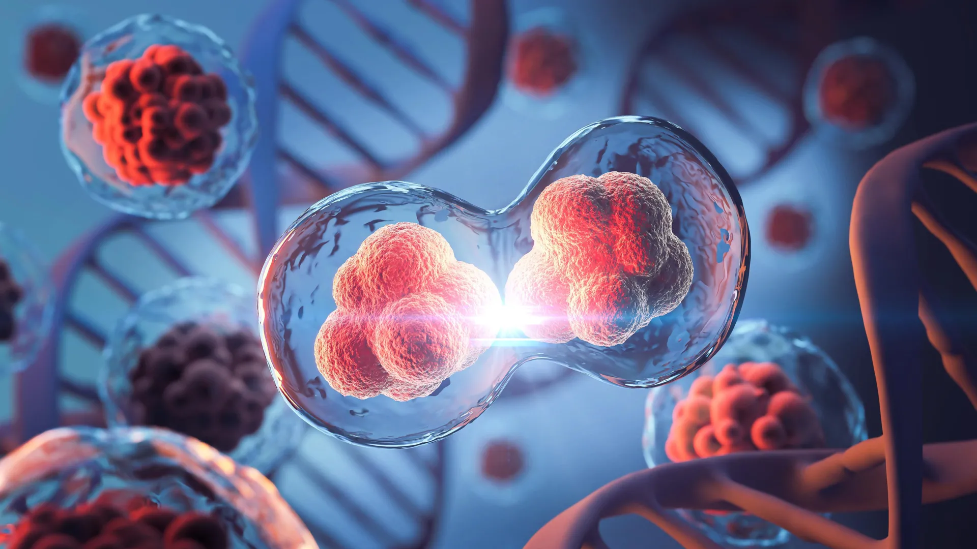
    Before a cell can split into two, it must first copy all of its chromosomes so each new cell inherits a complete set of genetic material. For years, researchers believed that as this process unfolded, the genome’s intricate three-dimensional shape temporarily vanished.

    After division, scientists thought, the DNA would slowly rebuild its complex, folded structure, which is key to regulating which genes are active in a given cell.

    Now, new research from MIT reveals that this long-accepted model isn’t quite right. Using a powerful, high-resolution genome mapping method, the team found that small 3D loops — connections between regulatory DNA elements and genes — remain intact even during cell division, a stage known as mitosis.

    “This study really helps to clarify how we should think about mitosis. In the past, mitosis was thought of as a blank slate, with no transcription and no structure related to gene activity. And we now know that that’s not quite the case,” says Anders Sejr Hansen, an associate professor of biological engineering at MIT. “What we see is that there’s always structure. It never goes away.”

    DNA Loops That Help Cells “Remember”

    The researchers also discovered that these DNA loops actually strengthen as chromosomes condense in preparation for division. This tightening brings distant regulatory elements closer together, encouraging them to stick. According to the team, this could allow cells to “remember” which genetic interactions existed before division and reestablish them afterward.

    “The findings help to bridge the structure of the genome to its function in managing how genes are turned on and off, which has been an outstanding challenge in the field for decades,” says Viraat Goel PhD ’25, the lead author of the study.

    Hansen and Edward Banigan, a research scientist at MIT’s Institute for Medical Engineering and Science, are senior authors of the study, published in Nature Structural and Molecular Biology. Co-authors include MIT professors Leonid Mirny and Gerd Blobel of the University of Pennsylvania’s Perelman School of Medicine.

    Mapping the Hidden Architecture of DNA

    Over the last two decades, scientists have uncovered that DNA in the cell nucleus organizes itself into 3D loops. Many of these loops allow genes to interact with distant regulatory regions — sometimes millions of base pairs apart — while others form during mitosis to pack chromosomes tightly.

    Most of this mapping has relied on a tool called Hi-C, a technique co-developed by MIT researchers and Job Dekker at the University of Massachusetts Chan Medical School. Hi-C works by cutting DNA into small fragments and linking together those that are near each other in the cell nucleus before sequencing them to identify which regions interact.

    While effective, Hi-C lacks the resolution needed to detect fine-scale interactions between genes and regulatory sequences known as enhancers. Enhancers are short DNA segments that activate genes by binding to promoters, the regions where transcription begins.

    A Breakthrough Tool: Region-Capture Micro-C

    In 2023, Hansen and colleagues developed a next-generation technique that can map genome structures with up to 1,000 times greater precision. The method, called Region-Capture Micro-C (RC-MC), uses a different enzyme to cut DNA into evenly sized fragments and focuses on a smaller portion of the genome. This allows researchers to create highly detailed 3D maps of targeted DNA regions.

    Using RC-MC, the team identified a new structural feature they named “microcompartments.” These are tiny, densely connected loops that form when nearby enhancers and promoters attach to one another.

    Earlier work had shown that microcompartments formed by a different mechanism than larger 3D genome structures, but the team didn’t yet understand how. To explore this, they decided to examine what happens to these structures as cells go through mitosis. During this phase, chromosomes compact dramatically to ensure they can be duplicated and evenly distributed between daughter cells. As this occurs, large genome domains known as A/B compartments and topologically associating domains (TADs) typically disappear.

    Unexpected Stability During Cell Division

    The researchers expected microcompartments to vanish as well. To test this, they monitored cells throughout the entire division cycle to see how these loops behaved before and after mitosis.

    “During mitosis, it has been thought that almost all gene transcription is shut off. And before our paper, it was also thought that all 3D structure related to gene regulation was lost and replaced by compaction. It’s a complete reset every cell cycle,” Hansen says.

    To their surprise, the loops didn’t disappear. In fact, they became even more pronounced as cells divided.

    “We went into this study thinking, well, the one thing we know for sure is that there’s no regulatory structure in mitosis, and then we accidentally found structure in mitosis,” Hansen says.

    Using their technique, the researchers also confirmed that larger structures such as A/B compartments and TADs do disappear during mitosis, as had been seen before.

    “This study leverages the unprecedented genomic resolution of the RC-MC assay to reveal new and surprising aspects of mitotic chromatin organization, which we have overlooked in the past using traditional 3C-based assays. The authors reveal that, contrary to the well-described dramatic loss of TADs and compartmentalization during mitosis, fine-scale “microcompartments” — nested interactions between active regulatory elements — are maintained or even transiently strengthened,” says Effie Apostolou, an associate professor of molecular biology in medicine at Weill Cornell Medicine, who was not involved in the study.

    Explaining a Mysterious Burst of Gene Activity

    The discovery may also explain a long-observed burst in gene transcription that occurs near the end of mitosis. Since the 1960s, scientists believed transcription stopped entirely during cell division. However, studies in 2016 and 2017 revealed a brief spike of gene activity before it shuts down again.

    In their new study, the MIT team found that during mitosis, microcompartments are more likely to be found near the genes that spike during cell division. They also discovered that these loops appear to form as a result of the genome compaction that occurs during mitosis. This compaction brings enhancers and promoters closer together, allowing them to stick together to form microcompartments.

    Once formed, the loops that constitute microcompartments may activate gene transcription somewhat by accident, which is then shut off by the cell. When the cell finishes dividing, entering a state known as G1, many of these small loops become weaker or disappear.

    “It almost seems like this transcriptional spiking in mitosis is an undesirable accident that arises from generating a uniquely favorable environment for microcompartments to form during mitosis,” Hansen says. “Then, the cell quickly prunes and filters many of those loops out when it enters G1.”

    Because chromosome compaction can also be influenced by a cell’s size and shape, the researchers are now exploring how variations in those features affect the structure of the genome and in turn, gene regulation.

    “We are thinking about some natural biological settings where cells change shape and size, and whether we can perhaps explain some 3D genome changes that previously lack an explanation,” Hansen says. “Another key question is how does the cell then pick what are the microcompartments to keep and what are the microcompartments to remove when you enter G1, to ensure fidelity of gene expression?”

    The research was funded in part by the National Institutes of Health, a National Science Foundation CAREER Award, the Gene Regulation Observatory of the Broad Institute, a Pew-Steward Scholar Award for Cancer Research, the Mathers Foundation, the MIT Westaway Fund, the Bridge Project of the Koch Institute and Dana-Farber/Harvard Cancer Center, and the Koch Institute Support (core) Grant from the National Cancer Institute.
  • Scientists just found a hidden factor behind Earth’s methane surge

    Scientists just found a hidden factor behind Earth’s methane surge

    Roughly two-thirds of all emissions of atmospheric methane — a highly potent greenhouse gas that is warming planet Earth — come from microbes that live in oxygen-free environments like wetlands, rice fields, landfills and the guts of cows.

    Tracking atmospheric methane to its specific sources and quantifying their importance remains a challenge, however. Scientists are pretty good at tracing the sources of the main greenhouse gas, carbon dioxide, to focus on mitigating these emissions. But to trace methane’s origins, scientists often have to measure the isotopic composition of methane’s component atoms, carbon and hydrogen, to use as a fingerprint of various environmental sources.

    A new paper by researchers at the University of California, Berkeley, reveals how the activity of one of the main microbial enzymes involved in producing methane affects this isotope composition. The finding could change how scientists calculate the contributions of different environmental sources to Earth’s total methane budget.

    “When we integrate all the sources and sinks of carbon dioxide into the atmosphere, we kind of get the number that we’re expecting from direct measurement in the atmosphere. But for methane, large uncertainties in fluxes exist — within tens of percents for some of the fluxes — that challenge our ability to precisely quantify the relative importance and changes in time of the sources,” said UC Berkeley postdoctoral fellow Jonathan Gropp, who is first author of the paper. “To quantify the actual sources of methane, you need to really understand the isotopic processes that are used to constrain these fluxes.”

    Gropp teamed up with a molecular biologist and a geochemist at UC Berkeley to, for the first time, employ CRISPR to manipulate the activity of this key enzyme to reveal how these methanogens interact with their food supply to produce methane.

    “It is well understood that methane levels are rising, but there is a lot of disagreement on the underlying cause,” said co-author Dipti Nayak, UC Berkeley assistant professor of molecular and cell biology. “This study is the first time the disciplines of molecular biology and isotope biogeochemistry have been fused to provide better constraints on how the biology of methanogens controls the isotopic composition of methane.”

    Many elements have heavier or lighter versions, called isotopes, that are found in small proportions in nature. Humans are about 99% carbon-12 and 1% carbon-13, which is slightly heavier because it has an extra neutron in its nucleus. The hydrogen in water is 99.985% hydrogen-1 and 0.015% deuterium or hydrogen-2, which is twice as heavy because it has a neutron in its nucleus.

    The natural abundances of isotopes are reflected in all biologically produced molecules and variations can be used to study and fingerprint various biological metabolisms.

    “Over the last 70 years, people have shown that methane produced by different organisms and other processes can have distinctive isotopic fingerprints,” said geochemist and co-author Daniel Stolper, UC Berkeley associate professor of earth and planetary science. “Natural gas from oil deposits often looks one way. Methane made by the methanogens within cow guts looks another way. Methane made in deep sea sediments by microorganisms has a different fingerprint. Methanogens can consume or ‘eat’, if you will, a variety of compounds including methanol, acetate or hydrogen; make methane; and generate energy from the process. Scientists have commonly assumed that the isotopic fingerprint depends on what the organisms are eating, which often varies from environment to environment, creating our ability to link isotopes to methane origins.”

    “I think what’s unique about the paper is, we learned that the isotopic composition of microbial methane isn’t just based on what methanogens eat,” Nayak said. “What you ‘eat’ matters, of course, but the amount of these substrates and the environmental conditions matter too, and perhaps more importantly, how microbes react to those changes.”

    “Microbes respond to the environment by manipulating their gene expression, and then the isotopic compositions change as well,” Gropp said. “This should cause us to think more carefully when we analyze data from the environment.”

    The paper will appear Aug. 14 in the journal Science.

    Vinegar- and alcohol-eating microbes

    Methanogens — microorganisms that are archaea, which are on an entirely separate branch of the tree of life from bacteria — are essential to ridding the world of dead and decaying matter. They ingest simple molecules — molecular hydrogen, acetate or methanol, for example — excreted by other organisms and produce methane gas as waste. This natural methane can be observed in the pale Will-o’-the-wisps seen around swamps and marshes at night, but it’s also released invisibly in cow burps, bubbles up from rice paddies and natural wetlands and leaks out of landfills. While most of the methane in the natural gas we burn formed in association with hydrocarbon generation, some deposits were originally produced by methanogens eating buried organic matter.

    The isotopic fingerprint of methane produced by methanogens growing on different “food” sources has been well established in laboratory studies, but scientists have found that in the complexity of the real world, methanogens don’t always produce methane with the same isotopic fingerprint as seen in the lab. For example, when grown in the lab, species of methanogens that eat acetate (essentially vinegar), methanol (the simplest alcohol), or molecular hydrogen (H2) produce methane, CH4, with a ratio of hydrogen and carbon isotopes different from the ratios observed in the environment.

    Gropp had earlier created a computer model of the metabolic network in methanogens to understand better how the isotope composition of methane is determined. When he got a fellowship to come to UC Berkeley, Stolper and Nayak proposed that he experimentally test his model. Stolper’s laboratory specializes in measuring isotope compositions to explore Earth’s history. Nayak studies methanogens and, as a postdoctoral fellow, found a way to use CRISPR gene editing in methanogens. Her group recently altered the expression of the key enzyme in methanogens that produces the methane — methyl-coenzyme M reductase (MCR) — so that its activity can be dialed down. Enzymes are proteins that catalyze chemical reactions.

    Experimenting with these CRISPR-edited microbes — in a common methanogen called Methanosarcina acetivorans growing on acetate and methanol — the researchers looked at how the isotopic composition of methane changed when the enzyme activity was reduced, mimicking what is thought to happen when the microbes are starved for their preferred food.

    They found that when MCR is at low concentrations, cells respond by altering the activity of many other enzymes in the cell, causing their inputs and outputs to accumulate and the rate of methane generation to slow so much that enzymes begin running both backwards and forwards. In reverse, these other enzymes remove a hydrogen from carbon atoms; running forward, they add a hydrogen. Together with MCR, they ultimately produce methane (CH4). Each forward and reverse cycle requires one of these enzymes to pull a hydrogen off of the carbon and add a new one ultimately sourced from water. As a result, the isotopic composition of methane’s four hydrogen molecules gradually comes to reflect that of the water, and not just their food source, which starts with three hydrogens.

    This is different from typical assumptions for growth on acetate and methanol that assume no exchange between hydrogen derived from water and that from the food source.

    “This isotope exchange we found changes the fingerprint of methane generated by acetate and methanol consuming methanogens vs. that typically assumed. Given this, it might be that we have underestimated the contribution of the acetate-consuming microbes, and they might be even more dominant than we have thought,” Gropp said. “We’re proposing that we at least should consider the cellular response of methanogens to their environment when studying isotopic composition of methane.”

    Beyond this study, the CRISPR technique for tuning production of enzymes in methanogens could be used to manipulate and study isotope effects in other enzyme networks broadly, which could help researchers answer questions about geobiology and the Earth’s environment today and in the past.

    “This opens up a pathway where modern molecular biology is married with isotope-geochemistry to answer environmental problems,” Stolper said. “There are an enormous number of isotopic systems associated with biology and biochemistry that are studied in the environment; I hope we can start looking at them in the way molecular biologists now are looking at these problems in people and other organisms — by controlling gene expression and looking at how the stable isotopes respond.”

    For Nayak, the experiments are also a big step in discovering how to alter methanogens to derail production of methane and redirect their energy to producing useful products instead of an environmentally destructive gas.

    “By reducing the amount of this enzyme that makes methane and by putting in alternate pathways that the cell can use, we can essentially give them another release valve, if you will, to put those electrons, which they were otherwise putting in carbon to make methane, into something else that would be more useful,” she said.

    Other co-authors of the paper are Markus Bill of Lawrence Berkeley National Laboratory and former UC Berkeley postdoc Rebekah Stein, and Max Lloyd, who is a professor at Penn State University. Gropp was supported by a fellowship from the European Molecular Biology Organization. Nayak and Stolper were funded, in part, by Alfred B. Sloan Research Fellowships. Nayak also is an investigator with the Chan-Zuckerberg Biohub.

  • New “evolution engine” creates super-proteins 100,000x faster

    New “evolution engine” creates super-proteins 100,000x faster

    In medicine and biotechnology, the ability to evolve proteins with new or improved functions is crucial, but current methods are often slow and laborious. Now, Scripps Research scientists have developed a synthetic biology platform that accelerates evolution itself — enabling researchers to evolve proteins with useful, new properties thousands of times faster than nature. The system, named T7-ORACLE, was described in Science on August 7, 2025, and represents a breakthrough in how researchers can engineer therapeutic proteins for cancer, neurodegeneration and essentially any other disease area.

    “This is like giving evolution a fast-forward button,” says co-senior author Pete Schultz, the President and CEO of Scripps Research, where he also holds the L.S. “Sam” Skaggs Presidential Chair. “You can now evolve proteins continuously and precisely inside cells without damaging the cell’s genome or requiring labor-intensive steps.”

    Directed evolution is a laboratory process that involves introducing mutations and selecting variants with improved function over multiple cycles. It’s used to tailor proteins with desired properties, such as highly selective, high-affinity antibodies, enzymes with new specificities or catalytic properties, or to investigate the emergence of resistance mutations in drug targets. However, traditional methods often require repeated rounds of DNA manipulation and testing with each round taking a week or more. Systems for continuous evolution — where proteins evolve inside living cells without manual intervention — aim to streamline this process by enabling simultaneous mutation and selection with each round of cell division (roughly 20 minutes for bacteria). But existing approaches have been limited by technical complexity or modest mutation rates.

    T7-ORACLE circumvents these bottlenecks by engineering E. coli bacteria — a standard model organism in molecular biology — to host a second, artificial DNA replication system derived from bacteriophage T7, a virus that infects bacteria and has been widely studied for its simple, efficient replication system. T7-ORACLE enables continuous hypermutation and accelerated evolution of biomacromolecules, and is designed to be broadly applicable to many protein targets and biological challenges. Conceptually, T7-ORACLE builds on and extends efforts on existing orthogonal replication systems — meaning they operate separately from the cell’s own machinery — such as OrthoRep in Saccharomyces cerevisiae (baker’s yeast) and EcORep in E. coli. In comparison to these systems, T7-ORACLE benefits from the combination of high mutagenesis, fast growth, high transformation efficiency, and the ease with which both the E. coli host and the circular replicon plasmid can be integrated into standard molecular biology workflows.

    The T-7 ORACLE orthogonal system targets only plasmid DNA (small, circular pieces of genetic material), leaving the cell’s host genome untouched. By engineering T7 DNA polymerase (a viral enzyme that replicates DNA) to be error-prone, the researchers introduced mutations into target genes at a rate 100,000 times higher than normal without damaging the host cells.

    “This system represents a major advance in continuous evolution,” says co-senior author Christian Diercks, an assistant professor of chemistry at Scripps Research. “Instead of one round of evolution per week, you get a round each time the cell divides — so it really accelerates the process.”

    To demonstrate the power of T7-ORACLE, the research team inserted a common antibiotic resistance gene, TEM-1 β-lactamase, into the system and exposed the E. coli cells to escalating doses of various antibiotics. In less than a week, the system evolved versions of the enzyme that could resist antibiotic levels up to 5,000 times higher than the original. This proof-of-concept demonstrated not only T7-ORACLE’s speed and precision, but also its real-world relevance by replicating how resistance develops in response to antibiotics.

    “The surprising part was how closely the mutations we saw matched real-world resistance mutations found in clinical settings,” notes Diercks. “In some cases, we saw new combinations that worked even better than those you would see in a clinic.”

    But Diercks emphasizes that the study isn’t focused on antibiotic resistance per se.

    “This isn’t a paper about TEM-1 β-lactamase,” he explains. “That gene was just a well-characterized benchmark to show how the system works. What matters is that we can now evolve virtually any protein, like cancer drug targets and therapeutic enzymes, in days instead of months.”

    The broader potential of T7-ORACLE lies in its adaptability as a platform for protein engineering. Although the system is built into E. coli, the bacterium serves primarily as a vessel for continuous evolution. Scientists can insert genes from humans, viruses or other sources into plasmids, which are then introduced into E. coli cells. T7-ORACLE mutates these genes, generating variant proteins that can be screened or selected for improved function. Because E. coli is easy to grow and widely used in labs, it provides a convenient, scalable system for evolving virtually any protein of interest.

    This could help scientists more rapidly evolve antibodies to target specific cancers, evolve more effective therapeutic enzymes, and design proteases that target proteins involved in cancer and neurodegenerative disease.

    “What’s exciting is that it’s not limited to one disease or one kind of protein,” says Diercks. “Because the system is customizable, you can drop in any gene and evolve it toward whatever function you need.”

    Moreover, T7-ORACLE works with standard E. coli cultures and widely used lab workflows, avoiding the complex protocols required by other continuous evolution systems.

    “The main thing that sets this apart is how easy it is to implement,” adds Diercks. “There’s no specialized equipment or expertise required. If you already work with E. coli, you can probably use this system with minimal adjustments.”

    T7-ORACLE reflects Schultz’s broader goal: to rebuild key biological processes — such as DNA replication, RNA transcription and protein translation — so they function independently of the host cell. This separation allows scientists to reprogram these processes without disrupting normal cellular activity. By decoupling fundamental processes from the genome, tools like T7-ORACLE help advance synthetic biology.

    “In the future, we’re interested in using this system to evolve polymerases that can replicate entirely unnatural nucleic acids: synthetic molecules that resemble DNA and RNA but with novel chemical properties,” says Diercks. “That would open up possibilities in synthetic genomics that we’re just beginning to explore.”

    Currently, the research team is focused on evolving human-derived enzymes for therapeutic use, and on tailoring proteases to recognize specific cancer-related protein sequences.

    “The T7-ORACLE approach merges the best of both worlds,” says Schultz. “We can now combine rational protein design with continuous evolution to discover functional molecules more efficiently than ever.”

    In addition to Diercks and Schultz, authors of the study, “An orthogonal T7 replisome for continuous hypermutation and accelerated evolution in E. coli,” are Philipp Sondermann, Cynthia Rong, Thomas G. Gillis, Yahui Ban, Celine Wang and David A. Dik of Scripps Research.

    This work was supported by funding from the National Institutes of Health (grant RGM145323A).

  • The EU should allow gene editing to make organic farming more sustainable, researchers say

    The EU should allow gene editing to make organic farming more sustainable, researchers say

    To achieve the European Green Deal’s goal of 25% organic agriculture by 2030, researchers argue that new genomic techniques (NGTs) should be allowed without pre-market authorization in organic as well as conventional food production. NGTs — also known as gene editing- — are classified under the umbrella of GMOs, but they involve more subtle genetic tweaks. In an opinion paper publishing May 30 in the Cell Press journal Cell Reports Sustainability, the researchers describe how NGTs could enable rapid development of crops that are climate resilient, produce higher yields, and require less fertilizers and pesticides.

    “This is an excellent opportunity to modernize European agriculture, to make it more science based, and to support the goal of improving sustainability inside the European Union,” says first author Alexandra Molitorisová, a food law researcher at the University of Bayreuth.

    Currently, 10% of EU farming areas are organic. Though organic farming can reduce carbon emissions and pollution from fertilizer and pesticides, Molitorisová’s team says that these benefits could be negated by biodiversity losses due to expanding agricultural land, since organic agriculture requires more land to grow the same amount of food.

    “The target of 25% organic land is unlikely to ensure sustainable food production in the EU if modern biotechnology, such as NGTs, is excluded from organic farming,” says Molitorisová.

    European institutions are currently debating how to regulate NGTs — which did not exist when the EU legislation on GMOs was adopted in 2001 — in response to a proposal from the European Commission that NGT usage be allowed in conventional but not organic farming.

    “Research suggests that NGTs are still something that European consumers are not completely aware of — they just do not distinguish between NGTs and GMOs,” says senior author Kai Purnhagen, Professor of Food Law at the University of Bayreuth. “There are strong indications that consumers would be willing to accept these technologies if they yield substantial benefits, and the Commission’s proposal for new regulation allowing NGTs in conventional farming points in this direction.”

    Though NGT crops are still developed through genetic alteration, the processes usually don’t involve the insertion of DNA from non-plant species. This means that, in theory, identical crops could be developed using conventional breeding methods, though it would take decades rather than months. For these reasons, the researchers argue that NGTs and GMOs should be defined and regulated separately, including in organic production.

    “From the consumer’s perception of naturalness, the normal breeding process is between two crossable varieties, and that is also what happens with NGTs,” says Molitorisová. “So, if consumers understand the nature and benefits of this technology, it should be easier for them to accept it compared with GMOs, which might involve inserting a gene from a non-plant organism into a plant genome.”

    The researchers also note that the most common type of NGT, targeted mutagenesis, is very similar to mutagenesis — which uses chemical or radioactive substances to induce random genetic mutations and has never been subject to GMO regulation in the EU, even for organic farms.

    “If mutagenesis had not been exempted from GMO legislation, the estimation is that 80%-90% of the cereal products on the European market would have been subject to GMO labeling,” says Purnhagen.

    The team highlights that allowing NGTs in conventional but not organic agriculture creates a formidable hurdle in terms of NGT identification, labeling, and traceability.

    “At the moment, there are unresolved practical problems with the identification of NGTs inside of food, feed, or seeds,” says Molitorisová. “One rational alternative is to allow NGTs in organic production, because if NGT organisms are not identifiable, they are also technically unavoidable.”

    Ultimately, the researchers say that the decision to allow NGTs in organic farming should be made by the organic farming and consumer communities — for example, by way of citizens’ juries or food councils.

    “Organic consumers care about the environment and sustainability. For organic farmers, accepting this technology is a way to speak to those consumers,” says Purnhagen.

  • A high-fat diet sets off metabolic dysfunction in cells, leading to weight gain

    A high-fat diet sets off metabolic dysfunction in cells, leading to weight gain

    Consuming a high-fat diet can lead to a variety of health problems — not only weight gain but also an increased risk of diabetes and other chronic diseases.

    At the cellular level, hundreds of changes take place in response to a high-fat diet. MIT researchers have now mapped out some of those changes, with a focus on metabolic enzyme dysregulation that is associated with weight gain.

    Their study, conducted in mice, revealed that hundreds of enzymes involved in sugar, lipid, and protein metabolism are affected by a high-fat diet, and that these disruptions lead to an increase in insulin resistance and an accumulation of damaging molecules called reactive oxygen species. These effects were more pronounced in males than females.

    The researchers also showed that most of the damage could be reversed by giving the mice an antioxidant along with their high-fat diet.

    “Under metabolic stress conditions, enzymes can be affected to produce a more harmful state than what was initially there,” says Tigist Tamir, a former MIT postdoc. “Then what we’ve shown with the antioxidant study is that you can bring them to a different state that is less dysfunctional.”

    Tamir, who is now an assistant professor of biochemistry and biophysics at the University of North Carolina at Chapel Hill School of Medicine, is the lead author of the new study, which appears today in Molecular Cell. Forest White, the Ned C. and Janet C. Rice Professor of Biological Engineering and a member of the Koch Institute for Integrative Cancer Research at MIT, is the senior author of the paper.

    Metabolic networks

    In previous work, White’s lab has found that a high-fat diet stimulates cells to turn on many of the same signaling pathways that are linked to chronic stress. In the new study, the researchers wanted to explore the role of enzyme phosphorylation in those responses.

    Phosphorylation, or the addition of a phosphate group, can turn enzyme activity on or off. This process, which is controlled by enzymes called kinases, gives cells a way to quickly respond to environmental conditions by fine-tuning the activity of existing enzymes within the cell.

    Many enzymes involved in metabolism — the conversion of food into the building blocks of key molecules such as proteins, lipids, and nucleic acids — are known to undergo phosphorylation.

    The researchers began by analyzing databases of human enzymes that can be phosphorylated, focusing on enzymes involved in metabolism. They found that many of the metabolic enzymes that undergo phosphorylation belong to a class called oxidoreductases, which transfer electrons from one molecule to another. Such enzymes are key to metabolic reactions such as glycolysis — the breakdown of glucose into a smaller molecule known as pyruvate.

    Among the hundreds of enzymes the researchers identified are IDH1, which is involved in breaking down sugar to generate energy, and AKR1C1, which is required for metabolizing fatty acids. The researchers also found that many phosphorylated enzymes are important for the management of reactive oxygen species, which are necessary for many cell functions but can be harmful if too many of them accumulate in a cell.

    Phosphorylation of these enzymes can lead them to become either more or less active, as they work together to respond to the intake of food. Most of the metabolic enzymes identified in this study are phosphorylated on sites found in regions of the enzyme that are important for binding to the molecules that they act upon or for forming dimers — pairs of proteins that join together to form a functional enzyme.

    “Tigist’s work has really shown categorically the importance of phosphorylation in controlling the flux through metabolic networks. It’s fundamental knowledge that emerges from this systemic study that she’s done, and it’s something that is not classically captured in the biochemistry textbooks,” White says.

    Out of balance

    To explore these effects in an animal model, the researchers compared two groups of mice, one that received a high-fat diet and one that consumed a normal diet. They found that overall, phosphorylation of metabolic enzymes led to a dysfunctional state in which cells were in redox imbalance, meaning that their cells were producing more reactive oxygen species than they could neutralize. These mice also became overweight and developed insulin resistance.

    “In the context of continued high fat diet, what we see is a gradual drift away from redox homeostasis towards a more disease-like setting,” White says.

    These effects were much more pronounced in male mice than female mice. Female mice were better able to compensate for the high fat diet by activating pathways involved in processing fat and metabolizing it for other uses, the researchers found.

    “One of the things we learned is that the overall systemic effect of these phosphorylation events led to, especially in males, an increased imbalance in redox homeostasis. They were expressing a lot more stress and a lot more of the metabolic dysfunction phenotype compared to females,” Tamir says.

    The researchers also found that if they gave mice who were on a high-fat diet an antioxidant called BHA, many of these effects were reversed. These mice showed a significant decrease in weight gain and did not become prediabetic, unlike the other mice fed a high-fat diet.

    It appears that the antioxidant treatment leads cells back into a more balanced state, with fewer reactive oxygen species, the researchers say. Additionally, metabolic enzymes showed a systemic rewiring and changed state of phosphorylationin those mice.

    “They’re experiencing a lot of metabolic dysfunction, but if you co-administer something that counters that, then they have enough reserve to maintain some sort of normalcy,” Tamir says. “The study suggests that there is something biochemically happening in cells to bring them to a different state — not a normal state, just a different state in which now, at the tissue and organism levels, the mice are healthier.”

    In her new lab at the University of North Carolina, Tamir now plans to further explore whether antioxidant treatment could be an effective way to prevent or treat obesity-associated metabolic dysfunction, and what the optimal timing of such a treatment would be.

    The research was funded in part by the Burroughs Wellcome Fund, the National Cancer Institute, the National Institutes of Health, the Ludwig Center at MIT, and the MIT Center for Precision Cancer Medicine.

  • ‘Selfish’ genes called introners proven to be a major source of genetic complexity

    ‘Selfish’ genes called introners proven to be a major source of genetic complexity

    DNA is the genetic code that provides the biological instructions for every living species, but not every bit of DNA helps the species survive. Some pieces of DNA are more like parasites, along for the ride and their own survival.

    To translate DNA into proteins, the building blocks of life, many of these selfish DNA elements have to be removed from the genetic code. Doing so enables the body to produce the wide diversity of proteins that allow for complex life, but the process can also lead to health problems, like some kinds of cancer.

    University of California, Santa Cruz researchers are studying the ways that these genetic elements hide and make copies of themselves, so they can propagate within a species’ DNA, or even hop from one species to an unrelated one in a process called “horizontal gene transfer.”

    A new study in the journal Proceedings of the National Academy of Sciences proves that a type of genetic element called “introners” are the cause of many of these selfish genes spreading within and between species. It provides evidence for eight instances in which introners have transferred between unrelated species, the first proven examples of this phenomenon.

    These results help us understand how genomes evolved to become so complex, and how we might take advantage of that complexity in human health research.

    “[Introners are] a way that genome architectures and complexity arise, but not necessarily because there is natural selection that favors this complexity,” said Russ Corbett-Detig, senior author on the study and professor of biomolecular engineering at the Baskin School of Engineering. “A few may ultimately benefit the host, but most are just cheaters that found a really good way to hide in the genome.”

    Investigating introners

    Corbett-Detig and his former undergraduate student Landen Gozashti, now a postdoctoral fellow at UC Berkeley following a stellar Harvard Ph.D., have spent years studying introns, the segments of noncoding DNA that must be removed before proteins can be produced.

    They wanted to figure out why these non protein-coding bits of DNA are seen in varying amounts across all animals, plants, fungi, and protists, and how they have managed to so successfully replicate themselves and survive. It’s long been a mystery of how all these introns first came to exist within DNA, as most don’t seem to serve an evolutionary function.

    Scientists are interested in this as a way to further understand genome evolution, but also because introns allow for a crucial process called “alternative splicing.” Introns must be spliced out of the DNA sequence to create proteins, but this process can have variation and error, which means that different versions of a protein can be created from the same gene. Ultimately this means that an organism can be more complex, but it also introduces the risk of health problems if splicing breaks a gene. Many researchers, including those at the UC Santa Cruz Genomics Institute, are studying how alternative splicing can be studied to better understand genetic disease. This research enhances the basic science of that health-related work.

    In this study, the researchers have proven that introners are one of the main ways that new introns appear with a species’ DNA. Introners are a kind of transposable element, a “jumping gene” that can move from part of a genome to another, that have found a way to successfully make copies of introns throughout a genome. The team’s past work has suggested this, but their advanced methods of searching the DNA of a wide range of species has now allowed them to definitively confirm their hypothesis.

    The researchers searched for introners in the DNA of thousands of species, something only recently made possible due to ongoing coordinated efforts to sequence a wide range of biodiversity and make the data publicly available, like the Earth BioGenome Project and the Sanger Tree of Life.

    They found evidence for 1,093 families of introners among the 8,716 genomes they analyzed, suggesting that there are many kinds of introners out there capable of spreading introns through the genomes of various species.

    “Because transposons are insanely diverse and present in basically every eukaryote, it implies that this really can be a very general way that new introns arise in different lineages,” Corbett-Detig said.

    These introners most commonly appeared in species of algae, fungi, and diverse single-celled eukaryotes, with an example found in a sea urchin and a tunicate, a tubular marine invertebrate.

    Transfer between species

    Among the many genomes they analyzed, the researchers found the first direct evidence for horizontal gene transfer of introners. They found eight examples of an introner hopping out of the genome of one species and settling into the genome of another unrelated species that mating could not explain.

    In one case, the researchers found horizontal gene transfer between two species so unrelated that their last common ancestor was 1.6 billion years ago. In looking at the genomes of the two species — a sea sponge and a marine protist called a dinoflagellate — they found evidence that around 40 million years ago, an introner jumped from one of these species to the other.

    The researchers hypothesize that the introners could be hitching a ride on giant viruses in order to transfer between species.

    “That virus itself is a selfish element as well, so this is like a selfish element shuttling around on another selfish element,” Corbett-Detig said.

    Although eight examples of horizontal gene transfer may not seem like many, the researchers believe there would be many more if they kept looking within the 8.74 million species of eukaryotes that exist.

    “Given how little of eukaryotic diversity we’ve sampled, I promise you that if we sampled the rest of them, we’d find many more,” Corbett-Detig said.

  • ‘Fast-fail’ AI blood test could steer patients with pancreatic cancer away from ineffective therapies

    ‘Fast-fail’ AI blood test could steer patients with pancreatic cancer away from ineffective therapies

    An artificial intelligence technique for detecting DNA fragments shed by tumors and circulating in a patient’s blood, developed by Johns Hopkins Kimmel Cancer Center investigators, could help clinicians more quickly identify and determine if pancreatic cancer therapies are working.

    After testing the method, called ARTEMIS-DELFI, in blood samples from patients participating in two large clinical trials of pancreatic cancer treatments, researchers found that it could be used to identify therapeutic responses. ARTEMIS-DELFI and another method developed by investigators, called WGMAF, to study mutations were found to be better predictors of outcome than imaging or other existing clinical and molecular markers two months after treatment initiation. However, ARTEMIS-DELFI was determined to be the superior test as it was simpler and potentially more broadly applicable.

    A description of the work was published May 21 in Science Advances. It was partly supported by grants from the National Institutes of Health.

    Time is of the essence when treating patients with pancreatic cancer, explains senior study author Victor E. Velculescu, M.D., Ph.D., co-director of the cancer genetics and epigenetics program at the cancer center. Many patients with pancreatic cancer receive a diagnosis at a late stage, when cancer may progress rapidly.

    “Providing patients with more potential treatment options is especially vital as a growing number of experimental therapies for pancreatic cancer have become available,” Velculescu says. “We want to know as quickly as we can if the therapy is helping the patient or not. If it is not working, we want to be able to switch to another therapy.”

    Currently, clinicians use imaging tools to monitor cancer treatment response and tumor progression. However, these tools produce results that may not be timely and are less accurate for patients receiving immunotherapies, which can make the results more complicated to interpret. In the study, Velculescu and his colleagues tested two alternate approaches to monitoring treatment response in patients participating in the phase 2 CheckPAC trial of immunotherapy for pancreatic cancer.

    One approach, called WGMAF (tumor-informed plasma whole-genome sequencing), analyzed DNA from tumor biopsies as well as cell-free DNA in blood samples to detect a treatment response. The other, called ARTEMIS-DELFI (tumor-independent genome-wide cfDNA fragmentation profiles and repeat landscapes), used machine learning, a form of artificial intelligence, to scan millions of cell-free DNA fragments only in the patient’s blood samples. Both approaches were able to detect which patients were benefiting from the therapies. However, not all patients had tumor samples, and many patients’ tumor samples had only a small fraction of cancer cells compared to the overall tissue, which also contained normal pancreatic and other cells, thereby confounding the WGMAF test.

    The ARTEMIS-DELFI approach worked with more patients and was simpler logistically, Velculescu says. The team then validated that ARTEMIS-DELFI was an effective treatment response monitoring tool in a second clinical trial called the PACTO trial. The study confirmed that ARTEMIS-DELFI could identify which patients were responding as soon as four weeks after therapy started.

    “The ‘fast-fail’ ARTEMIS-DELFI approach may be particularly useful in pancreatic cancer where changing therapies quickly could be helpful in patients who do not respond to the initial therapy,” says lead study author Carolyn Hruban, who was a graduate student at Johns Hopkins during the study and is now a postdoctoral researcher at the Dana-Farber Cancer Institute. “It’s simpler, likely less expensive, and more broadly applicable than using tumor samples.”

    The next step for the team will be prospective studies that test whether the information provided by ARTEMIS-DELFI helps clinicians more efficiently find an effective therapy and improve patient outcomes. A similar approach could also be used to monitor other cancers. Earlier this year, members of the team published a study in Nature Communications showing that a variation of the cell-free fragmentation monitoring approach called DELFI-TF was helpful in assessing colon cancer therapy response.

    “Our cell-free DNA fragmentation analyses provide a real-time assessment of a patient’s therapy response that can be used to personalize care and improve patient outcomes,” Velculescu says.

    Other co-authors include Daniel C. Bruhm, Shashikant Koul, Akshaya V. Annapragada, Nicholas A. Vulpescu, Sarah Short, Kavya Boyapati, Alessandro Leal, Stephen Cristiano, Vilmos Adleff, Robert B. Scharpf, Zachariah H. Foda, and Jillian Phallen of Johns Hopkins; Inna M. Chen, Susann Theile, and Julia S. Johannsen of Copenhagen University Hospital Herlev and Gentofte, and the University of Copenhagen; and Bahar Alipanahi and Zachary L. Skidmore of Delfi Diagnostics.

    The study was supported by the Dr. Miriam and Sheldon G. Adelson Medical Research Foundation, SU2C Lung Cancer Interception Dream Team Grant; Stand Up to Cancer-Dutch Cancer Society International Translational Cancer Research Dream Team Grant, the Gray Foundation, the Honorable Tina Brozman Foundation, the Commonwealth Foundation, the Cole Foundation, a research grant from Delfi Diagnostics and National Institutes of Health grants CA121113,1T32GM136577, CA006973, CA233259, CA062924 and CA271896.

    Annapragada, Scharpf, and Velculescu are inventors on a patent submitted by Johns Hopkins University for genome-wide repeat and cell-free DNA in cancer (US patent application number 63/532,642). Annapragada, Bruhm, Adleff, Foda, Phallen and Scharpf are inventors on patent applications submitted by the university on related technology and licensed to Delfi Diagnostics. Phallen, Adleff, and Scharpf are founders of Delfi Diagnostics. Adleff and Scharpf are consultants for the company and Skidmore and Alipanahi are employees of the company. Velculescu is a founder of Delfi Diagnostics, member of its Board of Directors, and owns stock in the company. Johns Hopkins University owns equity in the company as well. Velculescu is an inventor on patent applications submitted by The Johns Hopkins University related to cancer genomic analyses and cell-free DNA that have been licensed to one or more entities, including Delfi Diagnostics, LabCorp, Qiagen, Sysmex, Agios, Genzyme, Esoterix, Ventana and ManaT Bio that result in royalties to the inventors and the University. These relationships are managed by Johns Hopkins in accordance with its conflict-of-interest policies.

  • Researchers find CRISPR is capable of even more than we thought

    Researchers find CRISPR is capable of even more than we thought

    Every living creature on Earth needs to protect itself from things that would do it harm. Bacteria are no different. And despite their relative simplicity, they deploy remarkably savvy defensive strategies against viral invaders. The most well-known is CRISPR-Cas9, adapted for human use as the first FDA-approved genetic editing technique.

    In the past year, researchers at Rockefeller’s Laboratory of Bacteriology, headed by Luciano Marraffini, and at the MSKCC’s Structural Biology Laboratory, headed by Dinshaw Patel, have been studying key immune components of some CRISPR systems called CARF effectors. These newly discovered weapons take different approaches to achieving the same goal: arresting cellular activity, which prevents a virus from spreading through the rest of the bacterial population.

    In a recent publication in Science, the scientists announce the newest CARF effector they’ve discovered, which they coined Cat1. Thanks to an unusually complex molecular structure, this protein can deplete a metabolite essential for cellular function. Left without fuel, the viral invader’s plans for a further onslaught are brought to a grinding halt.

    “The collective work of our labs is revealing just how effective — and different — these CARF effectors are,” says Marraffini. “The range of their molecular activities is quite amazing.”

    Multiple defense systems

    CRISPR is a mechanism in the adaptive immune systems of bacteria and other certain single-cell organisms that offers protection against viruses, called phages. The six types of CRISPR systems work roughly the same way: A CRISPR RNA identifies foreign genetic code, which triggers a cas enzyme to mediate an immune response, often snipping off the invader material.

    But an increasing body of evidence indicates that CRISPR systems deploy a wide variety of defensive strategies beyond genetic scissors. Marraffini’s lab has led the way on much of this research. In particular, they have been studying a class of molecules in CRISPR-Cas10 systems called CARF effectors, which are proteins that are activated upon phage infection of a bacterium.

    CARF effector immunity is believed to work by creating an inhospitable environment for viral replication. For example, the Cam1 CARF effector causes membrane depolarization of an infected cell, while Cad1 triggers a sort of molecular fumigation, flooding an infected cell with toxic molecules.

    Metabolic freeze

    For the current study, the researchers wanted to try to identify additional CARF effectors. They used Foldseek, a powerful structural homology search tool, to find Cat1.

    They found that Cat1 is alerted to the presence of a virus by the binding of secondary messenger molecules called cyclic tetra-adenylate, or cA4, which stimulate the enzyme to cleave an essential metabolite in the cell called NAD+.

    “Once a sufficient amount of NAD+ is cleaved, the cell enters a growth-arrest state,” says co-first author Christian Baca, a TPCB graduate student in the Marraffini lab. “With cellular function on pause, the phage can no longer propagate and spread to the rest of the bacterial population. In this way, Cat1 is similar to Cam1 and Cad1 in that they all provide population-level bacterial immunity.”

    Unique complexity

    But while its immune strategy may be similar to these other CARF effectors, its form is not, as co-first author Puja Majumder, a postdoctoral research scholar in the Patel Lab, revealed through detailed structural analysis using cryo-EM.

    She found that the Cat1 protein has a surprisingly complex structure in which Cat1 dimers are glued by cA4 signal molecule, forming long filaments upon viral infection, and trap the NAD+ metabolites within sticky molecular pockets. “Once the NAD+ metabolite is cleaved by Cat1 filaments, it’s not available for the cell to use,” Majumder explains.

    But the protein’s singular structural complexity doesn’t stop there, she adds. “The filaments interact with each other to form trigonal spiral bundles, and these bundles can then expand to form pentagonal spiral bundles,” she says. The purpose of these structural components remains to be investigated.

    Also unusual is the fact Cat1 often seems to work alone. “Normally in type III CRISPR systems, you have two activities that contribute to the immunity effect,” Baca says. “However, most of the bacteria that encode Cat1 seem to primarily rely on Cat1 for their immunity effect.”

    Marraffini says these findings pose intriguing new questions. “While I think we’ve proven the big picture — that CARF effectors are great at preventing phage replication — we still have a lot to learn about the details of how they do it. It will be fascinating to see where this work leads us next.”

  • Artificial intelligence and genetics can help farmers grow corn with less fertilizer

    Artificial intelligence and genetics can help farmers grow corn with less fertilizer

    New York University scientists are using artificial intelligence to determine which genes collectively govern nitrogen use efficiency in plants such as corn, with the goal of helping farmers improve their crop yields and minimize the cost of nitrogen fertilizers.

    “By identifying genes-of-importance to nitrogen utilization, we can select for or even modify certain genes to enhance nitrogen use efficiency in major US crops like corn,” said Gloria Coruzzi, the Carroll & Milton Petrie Professor in NYU’s Department of Biology and Center for Genomics and Systems Biology and the senior author of the study, which appears in the journal The Plant Cell.

    In the last 50 years, farmers have been able to grow larger crop yields thanks to major improvements in plant breeding and fertilizers, including how efficiently crops uptake and use nitrogen, the key component of fertilizers.

    Still, most crops only use roughly 55 percent of the nitrogen in fertilizer that farmers apply to their fields, while the remainder ends up in the surrounding soil. When nitrogen seeps into groundwater, it can contaminate drinking water and cause harmful algae blooms in lakes, rivers, reservoirs, and warm ocean waters. Furthermore, the unused nitrogen that remains in the soil is converted by bacteria into nitrous oxide, a potent greenhouse gas that is 265 times more effective at trapping heat over a 100-year period than is carbon dioxide.

    The United States is the world’s leading producer of corn. This major cash crop requires large amounts of nitrogen to grow, but much of the fertilizer fed to corn is not taken up or used. Corn’s low nitrogen use efficiency presents a financial challenge for farmers, given the increasing costs of fertilizer — the majority of which is imported — and also risks harming the soil, water, air, and climate.

    To address this challenge in corn and other crops, NYU researchers have developed a novel process to improve nitrogen use efficiency that integrates plant genetics with machine learning, a type of artificial intelligence that detects patterns in data — in this case, to associate genes with a trait (nitrogen use efficiency).

    Using a model-to-crop approach, NYU researchers tracked the evolutionary history of corn genes that are shared with Arabidopsis, a small flowering weed often used as a model organism in plant biology due to the ease of studying it in the lab using the power of molecular genetic approaches. In a previous study published in Nature Communications, Coruzzi’s team identified genes whose responsiveness to nitrogen was conserved between corn and Arabidopsis and validated their role in plants.

    In The Plant Cell study, their most recent on this topic, the NYU researchers built upon their work in corn and Arabidopsis to identify how nitrogen use efficiency is governed by groups of genes — also known as “regulons” — that are activated or repressed by the same transcription factor (a regulatory protein).

    “Traits like nitrogen use efficiency or photosynthesis are never controlled by one single gene. The beauty of the machine learning process is it learns sets of genes that are collectively responsible for a trait, and can also identify the transcription factor or factors that control these sets of genes,” said Coruzzi.

    The researchers first used RNA sequencing to measure how genes in corn and Arabidopsis respond to nitrogen treatment. Using these data, they trained machine learning models to identify nitrogen-responsive genes conserved across corn and Arabidopsis varieties, as well as the transcription factors that regulate the genes-of-importance to nitrogen use efficiency (NUE). For each “NUE Regulon” — the transcription factor and corresponding set of regulated NUE genes — the researchers calculated a collective machine learning score and then ranked the top performers based on how well the combined expression levels could accurately predict how efficiently nitrogen is used in field-grown varieties of corn.

    For the top-ranked NUE Regulons, the researchers used cell-based studies in both corn and Arabidopsis to validate the machine learning predictions for the set of genes in the genome that are regulated by each transcription factor. These experiments confirmed NUE Regulons for two corn transcription factors (ZmMYB34/R3) that regulate 24 genes controlling nitrogen use as well as for a closely related transcription factor in Arabidopsis (AtDIV1), which regulates 23 target genes sharing a genetic history with corn that also control nitrogen use. When fed back into the machine learning models, these model-to-crop conserved NUE Regulons significantly enhanced the ability of AI to predict nitrogen use efficiency across field-grown corn varieties.

    Identifying NUE Regulons of collective genes and related transcription factors that govern nitrogen use will enable crop scientists to breed or engineer corn that needs less fertilizer.

    “By looking at corn hybrids at the seedling stage to see if expression of the identified genes-of-importance to nitrogen use efficiency is high, rather than planting them in the field and measuring their nitrogen use, we can use molecular markers to select the hybrids at the seedling stage that are most efficient in nitrogen use, and then plant those varieties,” said Coruzzi. “This will not only result in a cost savings for farmers, but also reduce the harmful effects of nitrogen pollution of groundwaters and nitrous oxide greenhouse gas emissions.”

    New York University has filed a patent application covering the research and findings described in this paper. Additional study authors include Ji Huang, Tim Jeffers, Nathan Doner, Hung-Jui Shih, Samantha Frangos, and Manpreet Singh Katari of NYU; Chia-Yi Cheng of NYU and National Taiwan University, and Matthew Brooks of the US Department of Agriculture’s Agricultural Research Service. The research was supported by the National Science Foundation Plant Genome Research Program (IOS-1339362) and the National Institutes of Health (R01-GM121753, F32GM116347).

  • AI-designed DNA controls genes in healthy mammalian cells for first time

    AI-designed DNA controls genes in healthy mammalian cells for first time

    A study published today in the journal Cell marks the first reported instance of generative AI designing synthetic molecules that can successfully control gene expression in healthy mammalian cells. Researchers at the Centre for Genomic Regulation (CRG) created an AI tool which dreams up DNA regulatory sequences not seen before in nature. The model can be told to create synthetic fragments of DNA with custom criteria, for example: ‘switch this gene on in stem cells which will turn into red-blood-cells but not platelets.’

    The model then predicts which combination of DNA letters (A, T, C, G) are needed for the gene expression patterns required in specific types of cells. Researchers can then chemically synthesise the roughly 250-letter DNA fragments and add them to a virus for delivery into cells.

    As a proof-of-concept, the authors of the study asked the AI to design synthetic fragments which activate a gene coding for a fluorescent protein in some cells while leaving gene expression patterns unaltered. They created the fragments from scratch and dropped them into mouse blood cells, where the sequence fused with the genome at random locations. The experiments worked exactly as predicted.

    “The potential applications are vast. It’s like writing software but for biology, giving us new ways of giving instructions to a cell and guiding how they develop and behave with unprecedented accuracy,” says Dr. Robert Frömel, first author of the study who carried out the work at the Centre for Genomic Regulation (CRG) in Barcelona.

    The study could lead to new ways for gene-therapy developers to boost or dampen the activity of genes only in the cells or tissues that need adjusting. It also paves the way for new strategies to fine-tune a patient’s genes and make treatments more effective and reduce side effects.

    The work marks an important milestone in in the field of generative biology. To date, advances in the field have largely benefited protein design, helping scientists create entirely new enzymes and antibodies faster than ever before. However, many human diseases stem from faulty gene expression that is cell-type specific, for which there might never be a perfect protein drug candidate.

    Gene expression is controlled by regulatory elements like enhancers, tiny fragments of DNA which switch genes on or off. To fix faulty gene expression, researchers can comb through genomes looking for naturally-existing enhancers that happen to suit their needs, limiting themselves to the sequences evolution has produced.

    AI-generated enhancers can help engineer ultra-selective switches that nature has not yet invented. They can be designed to have exactly the on/off patterns required in specific types of cells, a level of fine-tuning which is crucial for creating therapies that avoid unintended effects in healthy cells.

    However, the development of AI models requires lots of high-quality data, which has been historically lacking for enhancers. “To create a language model for biology, you have to understand the language cells speak. We set out to decipher these grammar rules for enhancers so that we can create entirely new words and sentences,” explains Dr. Lars Velten, corresponding author of the study and researcher at the Centre for Genomic Regulation (CRG).

    The authors of the study created huge volumes of biological data to build their AI model by carrying out thousands of experiments with lab models of blood formation. They studied both enhancers and transcription factors, proteins also involved in controlling gene expression.

    Until now, scientists studying enhancers and transcription factors typically used cancer cell lines because they are easier to work with. The researchers worked with healthy cells instead because it’s more representative of human biology. Their work helped uncover subtle mechanisms that shape our immune system and blood cell production.

    Over five years, the team synthesised more than 64,000 synthetic enhancers, each carefully designed to test different arrangements and strengths of binding sites for 38 different transcription factors. It’s the largest library of synthetic enhancers ever built in blood cells to date.

    Once inserted into the cells, the team tracked exactly how active each synthetic enhancer became across seven stages of blood-cell development. They discovered that while many enhancers activate genes in one type of cell, they repress genes in another.

    Most enhancers worked like a volume dial, turning gene activity up or down. Surprisingly, certain combinations acted like on/off switches. The scientists call this “negative synergy,” meaning two factors that usually turn a gene on individually could effectively shut that gene down when they occur together.

    The data from the experiments was crucial in setting out the design principles of the machine learning model. Once the model had enough measurements of how each synthetic enhancer changed gene activity in real cells, it could predict new designs that yield on/off outcomes, even if these enhancers had never existed in nature.

    The study was designed to determine if a technology can work in practice before committing to larger-scale research. The researchers have only scratched the surface. Both humans and mice have an estimated 1,600 transcription factors regulating their genomes.

    The work was carried out by Lars Velten, Robert Frömel, Julia Rühle, Aina Bernal Martínez, Chelsea Szu-Tu and Felix Pacheco Pastor, all members of Lars Velten’s research group at the Centre for Genomic Regulation. Rosa Martinez Corral from the Barcelona Collaboratorium, a joint initiative between the CRG and EMBL-Barcelona, also took part. The research was funded by an ERC Starting Grant from the European Union and a grant of the Spanish National Agency for Research.